



CFLAR Antibody (YA3531)
(Synonyms: CASH; FLIP; MRIT; CLARP; FLAME; Casper; FLAME1; c-FLIP; FLAME-1; I-FLICE; c-FLIPL; c-FLIPR; c-FLIPS; CASP8AP1)Based on 1 Customer Validation
CFLAR Antibody (YA3531) is a Mouse-derived and non-conjugated IgG1 monoclonal antibody, targeting to CFLAR.
-
Host:
Mouse
-
Isotype:
IgG
-
Application:
WB, IHC-P, FC, ELISA
-
Reactivity :
Human, Mouse
-
Formulation:
Supplied in PBS with 0.05% sodium azide
-
Conjugation:
Non-conjugated


To place orders, for customer services and technical support, please contact: MedChemExpress USA
Tel: 609-228-6898 E-mail: [email protected] [email protected]





Applications
| Application |
WB

WB: Western Blot
|
IHC-P
IHC-P: Immunohistochemistry-Paraffin
|
FC
FC: Flow Cytometry
|
ELISA
ELISA: Enzyme Linked Immunosorbent Assay
|
|---|---|---|---|---|
| Dilution Ratio | 1:500-1:2000 | 1:200-1:1000 | 1:200-1:400 | 1:10000 |


Product Details
CFLAR Antibody (YA3531) is a Mouse-derived and non-conjugated IgG1 monoclonal antibody, targeting to CFLAR.
-
Host Mouse

-
Clonality Monoclonal

-
Species ReactivityHuman, Mouse
-
Observed Molecular WeightObserved band size: 55 kDa
Note: Due to possible protein modifications or aggregation, the molecular weight should be confirmed by actual measurement, and the predicted value is for reference only.

-
Calculated Molecular Weight Predicted band size: 55 kDa

Purified recombinant fragment of human CFLAR (AA: 100-251) expressed in E. Coli.
affinity purified.
Non-conjugated
Unmodified
IgG


Product Properties
-
Appearance
Solution
-
Formulation
Supplied in PBS with 0.05% sodium azide
-
Concentration
Batch-dependent, Please check the COA for the concentration of each lot. Check Lot Concentration
-
Storage & Stability
Stored at -20°C for 1 year. Avoid repeated freeze / thaw cycles.
-
Shipping
Shipping with blue ice.


Verification Images
-
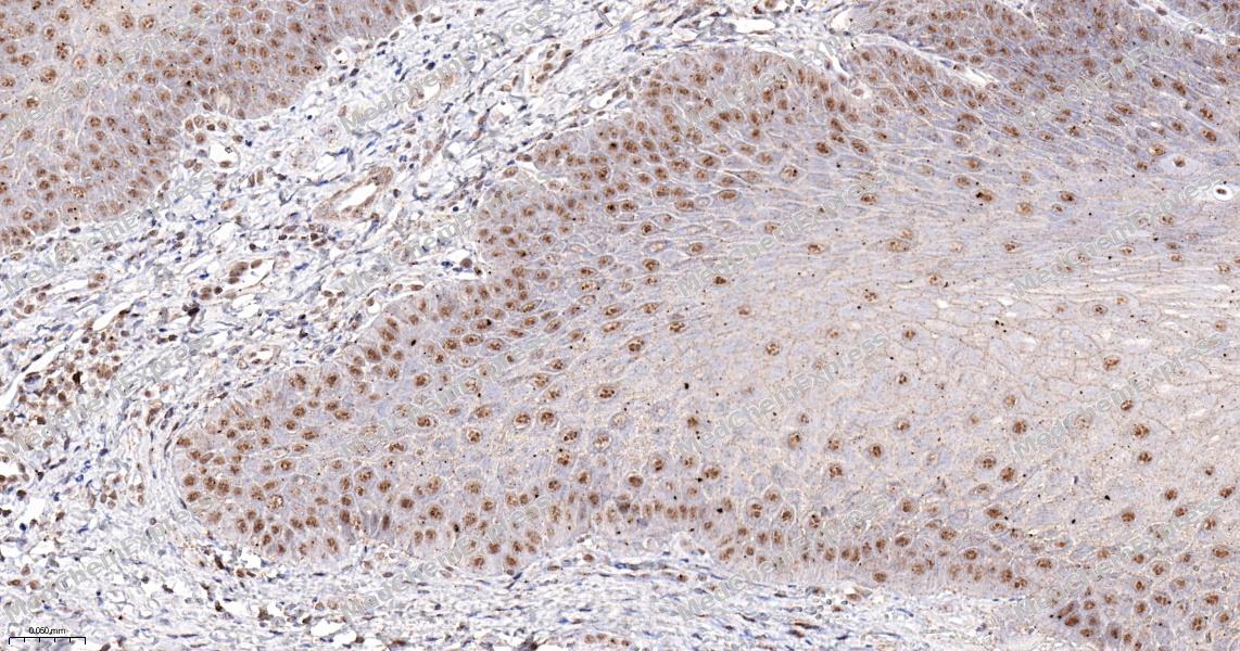 Immunohistochemical analysis of paraffin-embedded human endometrium tissue using CFLAR Antibody (HY-P83834, 1/500). The section was pretreated using heat mediated antigen retrieval with sodium citrate buffer (pH 6.0) for 8 minutes. The tissues were blocked with quick block buffer for 0.5 hours at room temperature, washed with PBS and PBST, and then incubated with the primary antibody overnight at 4℃. The detection was performed using an HRP conjugated compact polymer system. DAB was used as the chromogen. Tissues were counterstained with hematoxylin and mounted with DPX.
 -
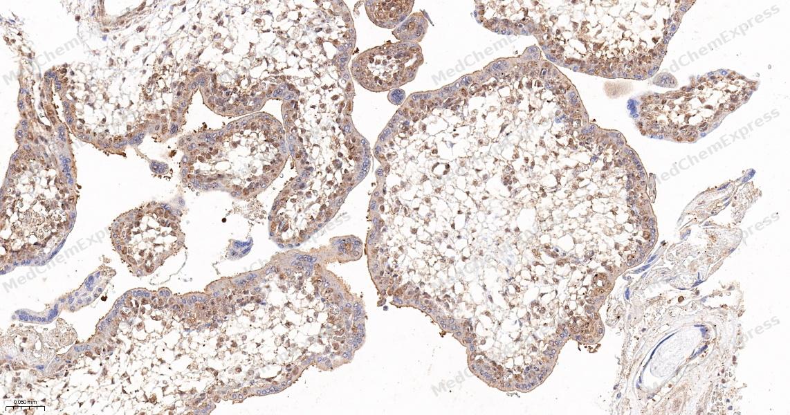 Immunohistochemical analysis of paraffin-embedded human small intestine tissue using CFLAR Antibody (HY-P83834, 1/500). The section was pretreated using heat mediated antigen retrieval with sodium citrate buffer (pH 6.0) for 8 minutes. The tissues were blocked with quick block buffer for 0.5 hours at room temperature, washed with PBS and PBST, and then incubated with the primary antibody overnight at 4℃. The detection was performed using an HRP conjugated compact polymer system. DAB was used as the chromogen. Tissues were counterstained with hematoxylin and mounted with DPX.
 -
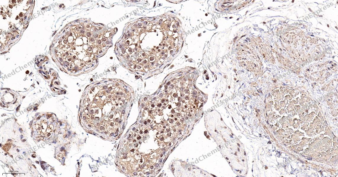 Immunohistochemical analysis of paraffin-embedded human tonsil tissue using CFLAR Antibody (HY-P83834, 1/500). The section was pretreated using heat mediated antigen retrieval with sodium citrate buffer (pH 6.0) for 8 minutes. The tissues were blocked with quick block buffer for 0.5 hours at room temperature, washed with PBS and PBST, and then incubated with the primary antibody overnight at 4℃. The detection was performed using an HRP conjugated compact polymer system. DAB was used as the chromogen. Tissues were counterstained with hematoxylin and mounted with DPX.
 -
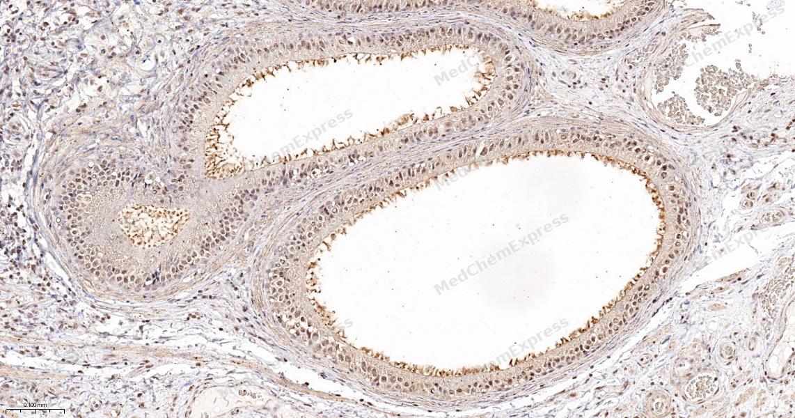 Immunohistochemical analysis of paraffin-embedded human soft tissue using CFLAR Antibody (HY-P83834, 1/500). The section was pretreated using heat mediated antigen retrieval with sodium citrate buffer (pH 6.0) for 8 minutes. The tissues were blocked with quick block buffer for 0.5 hours at room temperature, washed with PBS and PBST, and then incubated with the primary antibody overnight at 4℃. The detection was performed using an HRP conjugated compact polymer system. DAB was used as the chromogen. Tissues were counterstained with hematoxylin and mounted with DPX.
 -
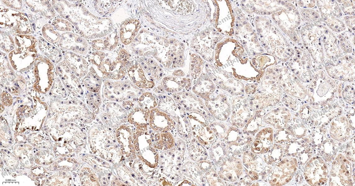 Immunohistochemical analysis of paraffin-embedded human colon cancer tissue using CFLAR Antibody (HY-P83834, 1/500). The section was pretreated using heat mediated antigen retrieval with sodium citrate buffer (pH 6.0) for 8 minutes. The tissues were blocked with quick block buffer for 0.5 hours at room temperature, washed with PBS and PBST, and then incubated with the primary antibody overnight at 4℃. The detection was performed using an HRP conjugated compact polymer system. DAB was used as the chromogen. Tissues were counterstained with hematoxylin and mounted with DPX.
 -
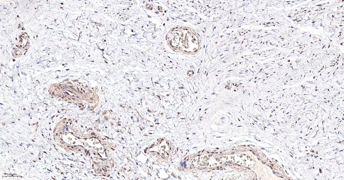 Immunohistochemical analysis of paraffin-embedded human endometrium cancer tissue using CFLAR Antibody (HY-P83834, 1/500). The section was pretreated using heat mediated antigen retrieval with sodium citrate buffer (pH 6.0) for 8 minutes. The tissues were blocked with quick block buffer for 0.5 hours at room temperature, washed with PBS and PBST, and then incubated with the primary antibody overnight at 4℃. The detection was performed using an HRP conjugated compact polymer system. DAB was used as the chromogen. Tissues were counterstained with hematoxylin and mounted with DPX.
 -
 Immunohistochemical analysis of paraffin-embedded human endometrium tissue using CFLAR Antibody (HY-P83834, 1/500). The section was pretreated using heat mediated antigen retrieval with sodium citrate buffer (pH 6.0) for 8 minutes. The tissues were blocked with quick block buffer for 0.5 hours at room temperature, washed with PBS and PBST, and then incubated with the primary antibody overnight at 4℃. The detection was performed using an HRP conjugated compact polymer system. DAB was used as the chromogen. Tissues were counterstained with hematoxylin and mounted with DPX.
 -
 Immunohistochemical analysis of paraffin-embedded human small intestine tissue using CFLAR Antibody (HY-P83834, 1/500). The section was pretreated using heat mediated antigen retrieval with sodium citrate buffer (pH 6.0) for 8 minutes. The tissues were blocked with quick block buffer for 0.5 hours at room temperature, washed with PBS and PBST, and then incubated with the primary antibody overnight at 4℃. The detection was performed using an HRP conjugated compact polymer system. DAB was used as the chromogen. Tissues were counterstained with hematoxylin and mounted with DPX.
 -
 Immunohistochemical analysis of paraffin-embedded human tonsil tissue using CFLAR Antibody (HY-P83834, 1/500). The section was pretreated using heat mediated antigen retrieval with sodium citrate buffer (pH 6.0) for 8 minutes. The tissues were blocked with quick block buffer for 0.5 hours at room temperature, washed with PBS and PBST, and then incubated with the primary antibody overnight at 4℃. The detection was performed using an HRP conjugated compact polymer system. DAB was used as the chromogen. Tissues were counterstained with hematoxylin and mounted with DPX.
 -
 Immunohistochemical analysis of paraffin-embedded human soft tissue using CFLAR Antibody (HY-P83834, 1/500). The section was pretreated using heat mediated antigen retrieval with sodium citrate buffer (pH 6.0) for 8 minutes. The tissues were blocked with quick block buffer for 0.5 hours at room temperature, washed with PBS and PBST, and then incubated with the primary antibody overnight at 4℃. The detection was performed using an HRP conjugated compact polymer system. DAB was used as the chromogen. Tissues were counterstained with hematoxylin and mounted with DPX.
 -
 Immunohistochemical analysis of paraffin-embedded human colon cancer tissue using CFLAR Antibody (HY-P83834, 1/500). The section was pretreated using heat mediated antigen retrieval with sodium citrate buffer (pH 6.0) for 8 minutes. The tissues were blocked with quick block buffer for 0.5 hours at room temperature, washed with PBS and PBST, and then incubated with the primary antibody overnight at 4℃. The detection was performed using an HRP conjugated compact polymer system. DAB was used as the chromogen. Tissues were counterstained with hematoxylin and mounted with DPX.
 -
 Immunohistochemical analysis of paraffin-embedded human endometrium cancer tissue using CFLAR Antibody (HY-P83834, 1/500). The section was pretreated using heat mediated antigen retrieval with sodium citrate buffer (pH 6.0) for 8 minutes. The tissues were blocked with quick block buffer for 0.5 hours at room temperature, washed with PBS and PBST, and then incubated with the primary antibody overnight at 4℃. The detection was performed using an HRP conjugated compact polymer system. DAB was used as the chromogen. Tissues were counterstained with hematoxylin and mounted with DPX.


Background
-
Function
CFLAR is an Apoptosis regulator protein which may function as a crucial link between cell survival and cell death pathways in mammalian cells. Acts as an inhibitor of TNFRSF6 mediated apoptosis. A proteolytic fragment (p43) is likely retained in the death-inducing signaling complex (DISC) thereby blocking further recruitment and processing of caspase-8 at the complex. Full length and shorter isoforms have been shown either to induce apoptosis or to reduce TNFRSF-triggered apoptosis. Lacks enzymatic (caspase) activity
-
Expression
Tissue_specificity:Widely expressed. Higher expression in skeletal muscle, pancreas, heart, kidney, placenta, and peripheral blood leukocytes. Also detected in diverse cell lines. Isoform 8 is predominantly expressed in testis and skeletal muscle
Induction:Repressed by IL2/interleukin-2 after TCR stimulation, during progression to the S phase of the cell cycle -
Isoforms & Post-Translational Modification
O15519 has 15 isomers: O15519-1: 55344 Da (predicted); O15519-2: 25379 Da (predicted); O15519-3: 41319 Da (predicted); O15519-4: 27055 Da (predicted); O15519-5: 51390 Da (predicted); O15519-6: 50661 Da (predicted); O15519-7: 39245 Da (predicted); O15519-8: 51330 Da (predicted); O15519-9: 30399 Da (predicted); O15519-10: 34384 Da (predicted); O15519-11: 52551 Da (predicted); O15519-12: 33273 Da (predicted); O15519-13: 23289 Da (predicted); O15519-14: 49778 Da (predicted); O15519-15: 44112 Da (predicted).
Proteolytically processed by CASP8 generating subunit p43 and p12 -
Subunit
TNFRSF6 stimulation triggers recruitment to the death-inducing signaling complex (DISC) formed by TNFRSF6, FADD and CASP8 (PubMed:9880531). A proteolytic fragment (p43) stays associated with the DISC (PubMed:9880531). Also interacts with FADD, CASP8, CASP3, TRAF1, TRAF2 and Bcl-X(L) (in vitro) (PubMed:9208847, PubMed:9326610). Interacts with RIPK1 (By similarity) (PubMed:9208847, PubMed:9326610, PubMed:9880531)
-
SwissProt ID
-
Synonyms
CASH; FLIP; MRIT; CLARP; FLAME; Casper; FLAME1; c-FLIP; FLAME-1; I-FLICE; c-FLIPL; c-FLIPR; c-FLIPS; CASP8AP1


Documentation
-
Data Sheet (234 KB)
-
SDS (252 KB)
- English - EN (252 KB)
- Français - FR (252 KB)
- Deutsch - DE (252 KB)
- Norwegian - NO (252 KB)
- Español - ES (252 KB)
- Swedish - SV (252 KB)
- Italian - IT (252 KB)
- Korean - KR (252 KB)
- Portuguese - PT (252 KB)
-
User Guide for Antibodies (1077 KB)

 Powered by Bioz
