



DNA PKcs Antibody (YA782)
(Synonyms: HYRC, HYRC1, PRKDC, DNA-dependent protein kinase catalytic subunit, DNA-PK catalytic subunit, DNA-PKcs, DNPK1, p460)
Based on 1 Customer Validation
DNA PKcs Antibody (YA782) is a Mouse-derived and non-conjugated IgG2a monoclonal antibody, targeting to DNA PKcs.
-
Host:
Mouse
-
Isotype:
IgG
-
Application:
WB, ICC/IF, IHC-P
-
Reactivity :
Human
-
Formulation:
Supplied in 1*PBS (pH7.4), 0.2% BSA and 40% Glycerol. Preservative: 0.05% Sodium Azide.
-
Conjugation:
Non-conjugated


To place orders, for customer services and technical support, please contact: MedChemExpress USA
Tel: 609-228-6898 E-mail: [email protected] [email protected]





Applications
| Application |
WB

WB: Western Blot
|
ICC/IF
ICC/IF: Immunocytochemistry/
Immunofluorescence |
IHC-P
IHC-P: Immunohistochemistry-Paraffin
|
|---|---|---|---|
| Dilution Ratio | 1:1000 | 1:100 | 1:500 |


Product Details
DNA PKcs Antibody (YA782) is a Mouse-derived and non-conjugated IgG2a monoclonal antibody, targeting to DNA PKcs.
-
Host Mouse

-
Clonality Monoclonal

-
Species ReactivityHuman
-
Observed Molecular WeightObserved band size: 469 kDa
Note: Due to possible protein modifications or aggregation, the molecular weight should be confirmed by actual measurement, and the predicted value is for reference only.

-
Calculated Molecular Weight Predicted band size: 469 kDa

Synthetic peptide corresponding to Human DNA-PKcs/PRKDC.AA range:3900-4128.
Endogenous
Protein A affinity purified.
Non-conjugated
Unmodified
IgG


Product Properties
-
Appearance
Solution
-
Formulation
Supplied in 1*PBS (pH7.4), 0.2% BSA and 40% Glycerol. Preservative: 0.05% Sodium Azide.
-
Concentration
Batch-dependent, Please check the COA for the concentration of each lot. Check Lot Concentration
-
Storage & Stability
Stored at -20°C for 1 year. Avoid repeated freeze / thaw cycles.
-
Shipping
Shipping with blue ice.


Verification Images
-
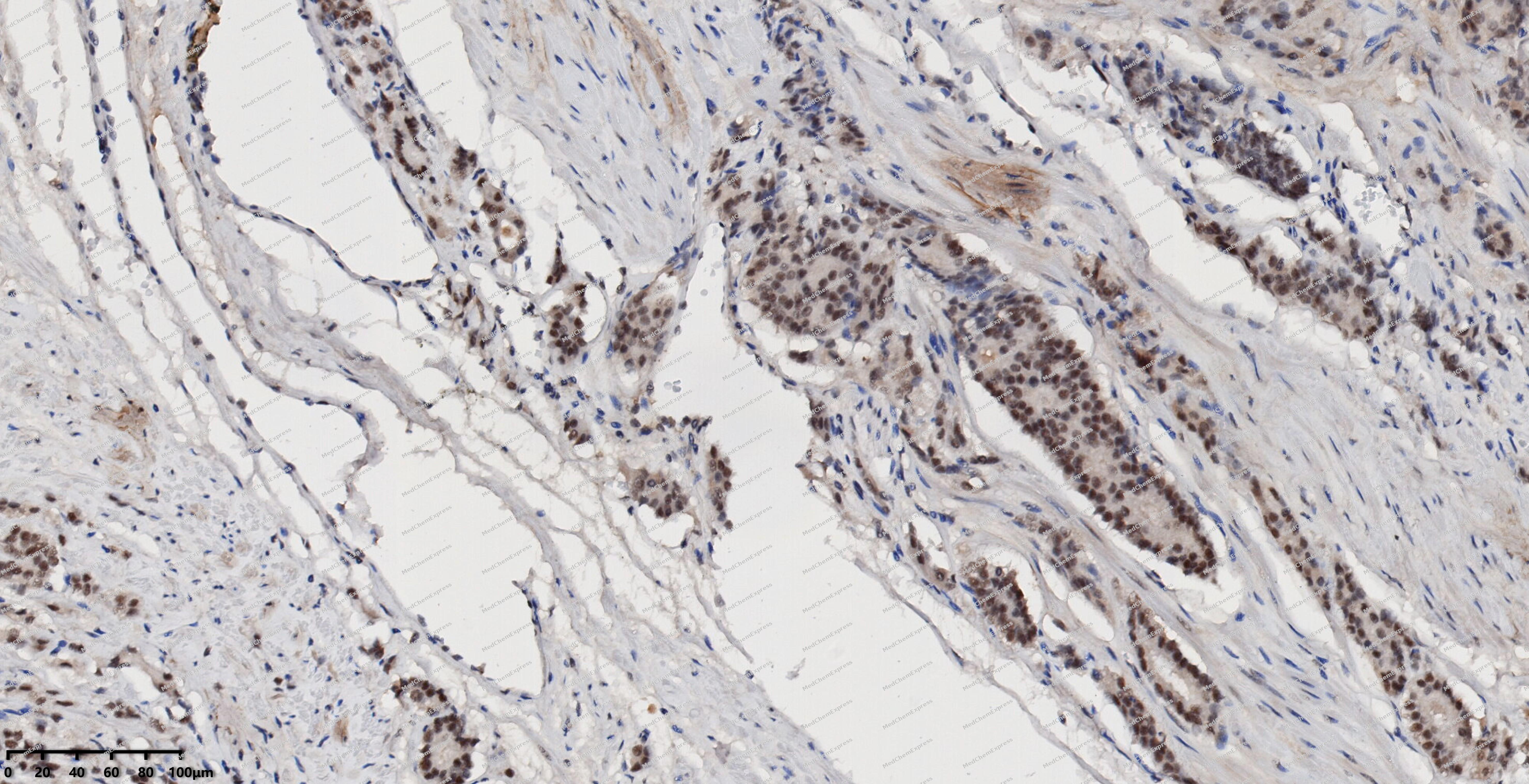 Immunohistochemical analysis of paraffin-embedded human prostate cancer using DNA PKcs antibody. The section was pre-treated using heat mediated antigen retrieval with EDTA (pH 9.0) for 14 minutes. The tissues were blocked in 5% BSA for 30 minutes at room temperature, washed with TBST, and then probed with the primary antibody (HY-P80104, 1/500) for 30 minutes at room temperature. The detection was performed using Polymer HRP-conjugated Goat Anti-Mouse/Rabbit lgG(H&L) secondary antibody (HY-P83652). DAB was used as the chromogen. Tissues were counterstained with hematoxylin and mounted with DPX.
 -
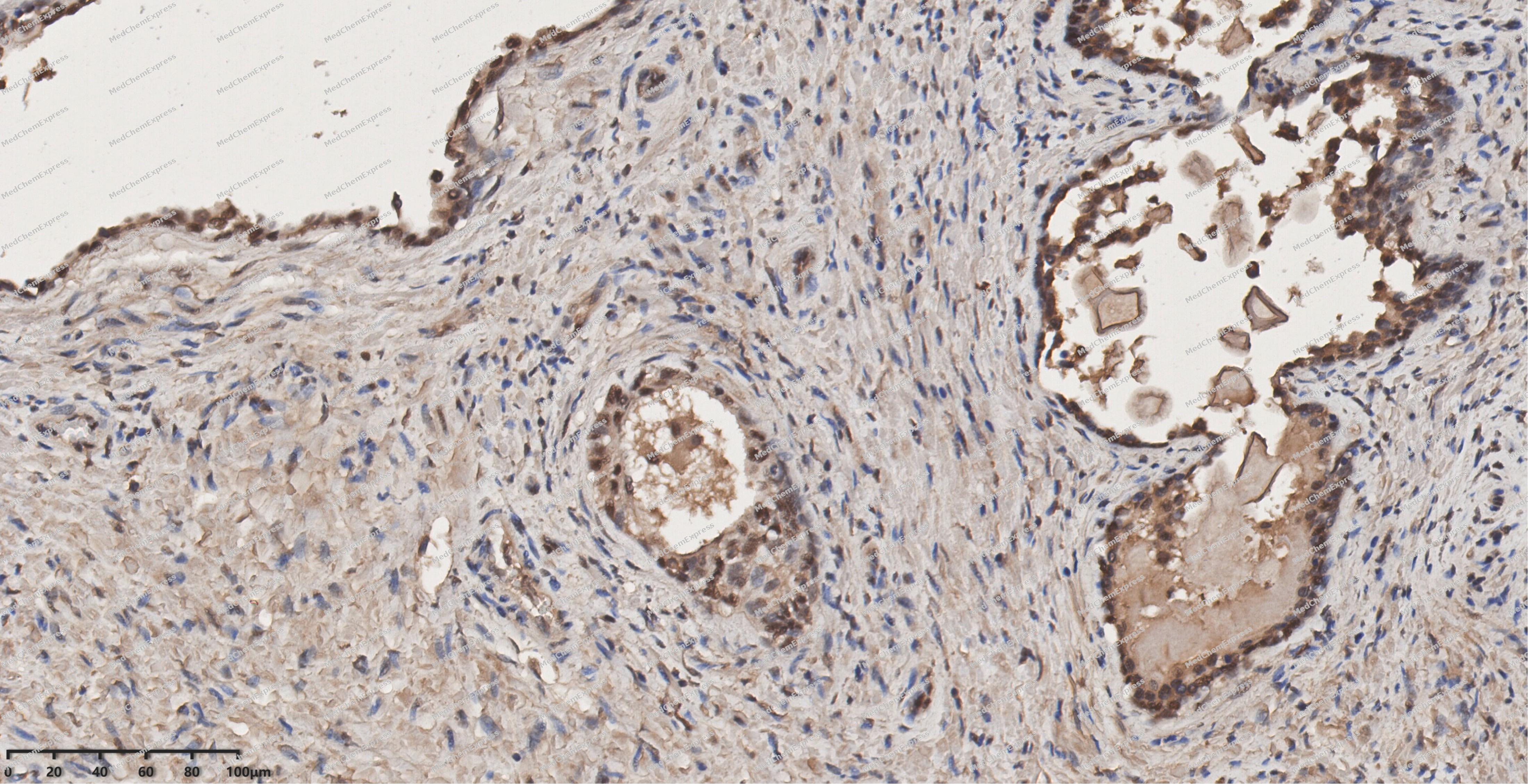 Immunohistochemical analysis of paraffin-embedded human prostate using DNA PKcs antibody. The section was pre-treated using heat mediated antigen retrieval with EDTA (pH 9.0) for 14 minutes. The tissues were blocked in 5% BSA for 30 minutes at room temperature, washed with TBST, and then probed with the primary antibody (HY-P80104, 1/500) for 30 minutes at room temperature. The detection was performed using Polymer HRP-conjugated Goat Anti-Mouse/Rabbit lgG(H&L) secondary antibody (HY-P83652). DAB was used as the chromogen. Tissues were counterstained with hematoxylin and mounted with DPX.
 -
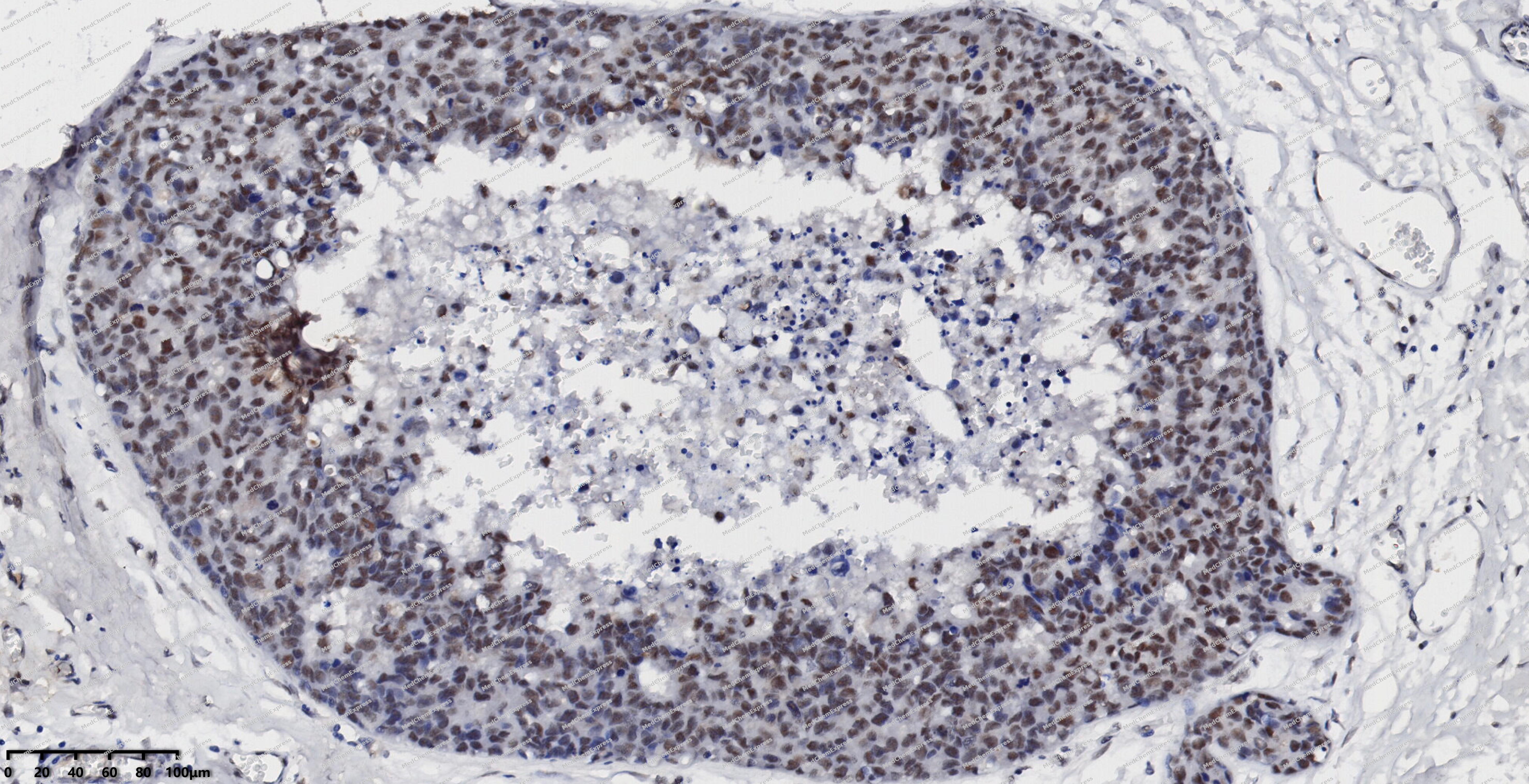 Immunohistochemical analysis of paraffin-embedded human breast cancer using DNA PKcs antibody. The section was pre-treated using heat mediated antigen retrieval with EDTA (pH 9.0) for 14 minutes. The tissues were blocked in 5% BSA for 30 minutes at room temperature, washed with TBST, and then probed with the primary antibody (HY-P80104, 1/500) for 30 minutes at room temperature. The detection was performed using Polymer HRP-conjugated Goat Anti-Mouse/Rabbit lgG(H&L) secondary antibody (HY-P83652). DAB was used as the chromogen. Tissues were counterstained with hematoxylin and mounted with DPX.
 -
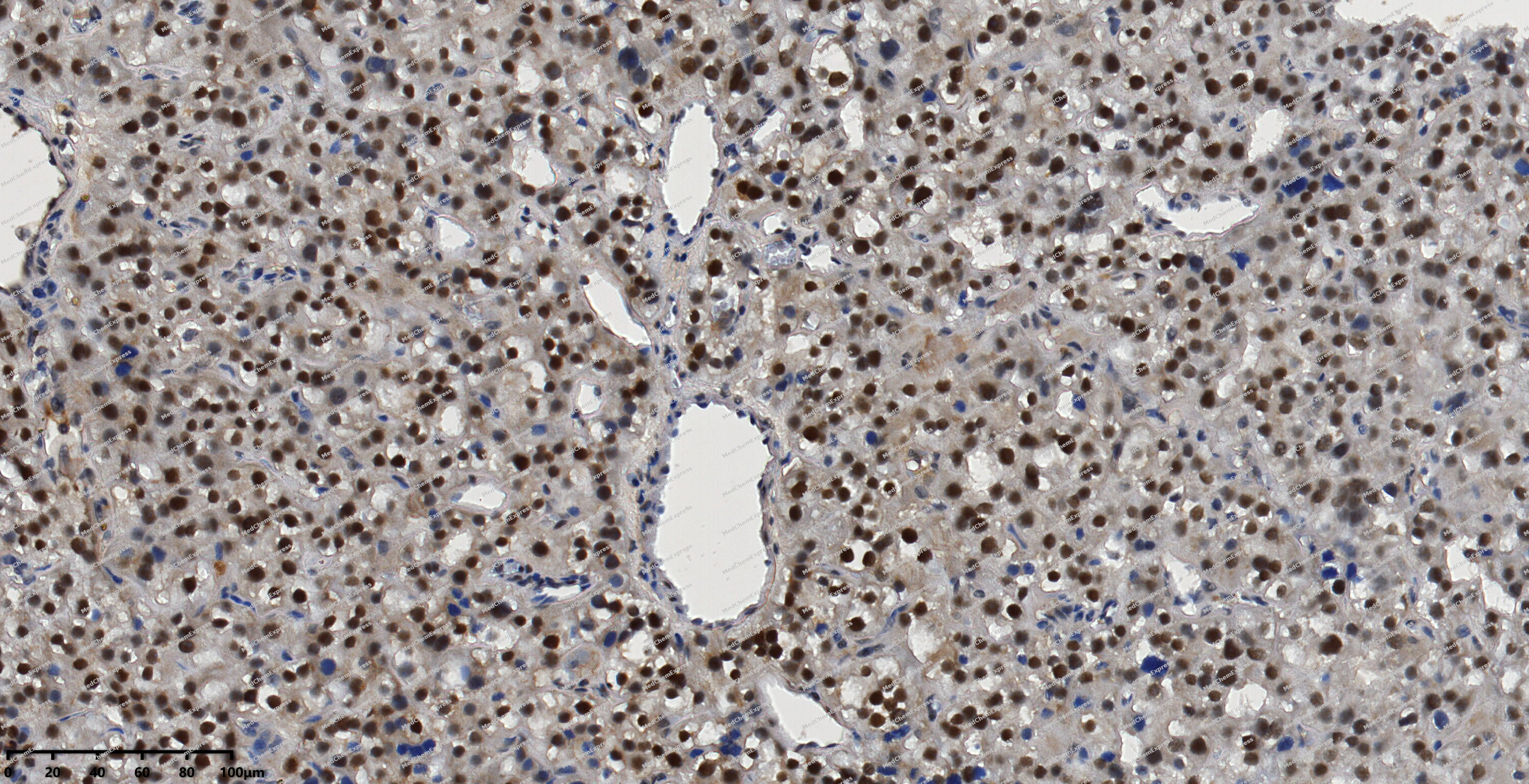 Immunohistochemical analysis of paraffin-embedded human thyroid cancer using DNA PKcs antibody. The section was pre-treated using heat mediated antigen retrieval with EDTA (pH 9.0) for 14 minutes. The tissues were blocked in 5% BSA for 30 minutes at room temperature, washed with TBST, and then probed with the primary antibody (HY-P80104, 1/500) for 30 minutes at room temperature. The detection was performed using Polymer HRP-conjugated Goat Anti-Mouse/Rabbit lgG(H&L) secondary antibody (HY-P83652). DAB was used as the chromogen. Tissues were counterstained with hematoxylin and mounted with DPX.
 -
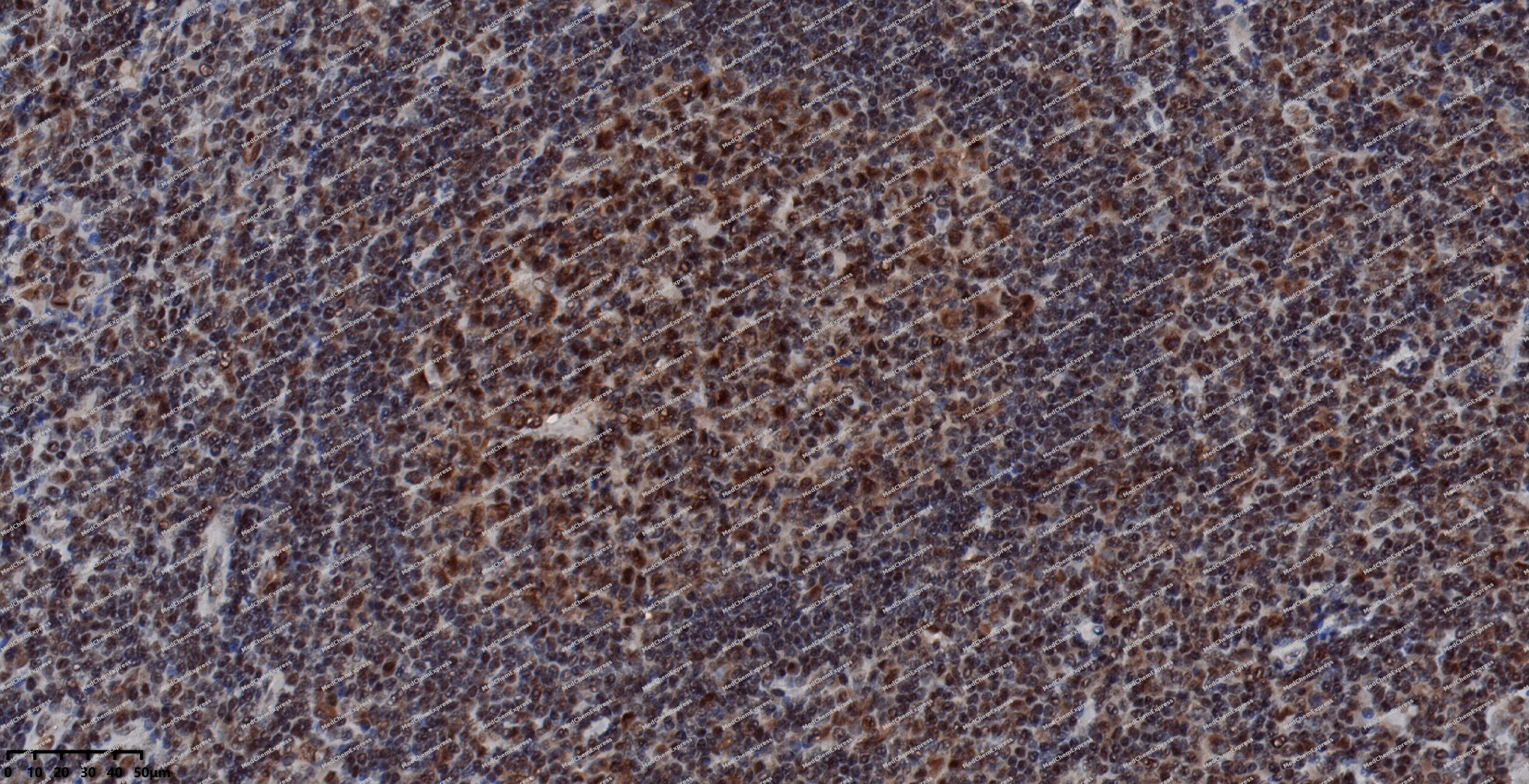 Immunohistochemical analysis of paraffin-embedded human tonsil using DNA PKcs antibody. The section was pre-treated using heat mediated antigen retrieval with EDTA (pH 9.0) for 14 minutes. The tissues were blocked in 5% BSA for 30 minutes at room temperature, washed with TBST, and then probed with the primary antibody (HY-P80104, 1/500) for 30 minutes at room temperature. The detection was performed using Polymer HRP-conjugated Goat Anti-Mouse/Rabbit lgG(H&L) secondary antibody (HY-P83652). DAB was used as the chromogen. Tissues were counterstained with hematoxylin and mounted with DPX.
 -
 Immunohistochemical analysis of paraffin-embedded human kidney using DNA PKcs antibody. The section was pre-treated using heat mediated antigen retrieval with EDTA (pH 9.0) for 14 minutes. The tissues were blocked in 5% BSA for 30 minutes at room temperature, washed with TBST, and then probed with the primary antibody (HY-P80104, 1/500) for 30 minutes at room temperature. The detection was performed using Polymer Polymer HRP-conjugated Goat Anti-Mouse lgG(H&L) (Ready to use) (HY-P89365 ). DAB was used as the chromogen. Tissues were counterstained with hematoxylin and mounted with DPX.
 -
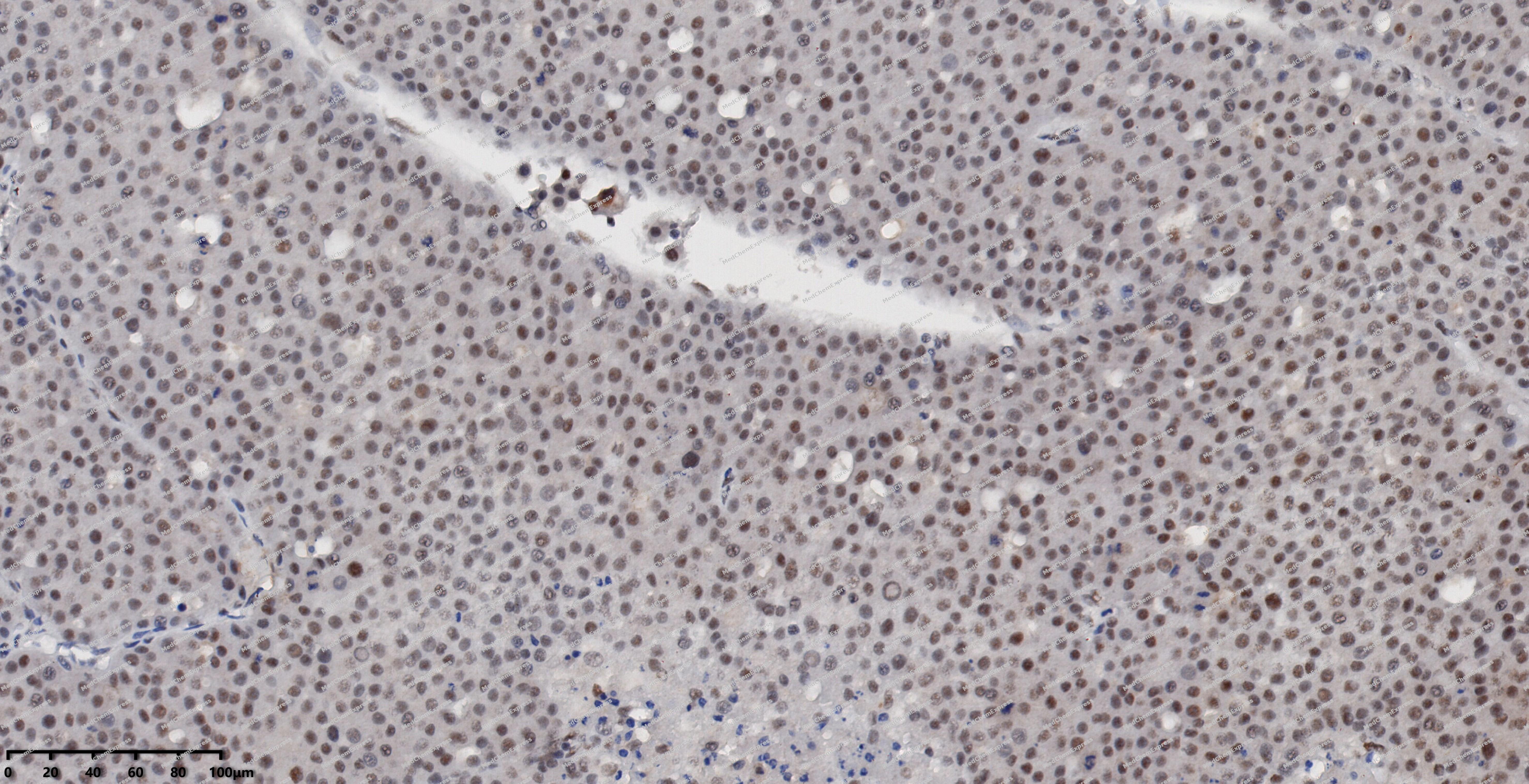 Immunohistochemical analysis of paraffin-embedded human liver cancer using DNA PKcs antibody. The section was pre-treated using heat mediated antigen retrieval with EDTA (pH 9.0) for 14 minutes. The tissues were blocked in 5% BSA for 30 minutes at room temperature, washed with TBST, and then probed with the primary antibody (HY-P80104, 1/500) for 30 minutes at room temperature. The detection was performed using Polymer HRP-conjugated Goat Anti-Mouse/Rabbit lgG(H&L) secondary antibody (HY-P83652). DAB was used as the chromogen. Tissues were counterstained with hematoxylin and mounted with DPX.
 -
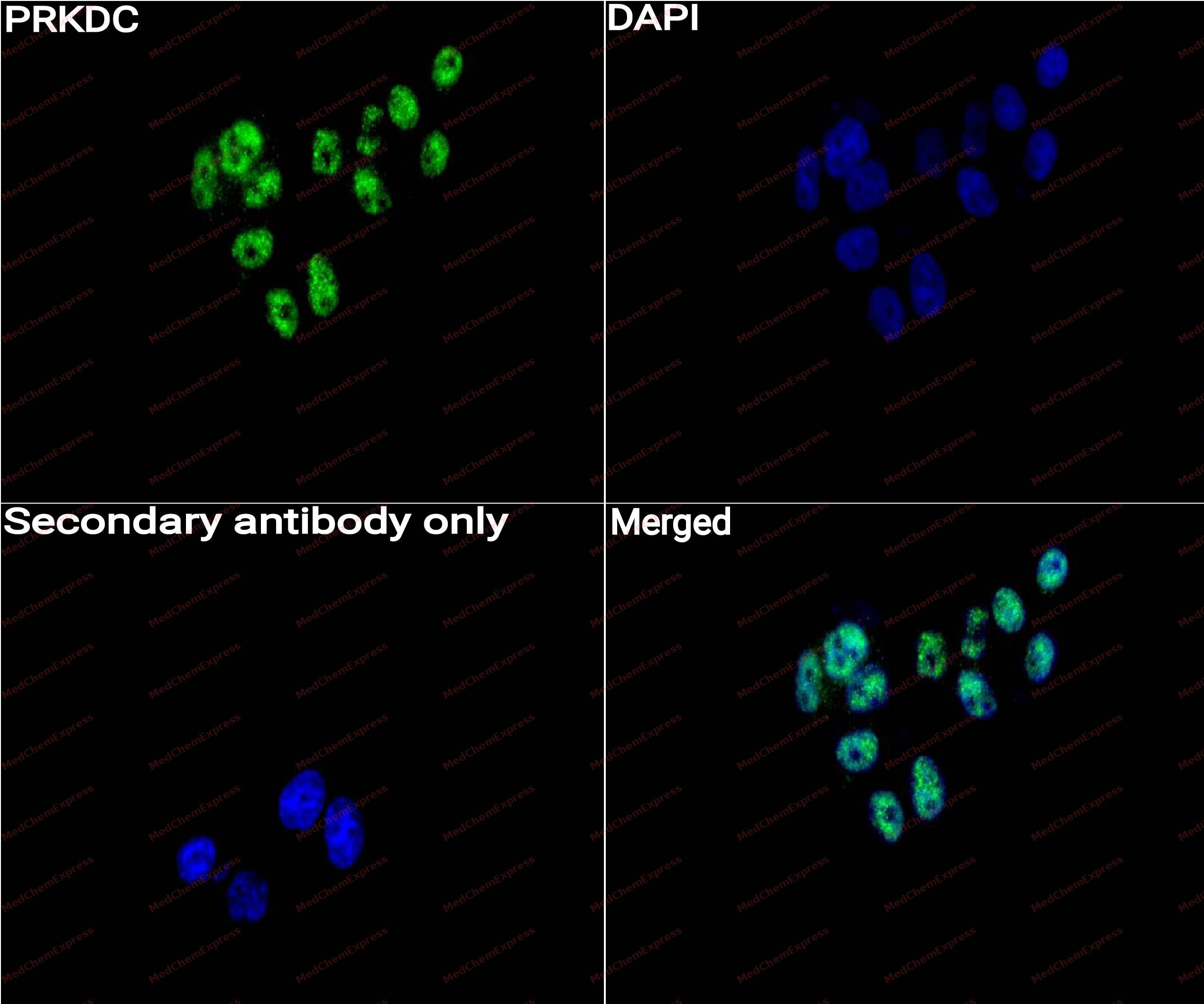 Immunocytochemistry analysis of HepG2 cells labeling PRKDC with PRKDC Antibody (HY-P80104) at 1/50 dilution. Cells were fixed in 4% paraformaldehyde for 15 minutes at room temperature, permeabilized with 0.1% Triton X-100 for 10 minutes at room temperature, then blocked with QuickBlock™ Blocking Buffer for Immunol Staining for 10 min at room temperature. Cells were then incubated with PRKDC Antibody (HY-P80104) at 1/50 dilution in QuickBlock™ Blocking Buffer for Immunol Staining at 4 ℃. Alexa Fluor® 488-conjugated AffiniPure Goat Anti-Mouse IgG H&L(HY-P8005, Green) was used as the secondary antibody at 1/1,000 dilution. PBS instead of the primary antibody was used as the secondary antibody only control. The Nuclear counterstain was DAPI (Blue).
 -
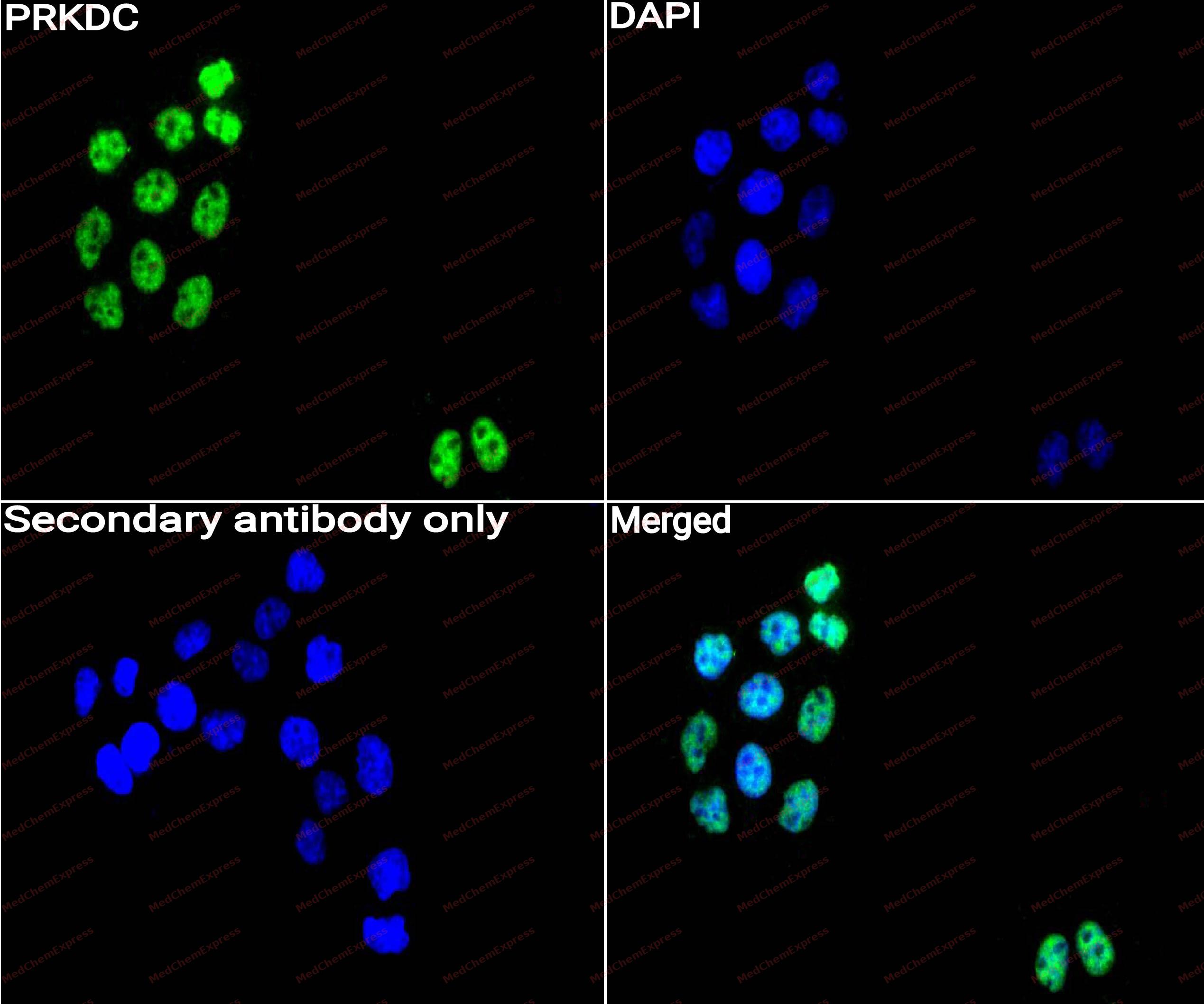 Immunocytochemistry analysis of HepG2 cells labeling PRKDC with PRKDC Antibody (HY-P80104) at 1/100 dilution. Cells were fixed in 4% paraformaldehyde for 15 minutes at room temperature, permeabilized with 0.1% Triton X-100 for 10 minutes at room temperature, then blocked with QuickBlock™ Blocking Buffer for Immunol Staining for 10 min at room temperature. Cells were then incubated with PRKDC Antibody (HY-P80104)at 1/100 dilution in QuickBlock™ Blocking Buffer for Immunol Staining at 4 ℃. Alexa Fluor® 488-conjugated AffiniPure Goat Anti-Mouse IgG H&L(HY-P8005,Green) was used as the secondary antibody at 1/1,000 dilution. PBS instead of the primary antibody was used as the secondary antibody only control. The Nuclear counterstain was DAPI (Blue).


Background
-
Function
DNA PKcs is a Serine/threonine-protein kinase that acts as a molecular sensor for DNA damage. Involved in DNA non-homologous end joining (NHEJ) required for double-strand break (DSB) repair and V(D)J recombination. Must be bound to DNA to express its catalytic properties. Promotes processing of hairpin DNA structures in V(D)J recombination by activation of the hairpin endonuclease artemis (DCLRE1C). Recruited by XRCC5 and XRCC6 to DNA ends and is required to (1) protect and align broken ends of DNA, thereby preventing their degradation, (2) and sequester the DSB for repair by NHEJ. Acts as a scaffold protein to aid the localization of DNA repair proteins to the site of damage. The assembly of the DNA-PK complex at DNA ends is also required for the NHEJ ligation step. Found at the ends of chromosomes, suggesting a further role in the maintenance of telomeric stability and the prevention of chromosomal end fusion. Also involved in modulation of transcription. As part of the DNA-PK complex, involved in the early steps of ribosome assembly by promoting the processing of precursor rRNA into mature 18S rRNA in the small-subunit processome. Binding to U3 small nucleolar RNA, recruits PRKDC and XRCC5/Ku86 to the small-subunit processome. Recognizes the substrate consensus sequence [ST]-Q. Phosphorylates 'Ser-139' of histone variant H2AX, thereby regulating DNA damage response mechanism. Phosphorylates ASF1A, DCLRE1C, c-Abl/ABL1, histone H1, HSPCA, c-jun/JUN, p53/TP53, PARP1, POU2F1, DHX9, FH, SRF, NHEJ1/XLF, XRCC1, XRCC4, XRCC5, XRCC6, WRN, MYC and RFA2. Can phosphorylate C1D not only in the presence of linear DNA but also in the presence of supercoiled DNA. Ability to phosphorylate p53/TP53 in the presence of supercoiled DNA is dependent on C1D. Acts as a regulator of the phosphatidylinositol 3-kinase/protein kinase B signal transduction by mediating phosphorylation of 'Ser-473' of protein kinase B (PKB/AKT1, PKB/AKT2, PKB/AKT3), promoting their activation. Contributes to the determination of the circadian period length by antagonizing phosphorylation of CRY1 'Ser-588' and increasing CRY1 protein stability, most likely through an indirect mechanism. Plays a role in the regulation of DNA virus-mediated innate immune response by assembling into the HDP-RNP complex, a complex that serves as a platform for IRF3 phosphorylation and subsequent innate immune response activation through the cGAS-STING pathway. Also regulates the cGAS-STING pathway by catalyzing phosphorylation of CGAS, thereby impairing CGAS oligomerization and activation. Also regulates the cGAS-STING pathway by mediating phosphorylation of PARP1[1][2][3][4][5][6][7][8][9][10][11][12][13][14][15][16][17][18][19][20][21][22][23][24][25][26][27][28][29][30][31][32][33][34][35][36].
-
Subcellular Localization
Nucleus; Nucleus, nucleolus; Cytoplasm, cytosol
-
Isoforms & Post-Translational Modification
P78527 has 2 isomers: P78527-1: 469089 Da (predicted); P78527-2: 465501 Da (predicted).
Autophosphorylated at two clusters, the T2609 cluster and the S2056 cluster (PubMed:33854234). Autophosphorylated on Ser-2056, Thr-2609, Thr-2638 and Thr-2647 (PubMed:12186630, PubMed:12231622, PubMed:14734805, PubMed:33854234). Ser-2056 and Thr-2609 are DNA damage-inducible phosphorylation sites (inducible with ionizing radiation, IR) dephosphorylated by PPP5C (PubMed:12186630, PubMed:12231622, PubMed:14734805). Autophosphorylation induces a conformational change that leads to remodeling of the DNA-PK complex, requisite for efficient end processing and DNA repair (PubMed:12186630, PubMed:12231622, PubMed:14734805). Autophosphorylation in trans within DNA-PK complexes loaded on DNA ends leads to the dissociation of PRKDC from DNA and the transition into the short-range NHEJ complex (PubMed:33854234). Autophosphorylation of the T2609 cluster is required for hematopoietic development and protein synthesis in erythrocytes precursors (By similarity);S-nitrosylated by GAPDH;Polyubiquitinated by RNF144A, leading to proteasomal degradation -
Subunit
DNA-PK is a heterotrimer of PRKDC and the Ku dimer (composed of XRCC6/Ku70 and XRCC5/Ku86) (PubMed:15758953, PubMed:25670504). Formation of this complex may be promoted by interaction with ILF3 (PubMed:9442054). Component of the core long-range non-homologous end joining (NHEJ) complex (also named DNA-PK complex) composed of PRKDC, LIG4, XRCC4, XRCC6/Ku70, XRCC5/Ku86 and NHEJ1/XLF (PubMed:15758953, PubMed:25670504, PubMed:33854234, PubMed:34352203). Additional component of the NHEJ complex includes PAXX (PubMed:25574025, PubMed:25941166). Following autophosphorylation, PRKDC dissociates from DNA (PubMed:33854234). Interacts with DNA-PKcs-interacting protein (KIP) with the region upstream the kinase domain (PubMed:9372844). PRKDC alone also interacts with and phosphorylates DCLRE1C, thereby activating the latent endonuclease activity of this protein (PubMed:11955432, PubMed:14744996, PubMed:15071507, PubMed:15456891, PubMed:15574326, PubMed:15811628, PubMed:15936993). Interacts with C1D (PubMed:9679063). Interacts with TTI1 and TELO2 (PubMed:20427287, PubMed:20801936, PubMed:20810650). Interacts with CIB1 (PubMed:9372844). Interacts with SETX (PubMed:23149945). Interacts with NR4A3; the DNA-dependent protein kinase complex DNA-PK phosphorylates and activates NR4A3 and prevents NR4A3 ubiquitination and degradation (PubMed:25852083). Interacts with BRAT1 (PubMed:22977523). Part of the HDP-RNP complex composed of at least HEXIM1, PRKDC, XRCC5, XRCC6, paraspeckle proteins (SFPQ, NONO, PSPC1, RBM14, and MATR3) and NEAT1 RNA (PubMed:28712728). Interacts with KAT5 (PubMed:32832608)
-
SwissProt ID
-
Synonyms
HYRC, HYRC1, PRKDC, DNA-dependent protein kinase catalytic subunit, DNA-PK catalytic subunit, DNA-PKcs, DNPK1, p460
-
Research Field
Epigenetics and Nuclear Signaling


Documentation
-
Data Sheet (270 KB)
-
SDS (252 KB)
- English - EN (252 KB)
- Français - FR (252 KB)
- Deutsch - DE (252 KB)
- Norwegian - NO (252 KB)
- Español - ES (252 KB)
- Swedish - SV (252 KB)
- Italian - IT (252 KB)
- Korean - KR (252 KB)
- Portuguese - PT (252 KB)
-
User Guide for Antibodies (1077 KB)
References
[1]. Ma Y, et al. Hairpin opening and overhang processing by an Artemis/DNA-dependent protein kinase complex in nonhomologous end joining and V(D)J recombination. Cell. 2002 Mar 22;108(6):781-94. [Content Brief]
[2]. Soubeyrand S, et al. Threonines 2638/2647 in DNA-PK are essential for cellular resistance to ionizing radiation. Cancer Res. 2003 Mar 15;63(6):1198-201. [Content Brief]
[3]. Wechsler T, et al. DNA-PKcs function regulated specifically by protein phosphatase 5. Proc Natl Acad Sci U S A. 2004 Feb 3;101(5):1247-52. [Content Brief]
[4]. Chen S, et al. Structural basis of long-range to short-range synaptic transition in NHEJ. Nature. 2021 May;593(7858):294-298. [Content Brief]
[5]. Chaplin AK, et al. Cryo-EM of NHEJ supercomplexes provides insights into DNA repair. Mol Cell. 2021 Aug 19;81(16):3400-3409.e3. [Content Brief]
[6]. Ma Y, et al. A biochemically defined system for mammalian nonhomologous DNA end joining. Mol Cell. 2004 Dec 3;16(5):701-13. [Content Brief]
[7]. Shao Z, et al. DNA-PKcs has KU-dependent function in rRNA processing and haematopoiesis. Nature. 2020 Mar;579(7798):291-296. [Content Brief]
[8]. Park EJ, et al. DNA-PK is activated by nucleosomes and phosphorylates H2AX within the nucleosomes in an acetylation-dependent manner. Nucleic Acids Res. 2003 Dec 1;31(23):6819-27. [Content Brief]
[9]. Reitsema T, et al. DNA-PK is responsible for enhanced phosphorylation of histone H2AX under hypertonic conditions. DNA Repair (Amst). 2005 Sep 28;4(10):1172-81. [Content Brief]
[10]. Chan DW, et al. DNA-dependent protein kinase phosphorylation sites in Ku 70/80 heterodimer. Biochemistry. 1999 Feb 9;38(6):1819-28. [Content Brief]
[11]. Ariumi Y, et al. Suppression of the poly(ADP-ribose) polymerase activity by DNA-dependent protein kinase in vitro. Oncogene. 1999 Aug 12;18(32):4616-25. [Content Brief]
[12]. Karmakar P, et al. Werner protein is a target of DNA-dependent protein kinase in vivo and in vitro, and its catalytic activities are regulated by phosphorylation. J Biol Chem. 2002 May 24;277(21):18291-302. [Content Brief]
[13]. Hsu HL, et al. Defining interactions between DNA-PK and ligase IV/XRCC4. DNA Repair (Amst). 2002 Mar 28;1(3):225-35. [Content Brief]
[14]. Yu Y, et al. DNA-PK phosphorylation sites in XRCC4 are not required for survival after radiation or for V(D)J recombination. DNA Repair (Amst). 2003 Nov 21;2(11):1239-52. [Content Brief]
[15]. Schild-Poulter C, et al. Down-regulation of histone H2B by DNA-dependent protein kinase in response to DNA damage through modulation of octamer transcription factor 1. Cancer Res. 2003 Nov 1;63(21):7197-205. [Content Brief]
[16]. Zhang S, et al. DNA-dependent protein kinase (DNA-PK) phosphorylates nuclear DNA helicase II/RNA helicase A and hnRNP proteins in an RNA-dependent manner. Nucleic Acids Res. 2004;32(1):1-10. [Content Brief]
[17]. Lee KJ, et al. Identification of DNA-PKcs phosphorylation sites in XRCC4 and effects of mutations at these sites on DNA end joining in a cell-free system. DNA Repair (Amst). 2004 Mar 4;3(3):267-76. [Content Brief]
[18]. Iijima S, et al. DNA-activated protein kinase in Raji Burkitt's lymphoma cells. Phosphorylation of c-Myc oncoprotein. Eur J Biochem. 1992 Jun 1;206(2):595-603. [Content Brief]
[19]. Lévy N, et al. XRCC1 is phosphorylated by DNA-dependent protein kinase in response to DNA damage. Nucleic Acids Res. 2006;34(1):32-41. [Content Brief]
[20]. Yu Y, et al. DNA-PK and ATM phosphorylation sites in XLF/Cernunnos are not required for repair of DNA double strand breaks. DNA Repair (Amst). 2008 Oct 1;7(10):1680-92. [Content Brief]
[21]. Carter T, et al. A DNA-activated protein kinase from HeLa cell nuclei. Mol Cell Biol. 1990 Dec;10(12):6460-71. [Content Brief]
[22]. Lees-Miller SP, et al. The human double-stranded DNA-activated protein kinase phosphorylates the 90-kDa heat-shock protein, hsp90 alpha at two NH2-terminal threonine residues. J Biol Chem. 1989 Oct 15;264(29):17275-80. [Content Brief]
[23]. Jiang Y, et al. Local generation of fumarate promotes DNA repair through inhibition of histone H3 demethylation. Nat Cell Biol. 2015 Sep;17(9):1158-68. [Content Brief]
[24]. Sharma MK, et al. In cellulo phosphorylation of XRCC4 Ser320 by DNA-PK induced by DNA damage. J Radiat Res. 2016 Mar;57(2):115-20. [Content Brief]
[25]. Morchikh M, et al. HEXIM1 and NEAT1 Long Non-coding RNA Form a Multi-subunit Complex that Regulates DNA-Mediated Innate Immune Response. Mol Cell. 2017 Aug 3;67(3):387-399.e5. [Content Brief]
[26]. Huang TH, et al. The Histone Chaperones ASF1 and CAF-1 Promote MMS22L-TONSL-Mediated Rad51 Loading onto ssDNA during Homologous Recombination in Human Cells. Mol Cell. 2018 Mar 1;69(5):879-892.e5. [Content Brief]
[27]. Amiri Moghani AR, et al. In cellulo phosphorylation of DNA double-strand break repair protein XRCC4 on Ser260 by DNA-PK. J Radiat Res. 2018 Nov 1;59(6):700-708. [Content Brief]
[28]. Liu SH, et al. The carboxyl-terminal transactivation domain of human serum response factor contains DNA-activated protein kinase phosphorylation sites. J Biol Chem. 1993 Oct 5;268(28):21147-54. [Content Brief]
[29]. Bannister AJ, et al. c-Jun is phosphorylated by the DNA-dependent protein kinase in vitro; definition of the minimal kinase recognition motif. Nucleic Acids Res. 1993 Mar 11;21(5):1289-95. [Content Brief]
[30]. Niu H, et al. Mapping of amino acid residues in the p34 subunit of human single-stranded DNA-binding protein phosphorylated by DNA-dependent protein kinase and Cdc2 kinase in vitro. J Biol Chem. 1997 May 9;272(19):12634-41. [Content Brief]
[31]. Jin S, et al. Double-strand break repair by Ku70 requires heterodimerization with Ku80 and DNA binding functions. EMBO J. 1997 Nov 17;16(22):6874-85. [Content Brief]
[32]. Yavuzer U, et al. DNA end-independent activation of DNA-PK mediated via association with the DNA-binding protein C1D. Genes Dev. 1998 Jul 15;12(14):2188-99. [Content Brief]
[33]. Shieh SY, et al. DNA damage-induced phosphorylation of p53 alleviates inhibition by MDM2. Cell. 1997 Oct 31;91(3):325-34. [Content Brief]
[34]. Feng J, et al. Identification of a PKB/Akt hydrophobic motif Ser-473 kinase as DNA-dependent protein kinase. J Biol Chem. 2004 Sep 24;279(39):41189-96. [Content Brief]
[35]. Sun X, et al. DNA-PK deficiency potentiates cGAS-mediated antiviral innate immunity. Nat Commun. 2020 Dec 3;11(1):6182. [Content Brief]
[36]. Wang F, et al. Cytoplasmic PARP1 links the genome instability to the inhibition of antiviral immunity through PARylating cGAS. Mol Cell. 2022 Jun 2;82(11):2032-2049.e7. [Content Brief]

 Powered by Bioz
