



ETFA Antibody (YA3789)
(Synonyms: EMA; GA2; MADD)ETFA Antibody (YA3789) is a Mouse-derived and non-conjugated IgG2a monoclonal antibody, targeting to ETFA.
-
Host:
Mouse
-
Isotype:
IgG2a
-
Application:
WB, IHC-P, ICC/IF, FC, ELISA
-
Reactivity :
Human
-
Formulation:
Supplied in PBS with 0.05% sodium azide
-
Conjugation:
Non-conjugated


To place orders, for customer services and technical support, please contact: MedChemExpress USA
Tel: 609-228-6898 E-mail: [email protected] [email protected]





Applications
| Application |
WB

WB: Western Blot
|
IHC-P
IHC-P: Immunohistochemistry-Paraffin
|
ICC/IF
ICC/IF: Immunocytochemistry/
Immunofluorescence |
FC
FC: Flow Cytometry
|
ELISA
ELISA: Enzyme Linked Immunosorbent Assay
|
|---|---|---|---|---|---|
| Dilution Ratio | 1:500-1:2000 | 1:200-1:1000 | 1:200-1:1000 | 1:200-1:400 | 1:10000 |


Product Details
ETFA Antibody (YA3789) is a Mouse-derived and non-conjugated IgG2a monoclonal antibody, targeting to ETFA.
-
Host Mouse

-
Clonality Monoclonal

-
Species ReactivityHuman
-
Observed Molecular WeightObserved band size: 35 kDa
Note: Due to possible protein modifications or aggregation, the molecular weight should be confirmed by actual measurement, and the predicted value is for reference only.

-
Calculated Molecular Weight Predicted band size: 35 kDa

Purified recombinant fragment of human ETFA (AA: 134-333) expressed in E. Coli.
affinity purified.
Non-conjugated
Unmodified
IgG2a


Product Properties
-
Appearance
Solution
-
Formulation
Supplied in PBS with 0.05% sodium azide
-
Storage & Stability
Stored at -20°C for 1 year. Avoid repeated freeze / thaw cycles.
-
Shipping
Shipping with blue ice.


Verification Images
-
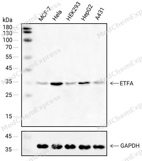 Western blot analysis of extracts from MCF-7(lane 1(40μg) ),Hela(lane 2(40μg) ),HEK293(lane 3(40μg) ),HepG2(lane 4(40μg) )and A431(lane 5(40μg) ) using ETFA antibody. Proteins were transferred to a NC membrane and blocked with 5% Skim milk in TBST for 2 hour at room temperature. The primary antibody ( 1/1000) and Loading control antibody (GAPDH,1/1000) was used in 5% Skim milk in TBST at 4°C overnight. Goat Anti-Mouse IgG-HRP Secondary Antibody (1/10,000) was used for 1 hour at room temperature.
 -
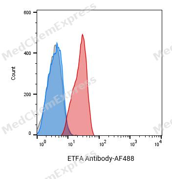 Flow cytometric analysis of 1X10^6 Hela cells labeling ETFA Antibody(red). Cells were fixed with 4% paraformaldehyde and permeabilised with 0.2% Triton X-100. Then stained with the primary antibody at 1/400 dilution overnight at 4℃. AF 488 Goat Anti-mouse IgG H&L was used as the secondary antibody at 1/1,000 dilution for 45 minutes at room temperature. Mouse IgG Isotype Control (blue) was used as the isotype control, cells without incubation with primary antibody were used as the unlabeled control (black).
 -
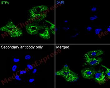 Immunofluorescence analysis of A549 cells labeling ETFA antibody at 1/50 dilution. Cells were fixed in 4% paraformaldehyde for 15 minutes at room temperature, permeabilized with 0.1% Triton X-100 in PBS for 15 minutes at room temperature, then blocked with 1% BSA in 10% negative goat serum for 1 hour at room temperature.Cells were then incubated with ETFA antibody at 1/50 dilution in 1% BSA in PBST overnight at 4 ℃. AF 488 Goat Anti-mouse IgG H&L (green) was used as the secondary antibody at 1/500 dilution. PBS instead of the primary antibody was used as the secondary antibody only control. The Nuclear counterstain was DAPI (Blue)
 -
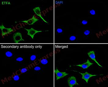 Immunofluorescence analysis of Sk-Br-3 cells labeling ETFA antibody at 1/50 dilution. Cells were fixed in 4% paraformaldehyde for 15 minutes at room temperature, permeabilized with 0.1% Triton X-100 in PBS for 15 minutes at room temperature, then blocked with 1% BSA in 10% negative goat serum for 1 hour at room temperature.Cells were then incubated with ETFA antibody at 1/50 dilution in 1% BSA in PBST overnight at 4 ℃. AF 488 Goat Anti-mouse IgG H&L (green) was used as the secondary antibody at 1/500 dilution. PBS instead of the primary antibody was used as the secondary antibody only control. The Nuclear counterstain was DAPI (Blue)


Background
-
Function
ETFA is a Heterodimeric electron transfer flavoprotein that accepts electrons from several mitochondrial dehydrogenases, including acyl-CoA dehydrogenases, glutaryl-CoA and sarcosine dehydrogenase. It transfers the electrons to the main mitochondrial respiratory chain via ETF-ubiquinone oxidoreductase (ETF dehydrogenase). Required for normal mitochondrial fatty acid oxidation and normal amino acid metabolism[1][2][3][4][5][6][7][8].
-
Subcellular Localization
Mitochondrion matrix
-
Isoforms & Post-Translational Modification
P13804 has 2 isomers: P13804-1: 35080 Da (predicted); P13804-2: 30026 Da (predicted).
The N-terminus is blocked -
Subunit
Heterodimer composed of ETFA and ETFB (PubMed:15159392, PubMed:15975918, PubMed:8962055, PubMed:9334218). Identified in a complex that contains ETFA, ETFB and ETFRF1 (PubMed:27499296).
-
SwissProt ID
-
Synonyms
EMA; GA2; MADD


Documentation

 Powered by Bioz
