



HIF-1α Antibody (YA7478)
(Synonyms: BHLHE78, MOP1, PASD8, HIF1A, Hypoxia-inducible factor 1-alpha, HIF-1-alpha, HIF1-alpha, ARNT-interacting protein, Basic-helix-loop-helix-PAS protein MOP1, Class E basic helix-loop-helix protein 78, Member of PAS protein 1, PAS domain-containing protein 8, bHLHe78)
Based on 1 Customer Validation
HIF-1α Antibody (YA7478) is a Rabbit-derived and non-conjugated IgG, Kappa monoclonal antibody, targeting to HIF-1α.
-
Host:
Rabbit
-
Isotype:
IgG
-
Application:
WB, ICC/IF, IP, ELISA
-
Reactivity :
Human, Mouse
-
Formulation:
Supplied in PBS (pH7.4) containing 50% glycerol, 0.05% Proclin 300, 0.05%BSA
-
Conjugation:
Non-conjugated


To place orders, for customer services and technical support, please contact: MedChemExpress USA
Tel: 609-228-6898 E-mail: [email protected] [email protected]





Applications
| Application |
WB

WB: Western Blot
|
ICC/IF
ICC/IF: Immunocytochemistry/
Immunofluorescence |
ELISA
ELISA: Enzyme Linked Immunosorbent Assay
|
IP
IP: Immunoprecipitation
|
|---|---|---|---|---|
| Dilution Ratio | 1:2000-1:10000 | 1:200-1:1000 | 1:5000-1:20000 | 1:50-1:200 |


Product Details
HIF-1α Antibody (YA7478) is a Rabbit-derived and non-conjugated IgG, Kappa monoclonal antibody, targeting to HIF-1α.
-
Host Rabbit

-
Clonality Monoclonal,Recombinant

-
Species ReactivityHuman, Mouse
-
Observed Molecular WeightObserved band size: 120 kDa
Note: Due to possible protein modifications or aggregation, the molecular weight should be confirmed by actual measurement, and the predicted value is for reference only.

-
Calculated Molecular Weight Predicted band size: 93 kDa

The exact sequence is proprietary to MCE.
Endogenous
Protein A affinity purified
Non-conjugated
Unmodified
IgG


Product Properties
-
Appearance
Solution
-
Formulation
Supplied in PBS (pH7.4) containing 50% glycerol, 0.05% Proclin 300, 0.05%BSA
-
Concentration
Batch-dependent, Please check the COA for the concentration of each lot. Check Lot Concentration
-
Storage & Stability
Stored at -20°C for 1 year. Avoid repeated freeze / thaw cycles.
-
Shipping
Shipping with blue ice.


Verification Images
-
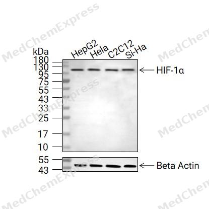 Western blot analysis of extracts from HepG2 (lane 2(20μg), Hela (lane 3(20μg), C2C12 (lane 4(20μg) and Si-Ha (lane 5(20μg) using HIF-1α Antibody (HY-P87793) Rabbit mAb. Proteins were transferred to a PVDF membrane and blocked with 5% non-fat milk in TBST for 2 hour at room temperature. The primary antibody (1/1000) and Loading control antibody (Beta Actin, HY-P80993, 1/10000) was used in 5% non-fat milk in TBST at 4°C overnight. Goat Anti-Rabbit IgG-HRP Secondary Antibody (1/10000) was used for 1 hour at room temperature.


Background
-
Function
HIF-1α (Hypoxia-Inducible Factor-1α) is a master transcription factor that coordinates cellular adaptation to reduced oxygen availability by regulating the expression of hypoxia-responsive genes involved in oxygen homeostasis, angiogenesis, and metabolic adaptation[1][2]. Mechanistically, HIF-1α activates transcriptional programs that increase glucose uptake, glycolytic flux, and mitochondrial adaptation, thereby enabling cells to maintain energy production under hypoxic conditions[3][4]. Through these metabolic and vascular responses, HIF-1α serves as a central regulator of hypoxia signaling and integrates crosstalk with pathways including PI3K-mTOR, NF-κB, and ERK signaling networks[5]. In disease settings, HIF-1α is frequently activated within the hypoxic tumor microenvironment, where it promotes angiogenesis, metabolic reprogramming, immune modulation, tumor progression, and resistance to therapy[6][7][8]. Experimental studies further demonstrate that HIF-1α regulates glycolytic gene expression and contributes to the adaptation of cancer cells to intratumoral hypoxia, making it a widely used molecular target in cancer biology research[3][4]. Compared with related isoforms, HIF-1α and HIF-2α share overlapping functions in hypoxic adaptation but exhibit distinct regulatory and biological roles; HIF-1α is more strongly associated with acute hypoxic responses and glycolytic regulation, whereas HIF-2α preferentially supports chronic hypoxia responses, vascular remodeling, and specific metabolic programs[9][10][11]. For experimental applications, HIF-1α remains an important pharmacological target, and small-molecule inhibitors are extensively investigated to dissect hypoxia-driven signaling and tumor progression mechanisms[12].
-
Subcellular Localization
Cytoplasm,Nucleus,Nucleus speckle
-
Expression
Tissue_Specificity: Expressed in most tissues with highest levels in kidney and heart. Overexpressed in the majority of common human cancers and their metastases, due to the presence of intratumoral hypoxia and as a result of mutations in genes encoding oncoproteins and tumor suppressors. A higher level expression seen in pituitary tumors as compared to the pituitary gland
Induction: Under reduced oxygen tension. Induced also by various receptor-mediated factors such as growth factors, cytokines, and circulatory factors such as PDGF, EGF, FGF2, IGF2, TGFB1, HGF, TNF, IL1B/interleukin-1 beta, angiotensin-2 and thrombin. However, this induction is less intense than that stimulated by hypoxia. Repressed by HIPK2 and LIMD1 -
Isoforms & Post-Translational Modification
Q16665 has three isomers: Q16665-1: 92670 Da (predicted); Q16665-2: 82746 Da (predicted); Q16665-3: 95634 Da (predicted).
S-nitrosylation of Cys-800 may be responsible for increased recruitment of p300 coactivator necessary for transcriptional activity of HIF-1 complex丨Requires phosphorylation for DNA-binding丨Sumoylated; with SUMO1 under hypoxia (PubMed:15465032, PubMed:15776016, PubMed:17610843)丨Acetylation of Lys-532 by ARD1 increases interaction with VHL and stimulates subsequent proteasomal degradation (PubMed:12464182)丨Polyubiquitinated; in normoxia, following hydroxylation and interaction with VHL丨In normoxia, is hydroxylated on Pro-402 and Pro-564 in the oxygen-dependent degradation domain (ODD) by EGLN1/PHD2 and EGLN2/PHD1 (PubMed:11292861, PubMed:11566883, PubMed:12351678, PubMed:15776016, PubMed:25974097)丨The iron and 2-oxoglutarate dependent 3-hydroxylation of asparagine is (S) stereospecific within HIF CTAD domains丨(Microbial infection) Glycosylated at Arg-18 by enteropathogenic E -
Subunit
Interacts with the ARNT; forms a heterodimer that binds core DNA sequence 5'-TACGTG-3' within the hypoxia response element (HRE) of target gene promoters (PubMed:10944113, PubMed:20699359)
-
SwissProt ID
-
Synonyms
BHLHE78, MOP1, PASD8, HIF1A, Hypoxia-inducible factor 1-alpha, HIF-1-alpha, HIF1-alpha, ARNT-interacting protein, Basic-helix-loop-helix-PAS protein MOP1, Class E basic helix-loop-helix protein 78, Member of PAS protein 1, PAS domain-containing protein 8, bHLHe78


Documentation
-
Data Sheet (262 KB)
-
SDS (252 KB)
- English - EN (252 KB)
- Français - FR (252 KB)
- Deutsch - DE (252 KB)
- Norwegian - NO (252 KB)
- Español - ES (252 KB)
- Swedish - SV (252 KB)
- Italian - IT (252 KB)
- Korean - KR (252 KB)
- Portuguese - PT (252 KB)
-
User Guide for Antibodies (1077 KB)
References
[1]. Loboda A, et al. HIF-1 versus HIF-2--is one more important than the other? Vascul Pharmacol. 2012 May-Jun;56(5-6):245-51. [Content Brief]
[2]. Shi YH, et al. Hypoxia-inducible factor-1 in tumour angiogenesis. World J Gastroenterol. 2004 Apr 15;10(8):1082-7. [Content Brief]
[3]. Semenza GL. HIF-1: upstream and downstream of cancer metabolism. Curr Opin Genet Dev. 2010 Feb;20(1):51-6. doi: 10.1016/j.gde.2009.10.009. Epub 2009 Nov 26. PMID: 19942427; PMCID: PMC2822127. et al. HIF-1: upstream and downstream of cancer metabolism. Curr Opin Genet Dev. 2010 Feb;20(1):51-6. [Content Brief]
[4]. Semenza GL. HIF-1 mediates metabolic responses to intratumoral hypoxia and oncogenic mutations. J Clin Invest. 2013 Sep;123(9):3664-71. doi: 10.1172/JCI67230. Epub 2013 Sep 3. PMID: 23999440; PMCID: PMC3754249. et al. HIF-1 mediates metabolic responses to intratumoral hypoxia and oncogenic mutations. J Clin Invest. 2013 Sep;123(9):3664-71. [Content Brief]
[5]. Luo Z, et al. Hypoxia signaling in human health and diseases: implications and prospects for therapeutics. Signal Transduct Target Ther. 2022 Jul 7;7(1):218. [Content Brief]
[6]. Chen Z, et al. Hypoxic microenvironment in cancer: molecular mechanisms and therapeutic interventions. Signal Transduct Target Ther. 2023 Feb 17;8(1):70. [Content Brief]
[7]. Petrova V, et al. The hypoxic tumour microenvironment. Oncogenesis. 2018 Jan 24;7(1):10. [Content Brief]
[8]. Bae T, et al. Hypoxia, oxidative stress, and the interplay of HIFs and NRF2 signaling in cancer. Exp Mol Med. 2024 Mar;56(3):501-514. [Content Brief]
[9]. Hoefflin R, et al. HIF-1α and HIF-2α differently regulate tumour development and inflammation of clear cell renal cell carcinoma in mice. Nat Commun. 2020 Aug 17;11(1):4111. [Content Brief]
[10]. Jaśkiewicz M, et al. The transition from HIF-1 to HIF-2 during prolonged hypoxia results from reactivation of PHDs and HIF1A mRNA instability. Cell Mol Biol Lett. 2022 Dec 8;27(1):109. [Content Brief]
[11]. Hu CJ, et al. Differential roles of hypoxia-inducible factor 1alpha (HIF-1alpha) and HIF-2alpha in hypoxic gene regulation. Mol Cell Biol. 2003 Dec;23(24):9361-74. [Content Brief]
[12]. Basheeruddin M, et al. Hypoxia-Inducible Factor 1-Alpha (HIF-1α) and Cancer: Mechanisms of Tumor Hypoxia and Therapeutic Targeting. Cureus. 2024 Oct 2;16(10):e70700. [Content Brief]

 Powered by Bioz
