



IL-10 Antibody (YA4644)
(Synonyms: Interleukin-10 precursor; Cytokine synthesis inhibitory factor; CSIF; Cytokine Synthesis Inhibitory Factor; IL 10; IL10A; Interleukin 10; Interleukin10; MGC126450; MGC126451; TGIF; IL10_HUMAN. 白介素10)Based on 1 Customer Validation
IL-10 Antibody (YA4644) is a Rabbit-derived and non-conjugated IgG monoclonal antibody, targeting to IL-10.
-
Host:
Rabbit
-
Isotype:
IgG
-
Application:
WB, IHC-P, ICC/IF
-
Reactivity :
Human, Mouse, Rat
-
Formulation:
Supplied in 1*TBS (pH7.4), 0.05% BSA, 40% Glycerol. Preservative: 0.05% Sodium Azide.
-
Conjugation:
Non-conjugated


To place orders, for customer services and technical support, please contact: MedChemExpress USA
Tel: 609-228-6898 E-mail: [email protected] [email protected]





Applications
| Application |
WB

WB: Western Blot
|
ICC/IF
ICC/IF: Immunocytochemistry/
Immunofluorescence |
IHC-P
IHC-P: Immunohistochemistry-Paraffin
|
|---|---|---|---|
| Dilution Ratio | 1:1000 | 1:500 | 1:1000 |


Product Details
IL-10 Antibody (YA4644) is a Rabbit-derived and non-conjugated IgG monoclonal antibody, targeting to IL-10.
-
Host Rabbit

-
Clonality Monoclonal,Recombinant

-
Species ReactivityHuman, Mouse, Rat
-
Observed Molecular WeightObserved band size: 21 kDa
Note: Due to possible protein modifications or aggregation, the molecular weight should be confirmed by actual measurement, and the predicted value is for reference only.

-
Calculated Molecular Weight Predicted band size: 21 kDa

Synthetic peptide within Human IL-10 aa 1-100.
affinity purified
Non-conjugated
Unmodified
IgG


Product Properties
-
Appearance
Solution
-
Formulation
Supplied in 1*TBS (pH7.4), 0.05% BSA, 40% Glycerol. Preservative: 0.05% Sodium Azide.
-
Concentration
Batch-dependent, Please check the COA for the concentration of each lot. Check Lot Concentration
-
Storage & Stability
Stored at -20°C for 1 year. Avoid repeated freeze / thaw cycles.
-
Shipping
Shipping with blue ice.


Verification Images
-
 Western blot analysis of extracts from Rat spleen tissue (lane2, 20μg), THP-1 (lane3, 20μg) and HL-60 (lane4, 20μg) using IL-10 Antibody (HY-P84952). Proteins were transferred to a PVDF membrane and blocked with 5% non-fat milk in TBST for 2 hour at room temperature. The primary antibody (1/1000) and Loading control antibody (Beta Actin, HY-P80993, 1/10,000) was used in 5% non-fat milk in TBST at 4°C overnight. Goat Anti-Rabbit IgG-HRP Secondary Antibody (HY-P8001,1/10,000) was used for 1 hour at room temperature.
 -
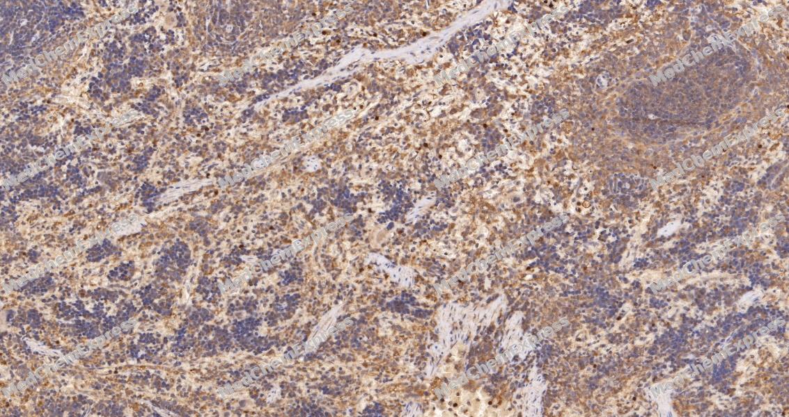 Immunohistochemical analysis of paraffin-embedded mouse spleen tissue using IL-10 Antibody. The section was pre-treated using heat mediated antigen retrieval with sodium citrate buffer (pH 6.0) for 8 minutes. The tissues were blocked in QuickBlock for 20 minutes at room temperature, washed with ddH2O and PBS, and then probed with the primary antibody at 1/100 dilution in 4℃ overnight. The detection was performed using an HRP conjugated compact polymer system. DAB was used as the chromogen. Tissues were counterstained with hematoxylin and mounted with DPX.
 -
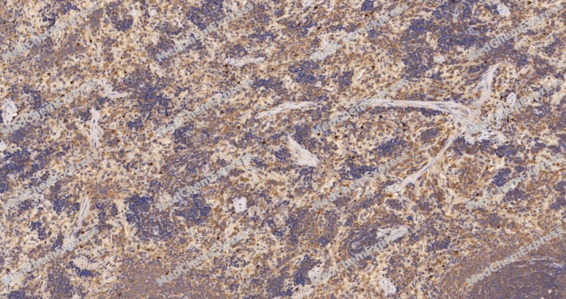 Immunohistochemical analysis of paraffin-embedded mouse spleen tissue using IL-10 Antibody. The section was pre-treated using heat mediated antigen retrieval with sodium citrate buffer (pH 6.0) for 8 minutes. The tissues were blocked in QuickBlock for 20 minutes at room temperature, washed with ddH2O and PBS, and then probed with the primary antibody at 1/100 dilution in 4℃ overnight. The detection was performed using an HRP conjugated compact polymer system. DAB was used as the chromogen. Tissues were counterstained with hematoxylin and mounted with DPX.


Background
-
Function
Interleukin-10 (IL-10) functions as a critical anti-inflammatory cytokine that modulates innate and adaptive immune responses[1][2]. Mechanistically, IL-10 activates the JAK-STAT signaling pathway, particularly STAT3, to suppress proinflammatory cytokine production and promote immunoregulatory gene expression[3][1]. In experimental models of bacterial sepsis, IL-10 partially restores mitochondrial function and oxidative phosphorylation in monocytes, counteracting pathogen-induced metabolic inhibition[1]. T cell-derived IL-10 production is regulated by metabolic pathways, including aryl hydrocarbon receptor (Ahr) and Bhlhe40, which control cytokine output and mucosal immune homeostasis in intestinal inflammation models[2]. Compared with related cytokines, IL-10 displays distinct anti-inflammatory potency and feedback regulation not shared by IL-6 or TNF-α[3][1]. Agonists and inhibitors targeting upstream metabolic or signaling modulators, such as dichloroacetate (DCA), can selectively enhance IL-10 production in Th1 cells, demonstrating translational potential for inflammatory bowel disease research[2]. Furthermore, IL-10 isoform-specific signaling contributes to differential regulation of oxidative phosphorylation genes, offering a mechanistic basis for isoform-focused experimental applications[1]. Collectively, these findings highlight IL-10 as a central regulator of immune homeostasis, with mechanistic specificity distinct from related cytokines and amenable to pharmacological modulation in experimental and disease-relevant contexts[3][1][2].
-
Subcellular Localization
Secreted
-
Expression
Tissue_specificity:It is generated from a variety of cell lines, including T cells, macrophages, mast cells, and other cell types.
Positive sample: THP-1 cell lysate, THP-1 treated with 800nM TPA overnight then treated with 100ng/mL LPS for 6 hours add 300ng/mL BFA for 3 hours cell lysate, mouse spleen tissue lysate, rat spleen tissue lysate, -
Subunit
Homodimer (PubMed:11485736, PubMed:15837194). Interacts with IL10RA and IL10RB (PubMed:16982608)
-
SwissProt ID
-
Synonyms
Interleukin-10 precursor; Cytokine synthesis inhibitory factor; CSIF; Cytokine Synthesis Inhibitory Factor; IL 10; IL10A; Interleukin 10; Interleukin10; MGC126450; MGC126451; TGIF; IL10_HUMAN. 白介素10


Documentation
-
Data Sheet (261 KB)
-
SDS (366 KB)
- English - EN (366 KB)
- Français - FR (366 KB)
- Deutsch - DE (366 KB)
- Norwegian - NO (366 KB)
- Español - ES (366 KB)
- Swedish - SV (366 KB)
- Italian - IT (366 KB)
- Korean - KR (366 KB)
- Portuguese - PT (366 KB)
-
User Guide for Antibodies (1077 KB)
References
[1]. Cheon H, et al. Prostaglandin E2 augments IL-10 signaling and function. J Immunol. 2006 Jul 15;177(2):1092-100. [Content Brief]
[2]. Gopinathan U, et al. Transcriptomic data from two primary cell models stimulating human monocytes suggest inhibition of oxidative phosphorylation and mitochondrial function by N. meningitidis which is partially up-regulated by IL-10. BMC Immunol. 2017 Oct 27;18(1):46. [Content Brief]
[3]. Liu H, et al. Metabolic regulation of T cell production of IL-10 and IL-22 protects against intestinal inflammation. Precis Clin Med. 2025 Oct 24;8(4):pbaf025. [Content Brief]

 Powered by Bioz
