



Keap1 Antibody (YA6097)
(Synonyms: Kelch-like ECH-associated protein 1 (Cytosolic inhibitor of Nrf2) (INrf2) (Kelch-like protein 19))
Based on 1 Customer Validation
Keap1 Antibody (YA6097) is a Rabbit-derived and non-conjugated IgG monoclonal antibody, targeting to Keap1.
-
Host:
Rabbit
-
Isotype:
IgG
-
Application:
WB, IHC-P, ICC/IF, IP, ELISA
-
Reactivity :
Human, Mouse, Rat
-
Formulation:
Supplied in PBS, 50% glycerol, 0.05% Proclin 300, 0.05%BSA
-
Conjugation:
Non-conjugated


To place orders, for customer services and technical support, please contact: MedChemExpress USA
Tel: 609-228-6898 E-mail: [email protected] [email protected]





Applications
| Application |
IHC-P

IHC-P: Immunohistochemistry-Paraffin
|
WB
WB: Western Blot
|
ICC/IF
ICC/IF: Immunocytochemistry/
Immunofluorescence |
ELISA
ELISA: Enzyme Linked Immunosorbent Assay
|
IP
IP: Immunoprecipitation
|
|---|---|---|---|---|---|
| Dilution Ratio | 1:200-1:1000 | 1:2000-1:10000 | 1:200-1:1000 | 1:5000-1:20000 | 1:50-1:200 |


Product Details
Keap1 Antibody (YA6097) is a Rabbit-derived and non-conjugated IgG monoclonal antibody, targeting to Keap1.
-
Host Rabbit

-
Clonality Monoclonal

-
Species ReactivityHuman, Mouse, Rat
-
Observed Molecular WeightObserved band size: 60-70 kDa
Note: Due to possible protein modifications or aggregation, the molecular weight should be confirmed by actual measurement, and the predicted value is for reference only.

-
Calculated Molecular Weight Predicted band size: 70 kDa

Protein A
Non-conjugated
Unmodified
IgG


Product Properties
-
Appearance
Liquid
-
Formulation
Supplied in PBS, 50% glycerol, 0.05% Proclin 300, 0.05%BSA
-
Concentration
Batch-dependent, Please check the COA for the concentration of each lot. Check Lot Concentration
-
Storage & Stability
Stored at -20°C for 1 year. Avoid repeated freeze / thaw cycles.
-
Shipping
Shipping with blue ice.


Verification Images
-
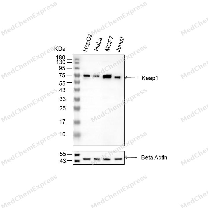 Western blot analysis of extracts from HepG2 (lane 2(20μg), HeLa (lane 3(20μg), MCF7 (lane 4(20μg) and Jurkat (lane 5(20μg) using Keap1 Antibody (HY-P86405) Rabbit mAb. Proteins were transferred to a PVDF membrane and blocked with 5% non-fat milk in TBST for 2 hour at room temperature. The primary antibody (1/1000)and Loading control antibody (Beta Actin, HY-P80993, 1/10000) was used in 5% non-fat milk in TBST at 4°C overnight. Goat Anti-Rabbit IgG-HRP Secondary Antibody (1/10000) was used for 1 hour at room temperature.
 -
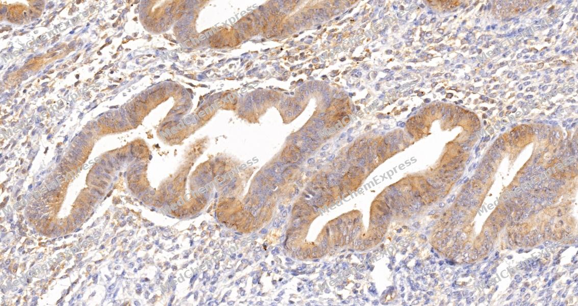 Immunohistochemical analysis of paraffin-embedded human endometrium tissue using Keap1 Antibody (HY-P86405, 1/500). The section was pretreated using heat mediated antigen retrieval with sodium citrate buffer (pH 6.0) for 8 minutes. The tissues were blocked with quick block buffer for 0.5 hours at room temperature, washed with PBS and PBST, and then incubated with the primary antibody overnight at 4℃. The detection was performed using an HRP conjugated compact polymer system. DAB was used as the chromogen. Tissues were counterstained with hematoxylin and mounted with DPX.
 -
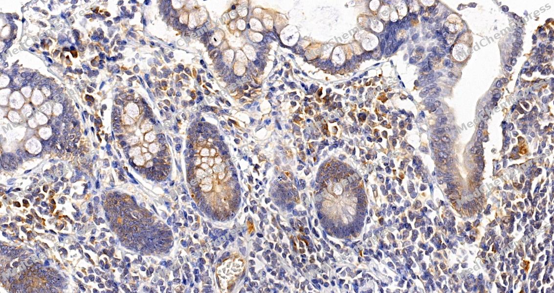 Immunohistochemical analysis of paraffin-embedded human small intestine tissue using Keap1 Antibody (HY-P86405, 1/500). The section was pretreated using heat mediated antigen retrieval with sodium citrate buffer (pH 6.0) for 8 minutes. The tissues were blocked with quick block buffer for 0.5 hours at room temperature, washed with PBS and PBST, and then incubated with the primary antibody overnight at 4℃. The detection was performed using an HRP conjugated compact polymer system. DAB was used as the chromogen. Tissues were counterstained with hematoxylin and mounted with DPX.
 -
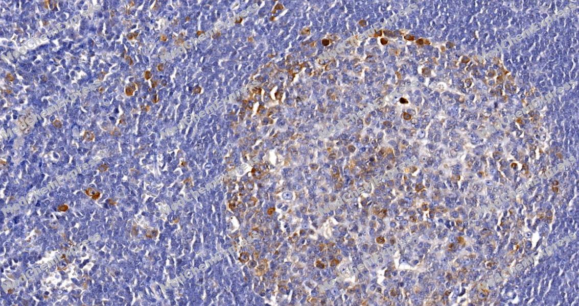 Immunohistochemical analysis of paraffin-embedded human tonsil tissue using Keap1 Antibody (HY-P86405, 1/500). The section was pretreated using heat mediated antigen retrieval with sodium citrate buffer (pH 6.0) for 8 minutes. The tissues were blocked with quick block buffer for 0.5 hours at room temperature, washed with PBS and PBST, and then incubated with the primary antibody overnight at 4℃. The detection was performed using an HRP conjugated compact polymer system. DAB was used as the chromogen. Tissues were counterstained with hematoxylin and mounted with DPX.
 -
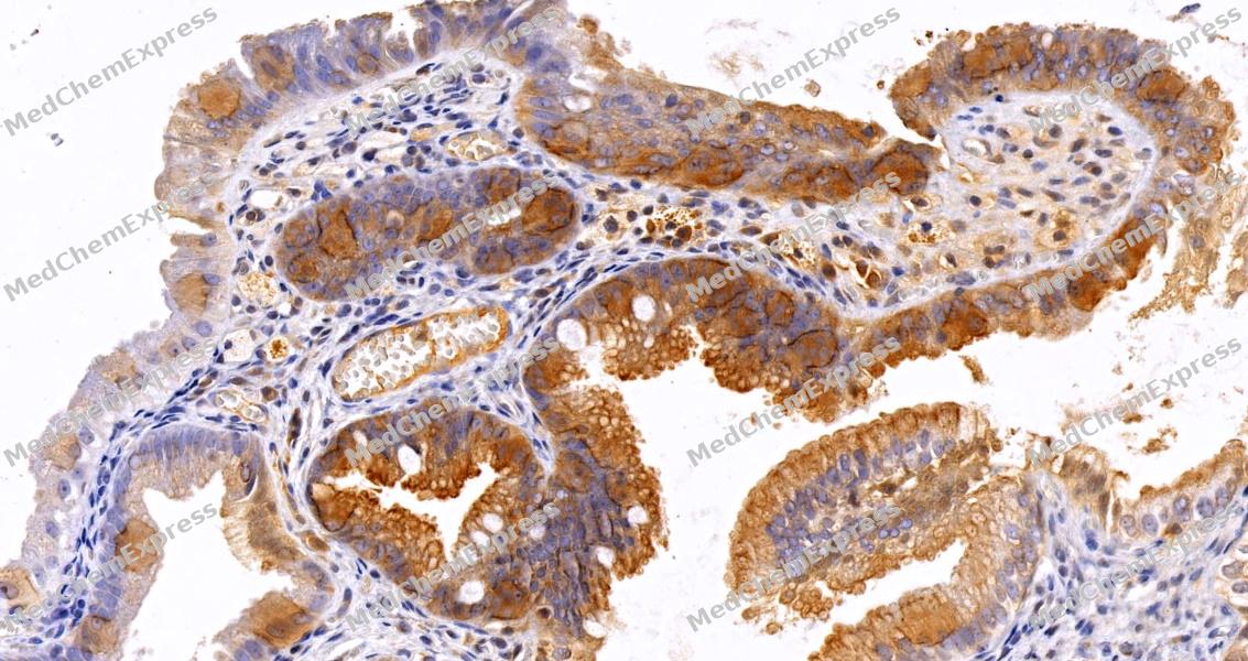 Immunohistochemical analysis of paraffin-embedded human gallbladder tissue using Keap1 Antibody (HY-P86405, 1/500). The section was pretreated using heat mediated antigen retrieval with sodium citrate buffer (pH 6.0) for 8 minutes. The tissues were blocked with quick block buffer for 0.5 hours at room temperature, washed with PBS and PBST, and then incubated with the primary antibody overnight at 4℃. The detection was performed using an HRP conjugated compact polymer system. DAB was used as the chromogen. Tissues were counterstained with hematoxylin and mounted with DPX.
 -
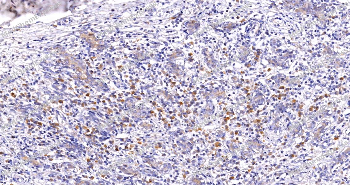 Immunohistochemical analysis of paraffin-embedded human breast tissue using Keap1 Antibody (HY-P86405, 1/500). The section was pretreated using heat mediated antigen retrieval with sodium citrate buffer (pH 6.0) for 8 minutes. The tissues were blocked with quick block buffer for 0.5 hours at room temperature, washed with PBS and PBST, and then incubated with the primary antibody overnight at 4℃. The detection was performed using an HRP conjugated compact polymer system. DAB was used as the chromogen. Tissues were counterstained with hematoxylin and mounted with DPX.
 -
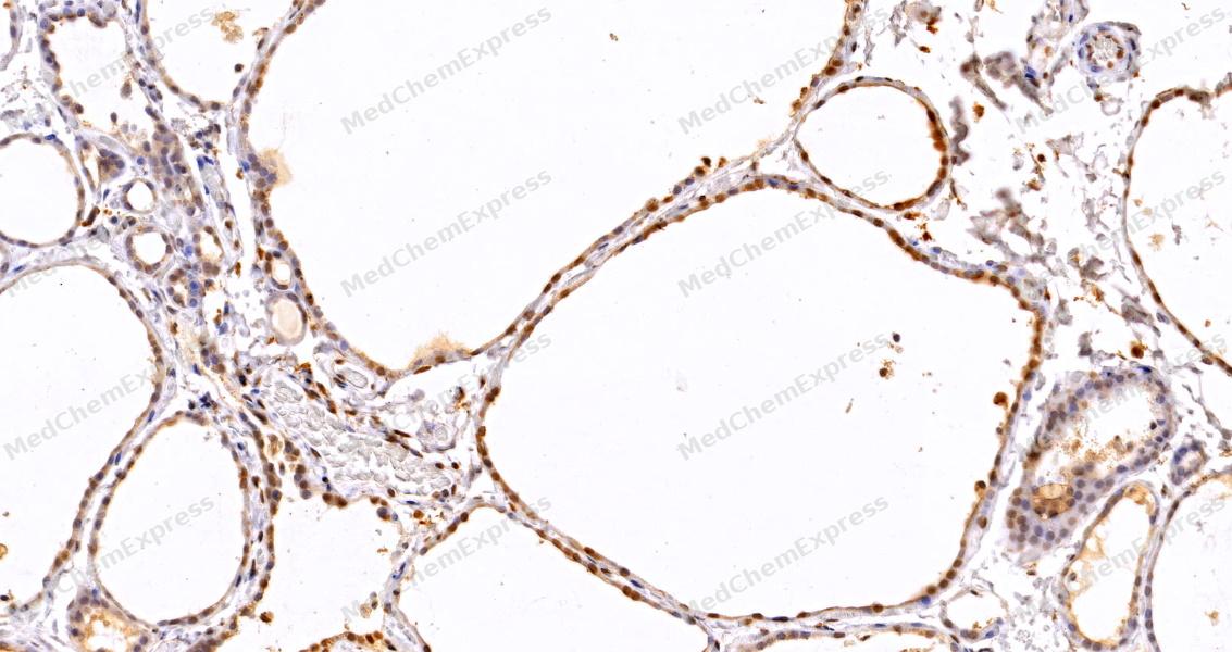 Immunohistochemical analysis of paraffin-embedded human thyroid gland tissue using Keap1 Antibody (HY-P86405, 1/500). The section was pretreated using heat mediated antigen retrieval with sodium citrate buffer (pH 6.0) for 8 minutes. The tissues were blocked with quick block buffer for 0.5 hours at room temperature, washed with PBS and PBST, and then incubated with the primary antibody overnight at 4℃. The detection was performed using an HRP conjugated compact polymer system. DAB was used as the chromogen. Tissues were counterstained with hematoxylin and mounted with DPX.
 -
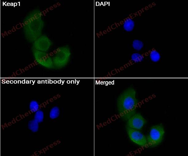 Immunocytochemistry analysis of HeLa cells labeling Keap1 Antibody (HY-P86405) at 1/200 dilution. Cells were fixed in 4% paraformaldehyde for 15 minutes at room temperature, permeabilized with 0.1% Triton X-100 for 10 minutes at room temperature, then blocked with BSA for Immunol Staining for 10 min at room temperature. Cells were then incubated with Keap1 Antibody (HY-P86405) at 1/50 dilution in BSA for Immunol Staining at 4 ℃ overnight. AF488-conjugated AffiniPure Goat Anti-Rabbit /Mouse IgG H&L(HY-P8002/HY-P8005, Green) was used as the secondary antibody at 1/1,000 dilution. PBS instead of the primary antibody was used as the secondary antibody only control. The Nuclear counterstain was DAPI (Blue).


Background
-
Function
Keap1 is a Substrate-specific adapter of a BCR (BTB-CUL3-RBX1) E3 ubiquitin ligase complex that regulates the response to oxidative stress by targeting NFE2L2/NRF2 for ubiquitination. KEAP1 acts as a key sensor of oxidative and electrophilic stress: in normal conditions, the BCR(KEAP1) complex mediates ubiquitination and degradation of NFE2L2/NRF2, a transcription factor regulating expression of many cytoprotective genes. In response to oxidative stress, different electrophile metabolites trigger non-enzymatic covalent modifications of highly reactive cysteine residues in KEAP1, leading to inactivate the ubiquitin ligase activity of the BCR(KEAP1) complex, promoting NFE2L2/NRF2 nuclear accumulation and expression of phase II detoxifying enzymes. In response to selective autophagy, KEAP1 is sequestered in inclusion bodies following its interaction with SQSTM1/p62, leading to inactivation of the BCR(KEAP1) complex and activation of NFE2L2/NRF2. The BCR(KEAP1) complex also mediates ubiquitination of SQSTM1/p62, increasing SQSTM1/p62 sequestering activity and degradation. The BCR(KEAP1) complex also targets BPTF and PGAM5 for ubiquitination and degradation by the proteasome[1][2][3][4][5][6][7][8][9][10][11][12][13][14].
-
Subcellular Localization
Cytoplasm; Nucleus
-
Expression
Tissue_specificity:It is widely expressed, with the highest concentration in skeletal muscle. -
Subunit
Component of the BCR(KEAP1) E3 ubiquitin ligase complex, at least composed of 2 molecules of CUL3, 2 molecules of KEAP1, and RBX1 (PubMed:15572695, PubMed:15601839, PubMed:15983046, PubMed:17127771, PubMed:18251510, PubMed:24896564). Interacts with NFE2L2/NRF2; the interaction is direct (PubMed:15379550, PubMed:15601839, PubMed:16006525, PubMed:16888629, PubMed:18387606). Forms a ternary complex with NFE2L2/NRF2 and PGAM5 (PubMed:17046835). Interacts with (phosphorylated) SQSTM1/p62; the interaction is direct and inactivates the BCR(KEAP1) complex by sequestering it in inclusion bodies, promoting its degradation (PubMed:20452972, PubMed:20495340). Interacts with NFE2L1 (PubMed:16687406). Interacts with BPTF and PTMA (PubMed:15657435). Interacts with MAP1LC3B (PubMed:24089205). Interacts indirectly with ENC1 (PubMed:19424503). Interacts with SESN1 and SESN2 (PubMed:23274085). Interacts with HSP90AA1 and HSP90AB1 (PubMed:26517842). Interacts with PGCKA1; this interaction prevents the ubiquitination of KEAP1 by TRIM25, thus protecting KEAP1 from degradation (PubMed:36882524)
-
SwissProt ID
-
Synonyms
Kelch-like ECH-associated protein 1 (Cytosolic inhibitor of Nrf2) (INrf2) (Kelch-like protein 19)


Documentation
-
Data Sheet (235 KB)
-
SDS (252 KB)
- English - EN (252 KB)
- Français - FR (252 KB)
- Deutsch - DE (252 KB)
- Norwegian - NO (252 KB)
- Español - ES (252 KB)
- Swedish - SV (252 KB)
- Italian - IT (252 KB)
- Korean - KR (252 KB)
- Portuguese - PT (252 KB)
-
User Guide for Antibodies (1077 KB)

 Powered by Bioz
