



Lipocalin 2 Antibody (YA992)
(Synonyms: LCN2; 24p3; MSFI; NGAL; Lipocalin-2; p25; Siderocalin; SV40 induced 24P3 protein; Uterocalin)Based on 1 publication(s) in Google Scholar
Lipocalin 2 Antibody (YA992) is a Rabbit-derived and non-conjugated IgG monoclonal antibody, targeting to Lipocalin 2.
-
Host:
Rabbit
-
Isotype:
IgG
-
Application:
WB, IHC-P, ICC/IF
-
Reactivity :
Human
-
Formulation:
Supplied in Rabbit IgG in 10mM phosphate buffered saline , pH 7.4, 150mM sodium chloride, 0.05% BSA, 0.02% sodium azide and 50% glycerol.
-
Conjugation:
Non-conjugated


To place orders, for customer services and technical support, please contact: MedChemExpress USA
Tel: 609-228-6898 E-mail: [email protected] [email protected]



Publications Citing Use of MedChemExpress (MCE) Lipocalin 2 Antibody (YA992)
More

Applications
| Application |
WB

WB: Western Blot
|
IHC-P
IHC-P: Immunohistochemistry-Paraffin
|
ICC/IF
ICC/IF: Immunocytochemistry/
Immunofluorescence |
|---|---|---|---|
| Dilution Ratio | 1:500-1:1000 | 1:50-1:100 | 1:50-1:200 |


Product Details
Lipocalin 2 Antibody (YA992) is a Rabbit-derived and non-conjugated IgG monoclonal antibody, targeting to Lipocalin 2.
-
Host Rabbit

-
Clonality Recombinant,Monoclonal

-
Species ReactivityHuman
-
Observed Molecular WeightObserved band size: 23 kDa
Note: Due to possible protein modifications or aggregation, the molecular weight should be confirmed by actual measurement, and the predicted value is for reference only.

-
Calculated Molecular Weight Predicted band size: 23 kDa

A synthesized peptide derived from human Lipocalin-2 aa50-100.
Endogenous
Affinity Chromatography
Non-conjugated
Unmodified
IgG


Product Properties
-
Appearance
Solution
-
Formulation
Supplied in Rabbit IgG in 10mM phosphate buffered saline , pH 7.4, 150mM sodium chloride, 0.05% BSA, 0.02% sodium azide and 50% glycerol.
-
Concentration
Batch-dependent, Please check the COA for the concentration of each lot. Check Lot Concentration
-
Storage & Stability
Stored at -20°C for 1 year. Avoid repeated freeze / thaw cycles.
-
Shipping
Shipping with blue ice.


Publications (1)
-
Journal Impact Factor
-
Most Recent
-
Int Immunopharmacol
SCD1 protects acinar cells from ferroptosis during severe acute pancreatitis by balancing lipid and redox homeostasis. [Abstract]2025 Sep 23:162:115171. PMID: 40617047


Verification Images
-
 Immunohistochemical analysis of paraffin-embedded human spleen tissue using Lipocalin 2 Antibody (HY-P81284A, 1/100). The section was pretreated using heat mediated antigen retrieval with sodium citrate buffer (pH 6.0) for 8 minutes. The tissues were blocked with quick block buffer for 0.5 hours at room temperature, washed with PBS and PBST, and then incubated with the primary antibody overnight at 4℃. The detection was performed using an HRP conjugated compact polymer system. DAB was used as the chromogen. Tissues were counterstained with hematoxylin and mounted with DPX.
 -
 Immunohistochemical analysis of paraffin-embedded human stomach tissue using Lipocalin 2 Antibody (HY-P81284A, 1/100). The section was pretreated using heat mediated antigen retrieval with sodium citrate buffer (pH 6.0) for 8 minutes. The tissues were blocked with quick block buffer for 0.5 hours at room temperature, washed with PBS and PBST, and then incubated with the primary antibody overnight at 4℃. The detection was performed using an HRP conjugated compact polymer system. DAB was used as the chromogen. Tissues were counterstained with hematoxylin and mounted with DPX.
 -
 Immunohistochemical analysis of paraffin-embedded human bone marrow tissue using Lipocalin 2 Antibody (HY-P81284A, 1/100). The section was pretreated using heat mediated antigen retrieval with sodium citrate buffer (pH 6.0) for 8 minutes. The tissues were blocked with quick block buffer for 0.5 hours at room temperature, washed with PBS and PBST, and then incubated with the primary antibody overnight at 4℃. The detection was performed using an HRP conjugated compact polymer system. DAB was used as the chromogen. Tissues were counterstained with hematoxylin and mounted with DPX.
 -
 Immunohistochemical analysis of paraffin-embedded rat stomach tissue using Lipocalin 2 Antibody (HY-P81284A, 1/100). The section was pretreated using heat mediated antigen retrieval with sodium citrate buffer (pH 6.0) for 8 minutes. The tissues were blocked with quick block buffer for 0.5 hours at room temperature, washed with PBS and PBST, and then incubated with the primary antibody overnight at 4℃. The detection was performed using an HRP conjugated compact polymer system. DAB was used as the chromogen. Tissues were counterstained with hematoxylin and mounted with DPX.
 -
 Immunohistochemical analysis of paraffin-embedded rat rectum tissue using Lipocalin 2 Antibody (HY-P81284A, 1/100). The section was pretreated using heat mediated antigen retrieval with sodium citrate buffer (pH 6.0) for 8 minutes. The tissues were blocked with quick block buffer for 0.5 hours at room temperature, washed with PBS and PBST, and then incubated with the primary antibody overnight at 4℃. The detection was performed using an HRP conjugated compact polymer system. DAB was used as the chromogen. Tissues were counterstained with hematoxylin and mounted with DPX.
 -
 Immunohistochemical analysis of paraffin-embedded rat colon tissue using Lipocalin 2 Antibody (HY-P81284A, 1/100). The section was pretreated using heat mediated antigen retrieval with sodium citrate buffer (pH 6.0) for 8 minutes. The tissues were blocked with quick block buffer for 0.5 hours at room temperature, washed with PBS and PBST, and then incubated with the primary antibody overnight at 4℃. The detection was performed using an HRP conjugated compact polymer system. DAB was used as the chromogen. Tissues were counterstained with hematoxylin and mounted with DPX.
 -
 Immunohistochemical analysis of paraffin-embedded rat stomach tissue using Lipocalin 2 Antibody (HY-P81284A, 1/50). The section was pretreated using heat mediated antigen retrieval with sodium citrate buffer (pH 6.0) for 8 minutes. The tissues were blocked with quick block buffer for 0.5 hours at room temperature, washed with PBS and PBST, and then incubated with the primary antibody overnight at 4℃. The detection was performed using an HRP conjugated compact polymer system. DAB was used as the chromogen. Tissues were counterstained with hematoxylin and mounted with DPX.
 -
 Immunohistochemical analysis of paraffin-embedded rat spleen tissue using Lipocalin 2 Antibody (HY-P81284A, 1/50). The section was pretreated using heat mediated antigen retrieval with sodium citrate buffer (pH 6.0) for 8 minutes. The tissues were blocked with quick block buffer for 0.5 hours at room temperature, washed with PBS and PBST, and then incubated with the primary antibody overnight at 4℃. The detection was performed using an HRP conjugated compact polymer system. DAB was used as the chromogen. Tissues were counterstained with hematoxylin and mounted with DPX.
 -
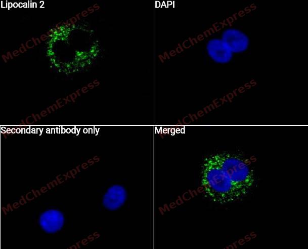 Immunocytochemistry analysis of A549 cells labeling Lipocalin 2 with Lipocalin 2 Antibody (HY-P81284A) at 1/50 dilution. Cells were fixed in 4% paraformaldehyde for 15 minutes at room temperature, permeabilized with 0.1% Triton X-100 for 10 minutes at room temperature, then blocked with QuickBlock™ Blocking Buffer for Immunol Staining for 10 min at room temperature. Cells were then incubated with Lipocalin 2 Antibody (HY-P81284A) at 1/50 dilution in QuickBlock™ Blocking Buffer for Immunol Staining at 4 ℃. Alexa Fluor® 488-conjugated AffiniPure Goat Anti-Rabbit IgG H&L(HY-P8002, Green) was used as the secondary antibody at 1/1,000 dilution. PBS instead of the primary antibody was used as the secondary antibody only control. The Nuclear counterstain was DAPI (Blue).
 -
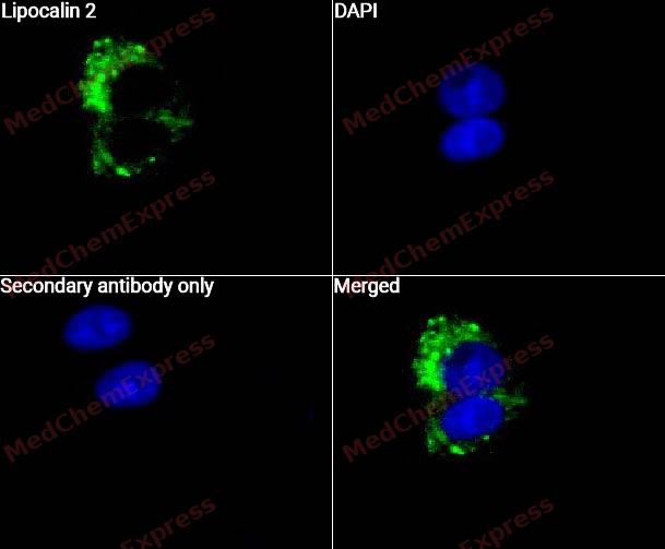 Immunocytochemistry analysis of A431 cells labeling Lipocalin 2 with Lipocalin 2 Antibody (HY-P81284A) at 1/50 dilution. Cells were fixed in 4% paraformaldehyde for 15 minutes at room temperature, permeabilized with 0.1% Triton X-100 for 10 minutes at room temperature, then blocked with QuickBlock™ Blocking Buffer for Immunol Staining for 10 min at room temperature. Cells were then incubated with Lipocalin 2 Antibody (HY-P81284A) at 1/50 dilution in QuickBlock™ Blocking Buffer for Immunol Staining at 4 ℃. Alexa Fluor® 488-conjugated AffiniPure Goat Anti-Rabbit IgG H&L(HY-P8002, Green) was used as the secondary antibody at 1/1,000 dilution. PBS instead of the primary antibody was used as the secondary antibody only control. The Nuclear counterstain was DAPI (Blue).


Background
-
Function
Lipocalin 2 is an Iron-trafficking protein involved in multiple processes such as apoptosis, innate immunity and renal development. Binds iron through association with 2,3-dihydroxybenzoic acid (2,3-DHBA), a siderophore that shares structural similarities with bacterial enterobactin, and delivers or removes iron from the cell, depending on the context. Iron-bound form (holo-24p3) is internalized following binding to the SLC22A17 (24p3R) receptor, leading to release of iron and subsequent increase of intracellular iron concentration. In contrast, association of the iron-free form (apo-24p3) with the SLC22A17 (24p3R) receptor is followed by association with an intracellular siderophore, iron chelation and iron transfer to the extracellular medium, thereby reducing intracellular iron concentration. Involved in apoptosis due to interleukin-3 (IL3) deprivation: iron-loaded form increases intracellular iron concentration without promoting apoptosis, while iron-free form decreases intracellular iron levels, inducing expression of the proapoptotic protein BCL2L11/BIM, resulting in apoptosis. Involved in innate immunity; limits bacterial proliferation by sequestering iron bound to microbial siderophores, such as enterobactin. Can also bind siderophores from M.tuberculosis[1][2][3][4][5].
-
Subcellular Localization
Secreted; Cytoplasmic granule lumen; Cytoplasmic vesicle lumen
-
Expression
Tissue_specificity:Detected in neutrophils (protein level) (PubMed:7683678, PubMed:8298140) . Expressed in bone marrow and tissues susceptible to microbial infection (PubMed:9339356) . Highly expressed in bone marrow, as well as in the uterus, prostate, salivary glands, stomach, appendix, colon, trachea, and lungs (PubMed:9339356) . Expressed in renal medullary tubules (PubMed:30418175) . Not detected in small intestine or peripheral blood leukocytes (PubMed:9339356) .
Induction:Expression is activated by the oncoprotein BCR-ABL; BCR-ABL misregulates expression via the JAK/STAT pathway and binding of STAT5A to the promoter (PubMed:19229297) . Induced by insulin (PubMed:30418175) -
Isoforms & Post-Translational Modification
P80188 has 2 isomers: P80188-1: 22588 Da (predicted); P80188-2: 22457 Da (predicted).
-
Subunit
Monomer (PubMed:1281792, PubMed:7683678). Homodimer; disulfide-linked (PubMed:7683678). Heterodimer; disulfide-linked with MMP9 (PubMed:7683678)
-
SwissProt ID
-
Synonyms
LCN2; 24p3; MSFI; NGAL; Lipocalin-2; p25; Siderocalin; SV40 induced 24P3 protein; Uterocalin
-
Research Field
Immunology
Documentation
-
Data Sheet (261 KB)
-
SDS (252 KB)
- English - EN (252 KB)
- Français - FR (252 KB)
- Deutsch - DE (252 KB)
- Norwegian - NO (252 KB)
- Español - ES (252 KB)
- Swedish - SV (252 KB)
- Italian - IT (252 KB)
- Korean - KR (252 KB)
- Portuguese - PT (252 KB)
-
User Guide for Antibodies (1077 KB)
References
[1]. Yang J, et al. An iron delivery pathway mediated by a lipocalin. Mol Cell. 2002 Nov;10(5):1045-56. [Content Brief]
[2]. Bao G, et al. Iron traffics in circulation bound to a siderocalin (Ngal)-catechol complex. Nat Chem Biol. 2010 Aug;6(8):602-9. [Content Brief]
[3]. Shields-Cutler RR, et al. Human Metabolome-derived Cofactors Are Required for the Antibacterial Activity of Siderocalin in Urine. J Biol Chem. 2016 Dec 9;291(50):25901-25910. [Content Brief]
[4]. Holmes MA, et al. Siderocalin (Lcn 2) also binds carboxymycobactins, potentially defending against mycobacterial infections through iron sequestration. Structure. 2005 Jan;13(1):29-41. [Content Brief]
[5]. Hoette TM, et al. Immune interference in Mycobacterium tuberculosis intracellular iron acquisition through siderocalin recognition of carboxymycobactins. ACS Chem Biol. 2011 Dec 16;6(12):1327-31. [Content Brief]

 Powered by Bioz
