



MST1 Antibody (YA2368)
(Synonyms: HGFL; MSP; MST1; NF15S2)Based on 1 publication(s) in Google Scholar
MST1 Antibody (YA2368) is a Rabbit-derived and non-conjugated IgG monoclonal antibody, targeting to MST1.
-
Host:
Rabbit
-
Isotype:
IgG
-
Application:
WB, IHC-P
-
Reactivity :
Human, Mouse, Rat
-
Formulation:
Supplied in 50mM Tris-Glycine(pH 7.4), 0.15M NaCl, 40%Glycerol, 0.01% sodium azide and 0.05% BSA.
-
Conjugation:
Non-conjugated


To place orders, for customer services and technical support, please contact: MedChemExpress USA
Tel: 609-228-6898 E-mail: [email protected] [email protected]



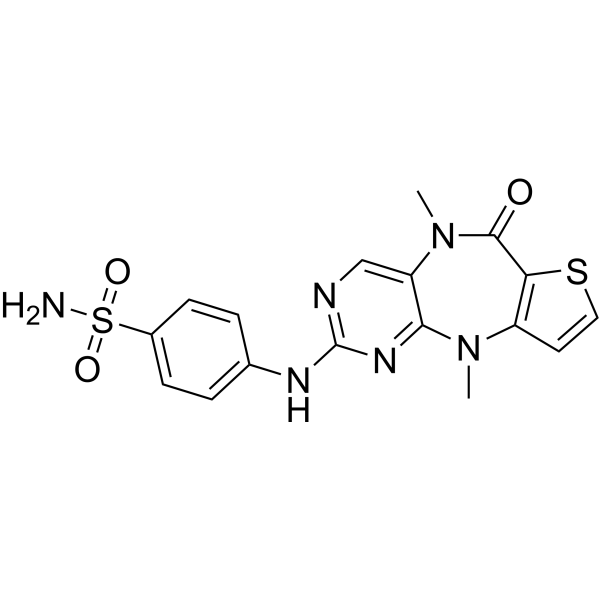
Publications Citing Use of MedChemExpress (MCE) MST1 Antibody (YA2368)
More

Applications
| Application |
WB

WB: Western Blot
|
IHC-P
IHC-P: Immunohistochemistry-Paraffin
|
|---|---|---|
| Dilution Ratio | 1:500-1:1000 | 1:50-1:100 |


Product Details
MST1 Antibody (YA2368) is a Rabbit-derived and non-conjugated IgG monoclonal antibody, targeting to MST1.
-
Host Rabbit

-
Clonality Recombinant,Monoclonal

-
Species ReactivityHuman, Mouse, Rat
-
Observed Molecular WeightObserved band size: 96 kDa
Note: Due to possible protein modifications or aggregation, the molecular weight should be confirmed by actual measurement, and the predicted value is for reference only.

-
Calculated Molecular Weight Predicted band size: 80 kDa

Entrez Gene: 4485 Human ; 15235 Mouse ; 24566 Rat
SwissProt: P26927 Human ; P26928 Mouse ;
OMIM: 142408 Human
A synthetic peptide of human MST1.
Endogenous
Affinity Chromatography
Non-conjugated
Unmodified
IgG


Product Properties
-
Appearance
Solution
-
Formulation
Supplied in 50mM Tris-Glycine(pH 7.4), 0.15M NaCl, 40%Glycerol, 0.01% sodium azide and 0.05% BSA.
-
Concentration
Batch-dependent, Please check the COA for the concentration of each lot. Check Lot Concentration
-
Storage & Stability
Stored at -20°C for 1 year. Avoid repeated freeze / thaw cycles.
-
Shipping
Shipping with blue ice.


Publications (1)
-
Journal Impact Factor
-
Most Recent
-
J Mol Histol
Dendrobine promotes osteogenesis-angiogenesis in postmenopausal osteoporosis by inhibiting Hippo signaling pathway. [Abstract]2026 May 13;57(3):164. PMID: 42126726


Verification Images
-
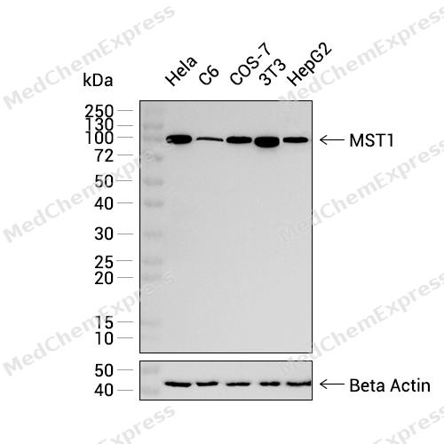 Western blot analysis was performed on extracts from Hela (lane 1, 20 μg), C6 (lane 2, 20 μg), COS-7 (lane 3, 20 μg), 3T3 (lane 4, 20 μg), and HepG2 (lane 5, 20 μg) using MST1 Rabbit mAb.Proteins were transferred to a PVDF membrane and blocked with 5% non - fat milk in TBST at 4°C overnight.The primary antibody (1:1000 dilution) and the loading control antibody (beta-Actin, HY-P83730, 1:5000 dilution) were incubated in 5% non-fat milk in TBST for 1 hour at 37°C.Goat Anti - Rabbit IgG - HRP Secondary Antibody (1:20000 dilution) was then applied for 40 minutes at 37°C.
 -
 Immunohistochemical analysis of paraffin-embedded human lymphoma tissue using MST1 Antibody (HY-P82623, 1/100). The section was pretreated using heat mediated antigen retrieval with sodium citrate buffer (pH 6.0) for 8 minutes. The tissues were blocked with quick block buffer for 0.5 hours at room temperature, washed with PBS and PBST, and then incubated with the primary antibody overnight at 4℃. The detection was performed using an HRP conjugated compact polymer system. DAB was used as the chromogen. Tissues were counterstained with hematoxylin and mounted with DPX.
 -
 Immunohistochemical analysis of paraffin-embedded human glioma tissue using MST1 Antibody (HY-P82623, 1/100). The section was pretreated using heat mediated antigen retrieval with sodium citrate buffer (pH 6.0) for 8 minutes. The tissues were blocked with quick block buffer for 0.5 hours at room temperature, washed with PBS and PBST, and then incubated with the primary antibody overnight at 4℃. The detection was performed using an HRP conjugated compact polymer system. DAB was used as the chromogen. Tissues were counterstained with hematoxylin and mounted with DPX.
 -
 Immunohistochemical analysis of paraffin-embedded human breast cancer tissue using MST1 Antibody (HY-P82623, 1/100). The section was pretreated using heat mediated antigen retrieval with sodium citrate buffer (pH 6.0) for 8 minutes. The tissues were blocked with quick block buffer for 0.5 hours at room temperature, washed with PBS and PBST, and then incubated with the primary antibody overnight at 4℃. The detection was performed using an HRP conjugated compact polymer system. DAB was used as the chromogen. Tissues were counterstained with hematoxylin and mounted with DPX.
 -
 Immunohistochemical analysis of paraffin-embedded human cervical cancer tissue using MST1 Antibody (HY-P82623, 1/100). The section was pretreated using heat mediated antigen retrieval with sodium citrate buffer (pH 6.0) for 8 minutes. The tissues were blocked with quick block buffer for 0.5 hours at room temperature, washed with PBS and PBST, and then incubated with the primary antibody overnight at 4℃. The detection was performed using an HRP conjugated compact polymer system. DAB was used as the chromogen. Tissues were counterstained with hematoxylin and mounted with DPX.
 -
 Immunohistochemical analysis of paraffin-embedded human thyroid cancer tissue using MST1 Antibody (HY-P82623, 1/100). The section was pretreated using heat mediated antigen retrieval with sodium citrate buffer (pH 6.0) for 8 minutes. The tissues were blocked with quick block buffer for 0.5 hours at room temperature, washed with PBS and PBST, and then incubated with the primary antibody overnight at 4℃. The detection was performed using an HRP conjugated compact polymer system. DAB was used as the chromogen. Tissues were counterstained with hematoxylin and mounted with DPX.
 -
 Immunohistochemical analysis of paraffin-embedded human lung cancer tissue using MST1 Antibody (HY-P82623, 1/100). The section was pretreated using heat mediated antigen retrieval with sodium citrate buffer (pH 6.0) for 8 minutes. The tissues were blocked with quick block buffer for 0.5 hours at room temperature, washed with PBS and PBST, and then incubated with the primary antibody overnight at 4℃. The detection was performed using an HRP conjugated compact polymer system. DAB was used as the chromogen. Tissues were counterstained with hematoxylin and mounted with DPX.
 -
 Immunohistochemical analysis of paraffin-embedded human endometrial cancer tissue using MST1 Antibody (HY-P82623, 1/100). The section was pretreated using heat mediated antigen retrieval with sodium citrate buffer (pH 6.0) for 8 minutes. The tissues were blocked with quick block buffer for 0.5 hours at room temperature, washed with PBS and PBST, and then incubated with the primary antibody overnight at 4℃. The detection was performed using an HRP conjugated compact polymer system. DAB was used as the chromogen. Tissues were counterstained with hematoxylin and mounted with DPX.
 -
 Immunohistochemical analysis of paraffin-embedded human rectum cancer tissue using MST1 Antibody (HY-P82623, 1/100). The section was pretreated using heat mediated antigen retrieval with sodium citrate buffer (pH 6.0) for 8 minutes. The tissues were blocked with quick block buffer for 0.5 hours at room temperature, washed with PBS and PBST, and then incubated with the primary antibody overnight at 4℃. The detection was performed using an HRP conjugated compact polymer system. DAB was used as the chromogen. Tissues were counterstained with hematoxylin and mounted with DPX.


Background
-
Function
MST1/STK4 protein is a stress-activated, pro-apoptotic kinase with diverse cellular functions, including a critical role in the Hippo signaling pathway. Following caspase-cleavage, MST1/STK4 enters the nucleus, inducing chromatin condensation and internucleosomal DNA fragmentation, contributing to apoptosis. In the Hippo pathway, MST1/STK4, along with its regulatory protein SAV1, forms a kinase cascade that phosphorylates and activates LATS1/2-MOB1 complex. This, in turn, phosphorylates and inactivates the YAP1 oncoprotein and WWTR1/TAZ, regulating genes involved in proliferation, cell death, and cell migration. MST1/STK4 is essential for repressing hepatocyte proliferation, inhibiting facultative adult liver stem cells, and preventing tumor formation. Additionally, it plays a role in histone modification during apoptosis, phosphorylates various substrates like FOXO3, MOBKL1A, MOBKL1B, RASSF2, TNNI3, and FOXO1, influencing diverse cellular processes such as transcription, cell death initiation, and signaling pathways like PKB/AKT1 and AR. Notably, MST1/STK4 acts as an inhibitor of PKB/AKT1, and its phosphorylation of SIRT1 promotes p53-dependent transcription and apoptosis in response to DNA damage. Furthermore, it intersects with PKB/AKT1 signaling to suppress AR activity by antagonizing the formation of AR-chromatin complexes.
-
Subcellular Localization
Secreted
-
Subunit
Dimer of an alpha chain and a beta chain linked by a disulfide bond. Interacts (via beta chain) with MST1R (via SEMA domain)
-
SwissProt ID
-
Synonyms
HGFL; MSP; MST1; NF15S2
-
Research Field
Cell Biology
Documentation
-
Data Sheet (232 KB)
-
SDS (251 KB)
- English - EN (251 KB)
- Français - FR (251 KB)
- Deutsch - DE (251 KB)
- Norwegian - NO (251 KB)
- Español - ES (251 KB)
- Swedish - SV (251 KB)
- Italian - IT (251 KB)
- Korean - KR (251 KB)
- Portuguese - PT (251 KB)
-
User Guide for Antibodies (1077 KB)

 Powered by Bioz
