



NOP10 Antibody (YA3116)
(Synonyms: DKCB1; NOLA3; NOP10P)
Based on 1 Customer Validation
NOP10 Antibody (YA3116) is a Rabbit-derived and non-conjugated IgG monoclonal antibody, targeting to NOP10.
-
Host:
Rabbit
-
Isotype:
IgG
-
Application:
WB, IHC-P
-
Reactivity :
Human
-
Formulation:
Supplied in 50mM Tris-Glycine(pH 7.4), 0.15M NaCl, 40% Glycerol, 0.01% Sodium azide and 0.05% BSA
-
Conjugation:
Non-conjugated



To place orders, for customer services and technical support, please contact: MedChemExpress USA
Tel: 609-228-6898 E-mail: [email protected] [email protected]





Applications
| Application |
WB

WB: Western Blot
|
IHC-P
IHC-P: Immunohistochemistry-Paraffin
|
|---|---|---|
| Dilution Ratio | 1:500-1:1000 | 1:50-1:100 |


Product Details
NOP10 Antibody (YA3116) is a Rabbit-derived and non-conjugated IgG monoclonal antibody, targeting to NOP10.
-
Host Rabbit

-
Clonality Recombinant,Monoclonal

-
Species ReactivityHuman
-
Observed Molecular WeightObserved band size: 8 kDa
Note: Due to possible protein modifications or aggregation, the molecular weight should be confirmed by actual measurement, and the predicted value is for reference only.

-
Calculated Molecular Weight Predicted band size: 8 kDa

A synthetic peptide of human NOP10 aa51-64.
Endogenous
Affinity Purified
Non-conjugated
Unmodified
IgG


Product Properties
-
Appearance
Liquid
-
Formulation
Supplied in 50mM Tris-Glycine(pH 7.4), 0.15M NaCl, 40% Glycerol, 0.01% Sodium azide and 0.05% BSA
-
Concentration
Batch-dependent, Please check the COA for the concentration of each lot. Check Lot Concentration
-
Storage & Stability
Stored at -20°C for 1 year. Avoid repeated freeze / thaw cycles.
-
Shipping
Shipping with blue ice.


Verification Images
-
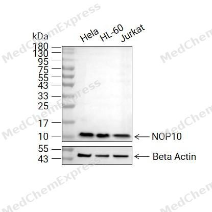 Western blot analysis of extracts from Hela (lane 2(20μg), HL-60 (lane 3(20μg), and Jurkat (lane 4(20μg) using NOP10 (HY-P83371) Rabbit mAb. Proteins were transferred to a PVDF membrane and blocked with 5% non-fat milk in TBST for 2 hour at room temperature. The primary antibody (1/1000) and Loading control antibody (Beta Actin, HY-P80438, 1/10000) was used in 5% non-fat milk in TBST at 4°C overnight. Goat Anti-Rabbit IgG-HRP Secondary Antibody (1/10000) was used for 1 hour at room temperature.
 -
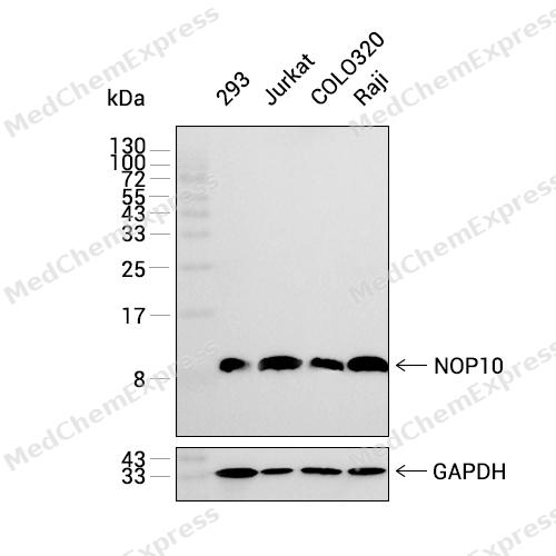
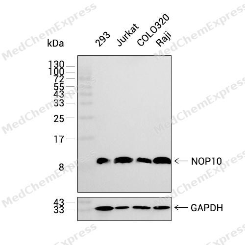 Western blot analysis was performed on extracts from 293 (lane 1, 15 μg), Jurkat (lane 2, 15 μg), COLO320 (lane 3, 15 μg), and Raji (lane 4, 15 μg) using NOP10 Rabbit mAb.Proteins were transferred to a PVDF membrane and blocked with 5% non-fat milk in TBST at 4°C overnight.The primary antibody (1:1000 dilution) and the loading control antibody (GAPDH, HY-P80137, 1:60000 dilution) were incubated in 5% non-fat milk in TBST for 1 hour at 37°C.Goat Anti-Rabbit IgG-HRP Secondary Antibody (1:20000 dilution) was then applied for 40 minutes at 37°C.
 -
 Immunohistochemical analysis of paraffin-embedded human endometrium tissue using NOP10 Antibody (HY-P83371, 1/100). The section was pretreated using heat mediated antigen retrieval with sodium citrate buffer (pH 6.0) for 8 minutes. The tissues were blocked with quick block buffer for 0.5 hours at room temperature, washed with PBS and PBST, and then incubated with the primary antibody overnight at 4℃. The detection was performed using an HRP conjugated compact polymer system. DAB was used as the chromogen. Tissues were counterstained with hematoxylin and mounted with DPX.
 -
 Immunohistochemical analysis of paraffin-embedded human lung tissue using NOP10 Antibody (HY-P83371, 1/100). The section was pretreated using heat mediated antigen retrieval with sodium citrate buffer (pH 6.0) for 8 minutes. The tissues were blocked with quick block buffer for 0.5 hours at room temperature, washed with PBS and PBST, and then incubated with the primary antibody overnight at 4℃. The detection was performed using an HRP conjugated compact polymer system. DAB was used as the chromogen. Tissues were counterstained with hematoxylin and mounted with DPX.
 -
 Immunohistochemical analysis of paraffin-embedded human salivary gland tissue using NOP10 Antibody (HY-P83371, 1/100). The section was pretreated using heat mediated antigen retrieval with sodium citrate buffer (pH 6.0) for 8 minutes. The tissues were blocked with quick block buffer for 0.5 hours at room temperature, washed with PBS and PBST, and then incubated with the primary antibody overnight at 4℃. The detection was performed using an HRP conjugated compact polymer system. DAB was used as the chromogen. Tissues were counterstained with hematoxylin and mounted with DPX.
 -
 Immunohistochemical analysis of paraffin-embedded human vagina tissue using NOP10 Antibody (HY-P83371, 1/100). The section was pretreated using heat mediated antigen retrieval with sodium citrate buffer (pH 6.0) for 8 minutes. The tissues were blocked with quick block buffer for 0.5 hours at room temperature, washed with PBS and PBST, and then incubated with the primary antibody overnight at 4℃. The detection was performed using an HRP conjugated compact polymer system. DAB was used as the chromogen. Tissues were counterstained with hematoxylin and mounted with DPX.
 -
 Immunohistochemical analysis of paraffin-embedded human kidney tissue using NOP10 Antibody (HY-P83371, 1/100). The section was pretreated using heat mediated antigen retrieval with sodium citrate buffer (pH 6.0) for 8 minutes. The tissues were blocked with quick block buffer for 0.5 hours at room temperature, washed with PBS and PBST, and then incubated with the primary antibody overnight at 4℃. The detection was performed using an HRP conjugated compact polymer system. DAB was used as the chromogen. Tissues were counterstained with hematoxylin and mounted with DPX.
 -
 Immunohistochemical analysis of paraffin-embedded human liver tissue using NOP10 Antibody (HY-P83371, 1/100). The section was pretreated using heat mediated antigen retrieval with sodium citrate buffer (pH 6.0) for 8 minutes. The tissues were blocked with quick block buffer for 0.5 hours at room temperature, washed with PBS and PBST, and then incubated with the primary antibody overnight at 4℃. The detection was performed using an HRP conjugated compact polymer system. DAB was used as the chromogen. Tissues were counterstained with hematoxylin and mounted with DPX.
 -
 Immunohistochemical analysis of paraffin-embedded human thyroid gland tissue using NOP10 Antibody (HY-P83371, 1/100). The section was pretreated using heat mediated antigen retrieval with sodium citrate buffer (pH 6.0) for 8 minutes. The tissues were blocked with quick block buffer for 0.5 hours at room temperature, washed with PBS and PBST, and then incubated with the primary antibody overnight at 4℃. The detection was performed using an HRP conjugated compact polymer system. DAB was used as the chromogen. Tissues were counterstained with hematoxylin and mounted with DPX.
 -
 Immunohistochemical analysis of paraffin-embedded human spleen tissue using NOP10 Antibody (HY-P83371, 1/100). The section was pretreated using heat mediated antigen retrieval with sodium citrate buffer (pH 6.0) for 8 minutes. The tissues were blocked with quick block buffer for 0.5 hours at room temperature, washed with PBS and PBST, and then incubated with the primary antibody overnight at 4℃. The detection was performed using an HRP conjugated compact polymer system. DAB was used as the chromogen. Tissues were counterstained with hematoxylin and mounted with DPX.


Background
-
Function
NOP10 is required for ribosome biogenesis and telomere maintenance. Part of the H/ACA small nucleolar ribonucleoprotein (H/ACA snoRNP) complex, which catalyzes pseudouridylation of rRNA. This involves the isomerization of uridine such that the ribose is subsequently attached to C5, instead of the normal N1. Each rRNA can contain up to 100 pseudouridine ('psi') residues, which may serve to stabilize the conformation of rRNAs. May also be required for correct processing or intranuclear trafficking of TERC, the RNA component of the telomerase reverse transcriptase (TERT) holoenzyme[1].
-
Subcellular Localization
Nucleus, nucleolus; Nucleus, Cajal body
-
Subunit
Part of the H/ACA small nucleolar ribonucleoprotein (H/ACA snoRNP) complex, which contains NHP2/NOLA2, GAR1/NOLA1, NOP10/NOLA3, and DKC1/NOLA4, which is presumed to be the catalytic subunit (PubMed:11074001). The complex contains a stable core formed by binding of one or two NOP10-DKC1 heterodimers to NHP2; GAR1 subsequently binds to this core via DKC1 (PubMed:11074001). The complex binds a box H/ACA small nucleolar RNA (snoRNA), which may target the specific site of modification within the RNA substrate (PubMed:16601202). During assembly, the complex contains NAF1 instead of GAR1/NOLA1 (PubMed:11074001, PubMed:16601202). The complex also interacts with TERC, which contains a 3'-terminal domain related to the box H/ACA snoRNAs (PubMed:11074001, PubMed:16601202). Specific interactions with snoRNAs or TERC are mediated by GAR1 and NHP2 (PubMed:11074001, PubMed:16601202). Associates with NOLC1/NOPP140 (PubMed:11074001, PubMed:16601202). H/ACA snoRNPs interact with the SMN complex, consisting of SMN1 or SMN2, GEMIN2/SIP1, DDX20/GEMIN3, and GEMIN4 (PubMed:11074001, PubMed:16601202). This is mediated by interaction between GAR1 and SMN1 or SMN2 (PubMed:11074001, PubMed:16601202). The SMN complex may be required for correct assembly of the H/ACA snoRNP complex (PubMed:11074001, PubMed:16601202). Component of the telomerase holoenzyme complex composed of one molecule of TERT, one molecule of WRAP53/TCAB1, two molecules of H/ACA ribonucleoprotein complex subunits DKC1, NOP10, NHP2 and GAR1, and a telomerase RNA template component (TERC) (PubMed:19179534, PubMed:20351177, PubMed:29695869). The telomerase holoenzyme complex is associated with TEP1, SMG6/EST1A and POT1 (PubMed:19179534)
-
SwissProt ID
-
Synonyms
DKCB1; NOLA3; NOP10P
-
Research Field
Epigenetics and Nuclear Signaling


Documentation
-
Data Sheet (234 KB)
-
SDS (251 KB)
- English - EN (251 KB)
- Français - FR (251 KB)
- Deutsch - DE (251 KB)
- Norwegian - NO (251 KB)
- Español - ES (251 KB)
- Swedish - SV (251 KB)
- Italian - IT (251 KB)
- Korean - KR (251 KB)
- Portuguese - PT (251 KB)
-
User Guide for Antibodies (1077 KB)

 Powered by Bioz
