



NR0B2 Antibody
(Synonyms: NR0B2; NR0B2_HUMAN; Nr0b2a; Nuclear receptor subfamily 0 group B member 2; Nuclear receptor subfamily 0, group B, member 2a; Orphan nuclear receptor SHP; SHP; SHP-1; Shp1; Small heterodimer partner.)
Based on 1 Customer Validation
NR0B2 Antibody is a Rabbit-derived and non-conjugated IgG polyclonal antibody, targeting to NR0B2.
-
Host:
Rabbit
-
Isotype:
IgG
-
Application:
WB, IHC-P, IHC-F, ICC/IF
-
Reactivity :
Mouse, Rat
-
Formulation:
Supplied in TBS (pH7.4) with 1% BSA, 0.02% Proclin300 and 50% Glycerol.
-
Conjugation:
Non-conjugated


To place orders, for customer services and technical support, please contact: MedChemExpress USA
Tel: 609-228-6898 E-mail: [email protected] [email protected]





Applications
| Application |
WB

WB: Western Blot
|
IHC-P
IHC-P: Immunohistochemistry-Paraffin
|
IHC-F
IHC-F: Immunohistochemistry-Frozen
|
ICC/IF
ICC/IF: Immunocytochemistry/
Immunofluorescence |
|---|---|---|---|---|
| Dilution Ratio | 1:500-2000 | 1:100-500 | 1:100-500 | 1:100-500 |


Product Details
NR0B2 Antibody is a Rabbit-derived and non-conjugated IgG polyclonal antibody, targeting to NR0B2.
-
Host Rabbit

-
Clonality Polyclonal

-
Species ReactivityMouse, Rat Predicted Reactivity: Human,Rabbit,Cow,Dog,Horse
Note: The predicted reactivity is for reference only and should not be considered a guarantee of product performance.
-
Observed Molecular WeightObserved band size: 28 kD
Note: Due to possible protein modifications or aggregation, the molecular weight should be confirmed by actual measurement, and the predicted value is for reference only.

-
Calculated Molecular Weight Predicted band size: 28 kDa

KLH conjugated synthetic peptide derived from human NR0B2: 31-130/257.
Protein A affinity purified.
Non-conjugated
Unmodified
IgG


Product Properties
-
Appearance
Solution
-
Formulation
Supplied in TBS (pH7.4) with 1% BSA, 0.02% Proclin300 and 50% Glycerol.
-
Concentration
Batch-dependent, Please check the COA for the concentration of each lot. Check Lot Concentration
-
Storage & Stability
Stored at -20°C for 1 year. Avoid repeated freeze / thaw cycles.
-
Shipping
Shipping with blue ice.


Verification Images
-
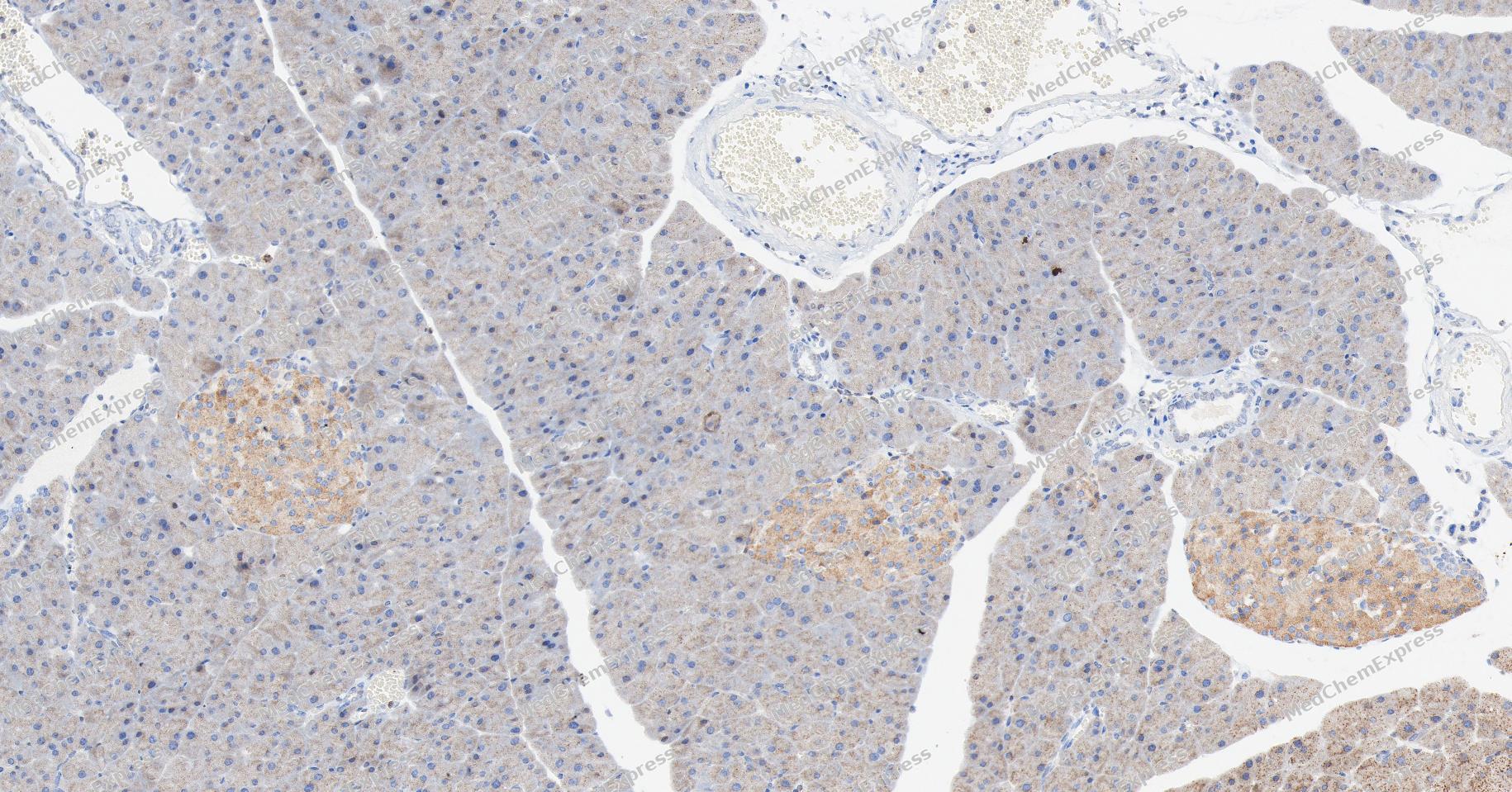 Immunohistochemical analysis of paraffin-embedded mouse pancreas tissue using NR0B2 Antibody. The section was pre-treated using heat mediated antigen retrieval with Tris/EDTA buffer (pH 9.0) for 20 minutes. The tissues were probed with the primary antibody (HY-P83732, 1/100) overnight at 4℃. The detection was performed using an HRP conjugated compact polymer system. DAB was used as the chromogen. Tissues were counterstained with hematoxylin and mounted with neutral balsam.
 -
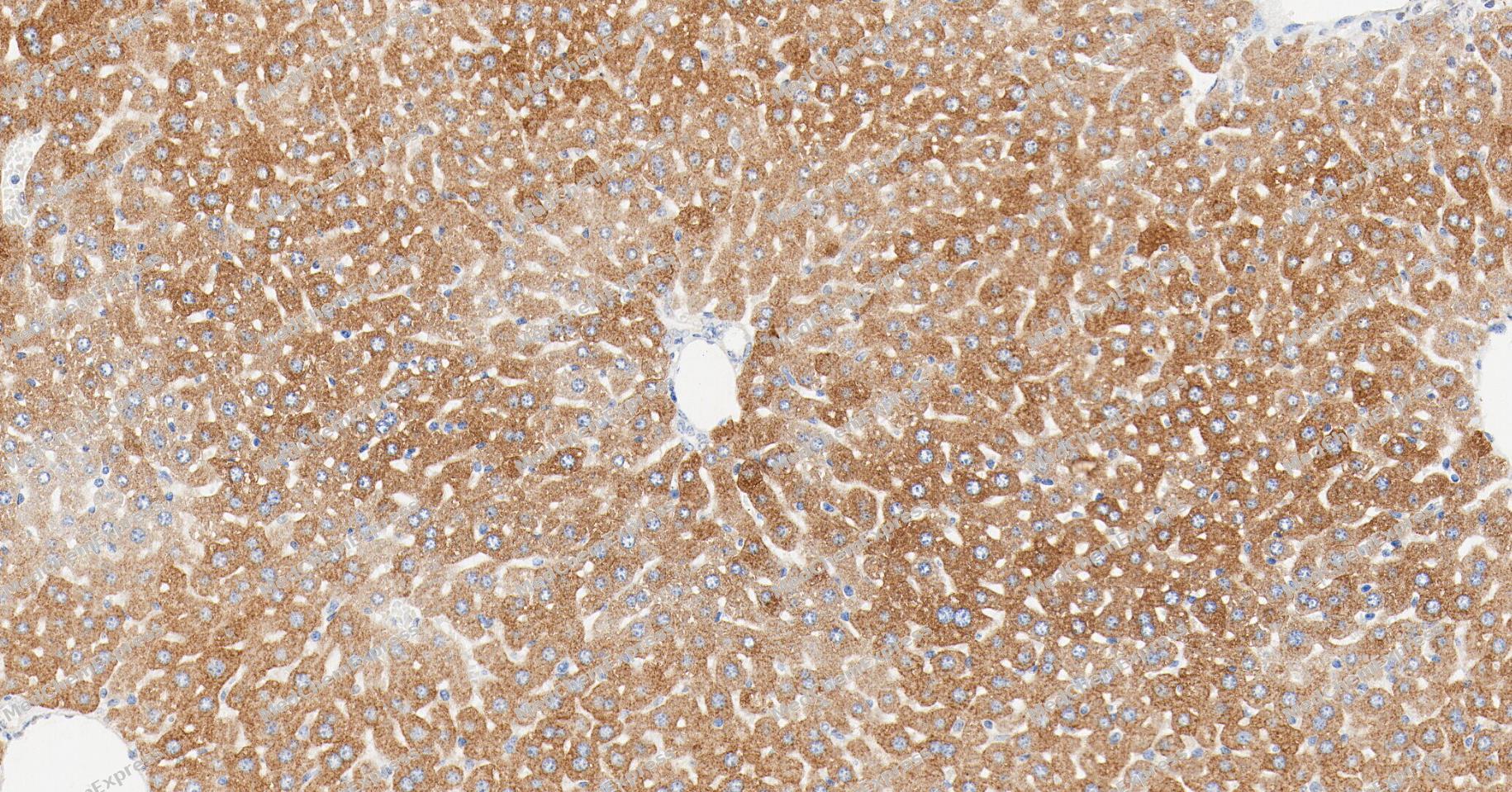 Immunohistochemical analysis of paraffin-embedded mouse liver tissue using NR0B2 Antibody. The section was pre-treated using heat mediated antigen retrieval with Tris/EDTA buffer (pH 9.0) for 20 minutes. The tissues were probed with the primary antibody (HY-P83732, 1/100) overnight at 4℃. The detection was performed using an HRP conjugated compact polymer system. DAB was used as the chromogen. Tissues were counterstained with hematoxylin and mounted with neutral balsam.
 -
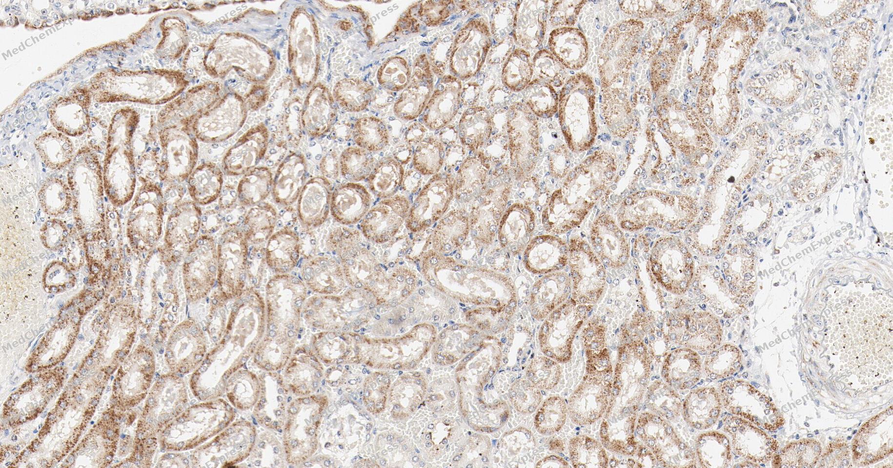 Immunohistochemical analysis of paraffin-embedded mouse kidney tissue using NR0B2 Antibody. The section was pre-treated using heat mediated antigen retrieval with Tris/EDTA buffer (pH 9.0) for 20 minutes. The tissues were probed with the primary antibody (HY-P83732, 1/100) overnight at 4℃. The detection was performed using an HRP conjugated compact polymer system. DAB was used as the chromogen. Tissues were counterstained with hematoxylin and mounted with neutral balsam.
 -
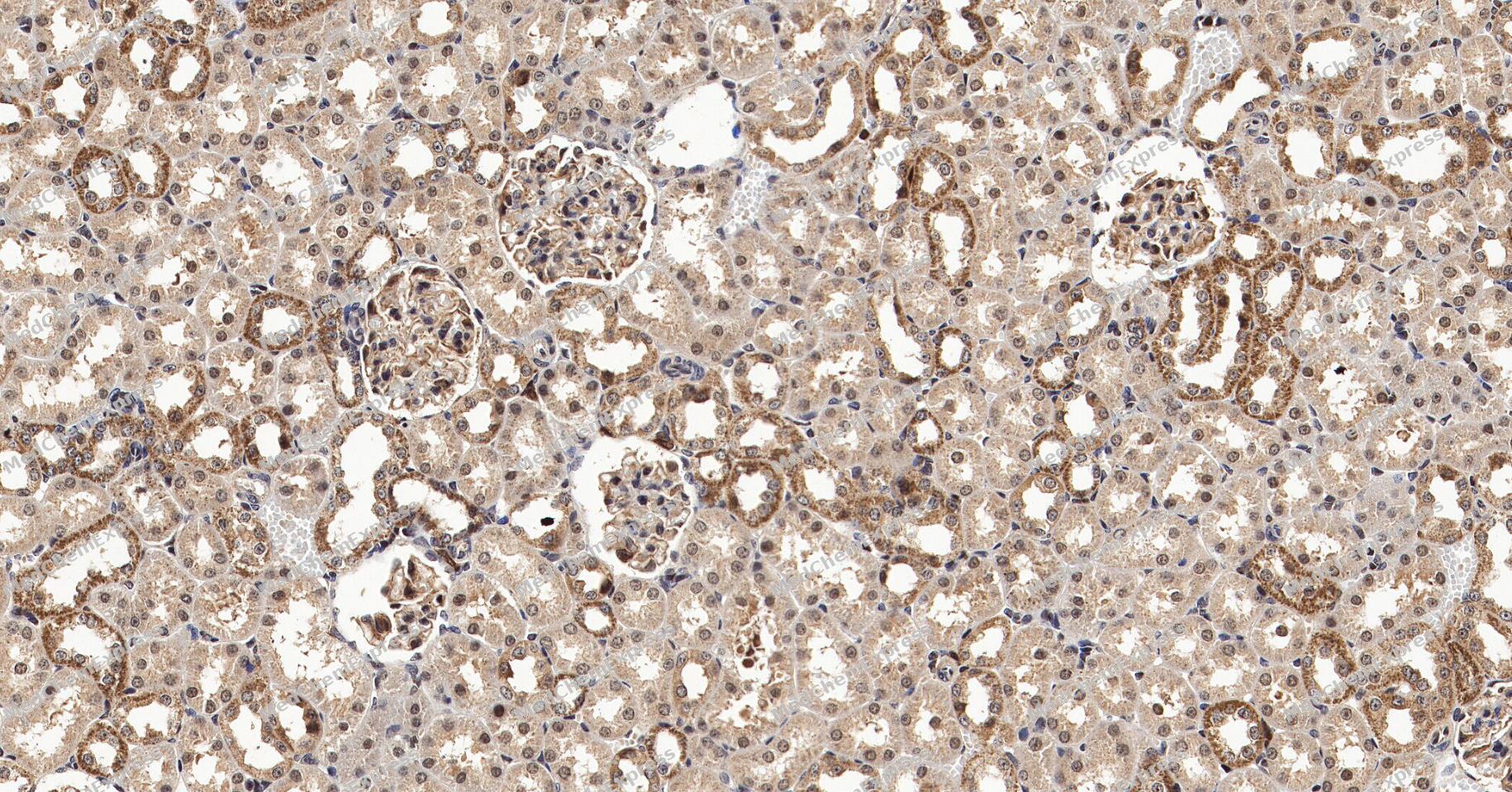 Immunohistochemical analysis of paraffin-embedded Rat kidney tissue using NR0B2 Antibody. The section was pre-treated using heat mediated antigen retrieval with Tris/EDTA buffer (pH 9.0) for 20 minutes. The tissues were probed with the primary antibody (HY-P83732, 1/100) overnight at 4℃. The detection was performed using an HRP conjugated compact polymer system. DAB was used as the chromogen. Tissues were counterstained with hematoxylin and mounted with neutral balsam.
 -
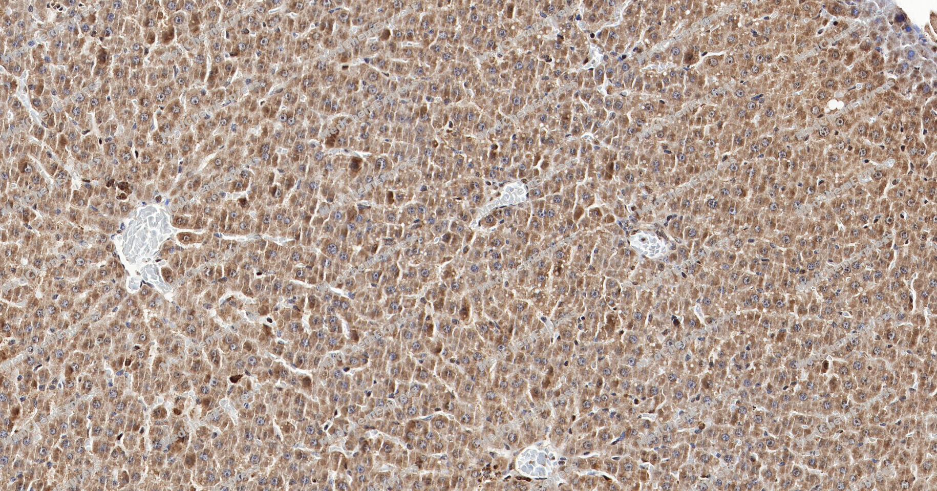 Immunohistochemical analysis of paraffin-embedded Rat liver tissue using NR0B2 Antibody. The section was pre-treated using heat mediated antigen retrieval with Tris/EDTA buffer (pH 9.0) for 20 minutes. The tissues were probed with the primary antibody (HY-P83732, 1/100) overnight at 4℃. The detection was performed using an HRP conjugated compact polymer system. DAB was used as the chromogen. Tissues were counterstained with hematoxylin and mounted with neutral balsam.
 -
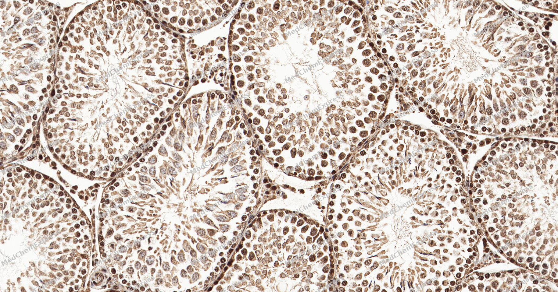 Immunohistochemical analysis of paraffin-embedded Rat testis tissue using NR0B2 Antibody. The section was pre-treated using heat mediated antigen retrieval with Tris/EDTA buffer (pH 9.0) for 20 minutes. The tissues were probed with the primary antibody (HY-P83732, 1/100) overnight at 4℃. The detection was performed using an HRP conjugated compact polymer system. DAB was used as the chromogen. Tissues were counterstained with hematoxylin and mounted with neutral balsam.
 -
 Immunocytochemistry analysis of A549 cells labeling NR0B2 with NR0B2 Antibody (HY-P83732) at 1/100 dilution. Cells were fixed in 4% paraformaldehyde for 15 minutes at room temperature, permeabilized with 0.1% Triton X-100 in PBS for 15 minutes at room temperature, then blocked with quick block buffer for 10 minutes at room temperature. Cells were then incubated with NR0B2 Antibody (HY-P83732) at 1/100 dilution in quick block buffer overnight at 4 ℃. AF488-conjugated Goat Anti-Rabbit IgG H&L (HY-P8002, Green) was used as the secondary antibody at 1/1,000 dilution. PBS instead of the primary antibody was used as the secondary antibody only control. The Nuclear counterstain was DAPI (Blue).
 -
 Immunocytochemistry analysis of A549 cells labeling NR0B2 with NR0B2 Antibody (HY-P83732) at 1/300 dilution. Cells were fixed in 4% paraformaldehyde for 15 minutes at room temperature, permeabilized with 0.1% Triton X-100 in PBS for 15 minutes at room temperature, then blocked with quick block buffer for 10 minutes at room temperature. Cells were then incubated with NR0B2 Antibody (HY-P83732) at 1/300 dilution in quick block buffer overnight at 4 ℃. AF488-conjugated Goat Anti-Rabbit IgG H&L (HY-P8002, Green) was used as the secondary antibody at 1/1,000 dilution. PBS instead of the primary antibody was used as the secondary antibody only control. The Nuclear counterstain was DAPI (Blue).


Background
-
Function
NR0B2 transcriptional regulator that acts as a negative regulator of receptor-dependent signaling pathways. Specifically inhibits transactivation of the nuclear receptor with which it interacts. Inhibits transcriptional activity of NEUROD1 on E-box-containing promoter by interfering with the coactivation function of the p300/CBP-mediated transcription complex for NEUROD1. Essential component of the liver circadian clock which via its interaction with NR1D1 and RORG regulates NPAS2-mediated hepatic lipid metabolism. Regulates the circadian expression of cytochrome P450 (CYP) enzymes. Represses: NR5A2 and HNF4A to down-regulate CYP2C38, NFLI3 to up-regulate CYP2A5, BHLHE41/HNF1A axis to up-regulate CYP1A2, CYP2E1 and CYP3A11, and NR1D1 to up-regulate CYP2B10, CYP4A10 and CYP4A14[1][2].
-
Subcellular Localization
Nucleus; Cytoplasm
-
Expression
Tissue_specificity:Liver. Low levels of expression were detected in heart and pancreas -
Subunit
Interacts (via N-terminus) with NEUROD1 (via N-terminus and C-terminus) (PubMed:14752053). Interacts with ID2 (PubMed:14752053).
-
SwissProt ID
-
Synonyms
NR0B2; NR0B2_HUMAN; Nr0b2a; Nuclear receptor subfamily 0 group B member 2; Nuclear receptor subfamily 0, group B, member 2a; Orphan nuclear receptor SHP; SHP; SHP-1; Shp1; Small heterodimer partner.


Documentation
-
Data Sheet (261 KB)
-
SDS (252 KB)
- English - EN (252 KB)
- Français - FR (252 KB)
- Deutsch - DE (252 KB)
- Norwegian - NO (252 KB)
- Español - ES (252 KB)
- Swedish - SV (252 KB)
- Italian - IT (252 KB)
- Korean - KR (252 KB)
- Portuguese - PT (252 KB)
-
User Guide for Antibodies (1077 KB)
References
[1]. Musille PM, et al. Antidiabetic phospholipid-nuclear receptor complex reveals the mechanism for phospholipid-driven gene regulation. Nat Struct Mol Biol. 2012 Apr 15;19(5):532-S2. [Content Brief]
[2]. Kim JY, et al. Orphan nuclear receptor small heterodimer partner, a novel corepressor for a basic helix-loop-helix transcription factor BETA2/neuroD. Mol Endocrinol. 2004 Apr;18(4):776-90. [Content Brief]

 Powered by Bioz
