



p21 Antibody (YA3460)
(Synonyms: CDKN1A; CAP20; CDKN1; CIP1; MDA6; PIC1; SDI1; WAF1; Cyclin-dependent kinase inhibitor 1; CDK-interacting protein 1; Melanoma differentiation-associated protein 6; MDA-6; p21)Based on 3 publication(s) in Google Scholar
p21 Antibody (YA3460) is a Rabbit-derived and non-conjugated IgG monoclonal antibody, targeting to p21.
-
Host:
Rabbit
-
Isotype:
IgG
-
Application:
WB, ICC/IF
-
Reactivity :
Human, Mouse, Rat
-
Formulation:
Supplied in Rabbit IgG in 10mM phosphate buffered saline , pH 7.4, 150mM sodium chloride, 0.05% BSA, 0.02% sodium azide and 50% glycerol.
-
Conjugation:
Non-conjugated


To place orders, for customer services and technical support, please contact: MedChemExpress USA
Tel: 609-228-6898 E-mail: [email protected] [email protected]



Publications Citing Use of MedChemExpress (MCE) p21 Antibody (YA3460)
More

Applications
| Application |
WB

WB: Western Blot
|
ICC/IF
ICC/IF: Immunocytochemistry/
Immunofluorescence |
|---|---|---|
| Dilution Ratio | 1:500-1:1000 | 1:50-1:200 |


Product Details
p21 Antibody (YA3460) is a Rabbit-derived and non-conjugated IgG monoclonal antibody, targeting to p21.
-
Host Rabbit

-
Clonality Monoclonal,Recombinant

-
Species ReactivityHuman, Mouse, Rat
-
Observed Molecular WeightObserved band size: 21 kDa
Note: Due to possible protein modifications or aggregation, the molecular weight should be confirmed by actual measurement, and the predicted value is for reference only.

-
Calculated Molecular Weight Predicted band size: 18 kDa

Entrez Gene: 1026 Human ; 12575 Mouse ; 114851 Rat
SwissProt: P38936 Human ; P39689 Mouse ;
OMIM: 116899 Human
A synthesized peptide derived from human p21 aa115-164.
affinity purified
Non-conjugated
Unmodified
IgG


Product Properties
-
Appearance
Liquid
-
Formulation
Supplied in Rabbit IgG in 10mM phosphate buffered saline , pH 7.4, 150mM sodium chloride, 0.05% BSA, 0.02% sodium azide and 50% glycerol.
-
Concentration
Batch-dependent, Please check the COA for the concentration of each lot. Check Lot Concentration
-
Storage & Stability
Stored at -20°C for 1 year. Avoid repeated freeze / thaw cycles.
-
Shipping
Shipping with blue ice.


Publications (3)
-
Journal Impact Factor
-
Most Recent
-
Genes Dis
Identifying C1orf122 as a potential HCC exacerbated biomarker dependently of SRPK1 regulates PI3K/AKT/GSK3β signaling pathway. [Abstract]2025 Jun 18;13(1):101721. PMID: 41141389 -
Cell Death Differ
hMTR4 promotes p53 protein degradation and tumor growth by accelerating rRNA processing and regulating the RPL5-MDM2 axis. [Abstract]2025 Jul 13. PMID: 40652043 -
Cancer Med
2025 Sep;14(17):e71146. PMID: 40856423


Verification Images
-
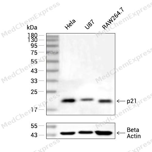 Western blot analysis of extracts from Hela (lane 2(20μg), U87 (lane 3(20μg) and RAW264.7 (lane 4(20μg) using p21 (HY-P80774A) Rabbit mAb. Proteins were transferred to a PVDF membrane and blocked with 5% non-fat milk in TBST for 2 hour at room temperature. The primary antibody (1/1000) and Loading control antibody (Beta Actin, HY-P80438, 1/10000) was used in 5% non-fat milk in TBST at 4°C overnight. Goat Anti-Mouse/Rabbit IgG-HRP Secondary Antibody (1/10000) was used for 1 hour at room temperature.
 -
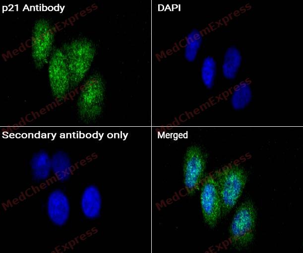 Immunocytochemistry analysis of MCF-7 cells labeling p21 with p21 antibody (HY-P80744A) at 1/50 dilution. Cells were fixed in 4% paraformaldehyde for 15 minutes at room temperature, permeabilized with 0.1% Triton X-100 for 10 minutes at room temperature, then blocked with QuickBlock™ Blocking Buffer for Immunol Staining for 10 min at room temperature. Cells were then incubated with p21 antibody (HY-P80744A) at 1/50 dilution in QuickBlock™ Blocking Buffer for Immunol Staining at 4 ℃. Alexa Fluor® 488-conjugated AffiniPure Goat Anti-Rabbit IgG H&L(HY-P8002, Green) was used as the secondary antibody at 1/1,000 dilution. PBS instead of the primary antibody was used as the secondary antibody only control. The Nuclear counterstain was DAPI (Blue).
 -
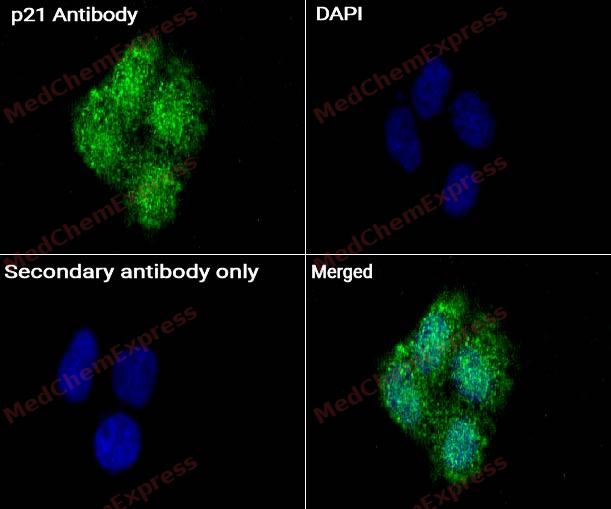 Immunocytochemistry analysis of MCF-7 cells labeling p21 with p21 antibody (HY-P80744A) at 1/100 dilution. Cells were fixed in 4% paraformaldehyde for 15 minutes at room temperature, permeabilized with 0.1% Triton X-100 for 10 minutes at room temperature, then blocked with QuickBlock™ Blocking Buffer for Immunol Staining for 10 min at room temperature. Cells were then incubated with p21 antibody (HY-P80744A) at 1/100 dilution in QuickBlock™ Blocking Buffer for Immunol Staining at 4 ℃. Alexa Fluor® 488-conjugated AffiniPure Goat Anti-Rabbit IgG H&L(HY-P8002, Green) was used as the secondary antibody at 1/1,000 dilution. PBS instead of the primary antibody was used as the secondary antibody only control. The Nuclear counterstain was DAPI (Blue).


Background
-
Function
p21 plays an important role in controlling cell cycle progression and DNA damage-induced G2 arrest. Involved in p53/TP53 mediated inhibition of cellular proliferation in response to DNA damage. Also involved in p53-independent DNA damage-induced G2 arrest mediated by CREB3L1 in astrocytes and osteoblasts. Binds to and inhibits cyclin-dependent kinase activity, preventing phosphorylation of critical cyclin-dependent kinase substrates and blocking cell cycle progression. Functions in the nuclear localization and assembly of cyclin D-CDK4 complex and promotes its kinase activity towards RB1. At higher stoichiometric ratios, inhibits the kinase activity of the cyclin D-CDK4 complex. Inhibits DNA synthesis by DNA polymerase delta by competing with POLD3 for PCNA binding. Negatively regulates the CDK4- and CDK6-driven phosphorylation of RB1 in keratinocytes, thereby resulting in the release of E2F1 and subsequent transcription of E2F1-driven G1/S phase promoting genes[1][2].
-
Subcellular Localization
Cytoplasm; Nucleus
-
Expression
Tissue_specificity:It is expressed in all adult tissues, but the expression level in the brain is 5 times lower.
Induction:Activated by p53/TP53, mezerein (antileukemic compound) and IFNB1. Repressed by HDAC1 -
Subunit
Interacts with HDAC1; the interaction is prevented by competitive binding of C10orf90/FATS to HDAC1 facilitating acetylation and protein stabilization of CDKN1A/p21 (By similarity). Interacts with MKRN1 (PubMed:19536131). Interacts with PSMA3 (PubMed:11350925). Interacts with PCNA (PubMed:11595739, PubMed:18703516, PubMed:18794347, PubMed:8861913). Component of the ternary complex, cyclin D-CDK4-CDKN1A. Interacts (via its N-terminal domain) with CDK4; the interaction promotes the assembly of the cyclin D-CDK4 complex, its nuclear translocation and promotes the cyclin D-dependent enzyme activity of CDK4 (PubMed:9106657). Binding to CDK2 leads to CDK2/cyclin E inactivation at the G1-S phase DNA damage checkpoint, thereby arresting cells at the G1-S transition during DNA repair (PubMed:19445729). Interacts with PIM1 (PubMed:12431783). Interacts with STK11 and NUAK1 (PubMed:25329316). Interacts wih DTL (PubMed:23213251). Interacts with isoform 1 and isoform 2 of TRIM39 (PubMed:23213251). Interacts with PKP3; the interaction sequesters CDKN1A to the cytoplasm thereby repressing its role as an inhibitor of CDK4- and CDK6-driven RB1 phosphorylation (By similarity)
-
SwissProt ID
-
Synonyms
CDKN1A; CAP20; CDKN1; CIP1; MDA6; PIC1; SDI1; WAF1; Cyclin-dependent kinase inhibitor 1; CDK-interacting protein 1; Melanoma differentiation-associated protein 6; MDA-6; p21
-
Research Field
Cell Biology
Documentation
-
Data Sheet (234 KB)
-
SDS (252 KB)
- English - EN (252 KB)
- Français - FR (252 KB)
- Deutsch - DE (252 KB)
- Norwegian - NO (252 KB)
- Español - ES (252 KB)
- Swedish - SV (252 KB)
- Italian - IT (252 KB)
- Korean - KR (252 KB)
- Portuguese - PT (252 KB)
-
User Guide for Antibodies (1077 KB)

 Powered by Bioz
