



PD-L1 Antibody (YA235)
(Synonyms: CD274, B7H1, PDCD1L1, PDCD1LG1, PDL1, Programmed cell death 1 ligand 1, PD-L1, PDCD1 ligand 1, Programmed death ligand 1, hPD-L1, B7 homolog 1, B7-H1)
Based on 1 Customer Validation
PD-L1 Antibody (YA235) is a Rabbit-derived and non-conjugated IgG monoclonal antibody, targeting to PD-L1.
-
Host:
Rabbit
-
Isotype:
IgG
-
Application:
WB, IHC-P
-
Reactivity :
Human
-
Formulation:
Supplied in 1*TBS (pH7.4), 0.05% BSA and 40% Glycerol. Preservative: 0.05% Sodium Azide.
-
Conjugation:
Non-conjugated


To place orders, for customer services and technical support, please contact: MedChemExpress USA
Tel: 609-228-6898 E-mail: [email protected] [email protected]





Applications
| Application |
WB

WB: Western Blot
|
IHC-P
IHC-P: Immunohistochemistry-Paraffin
|
|---|---|---|
| Dilution Ratio | 1:500-1:2000 | 1:50-1:200 |


Product Details
PD-L1 Antibody (YA235) is a Rabbit-derived and non-conjugated IgG monoclonal antibody, targeting to PD-L1.
-
Host Rabbit

-
Clonality Recombinant,Monoclonal

-
Species ReactivityHuman
-
Observed Molecular WeightObserved band size: 45 kDa
Note: Due to possible protein modifications or aggregation, the molecular weight should be confirmed by actual measurement, and the predicted value is for reference only.

-
Calculated Molecular Weight Predicted band size: 33 kDa;

Synthetic peptide corresponding to Human PD-L1.AA range:191-240.
Endogenous
Protein A affinity purified.
Non-conjugated
Unmodified
IgG


Product Properties
-
Appearance
Solution
-
Formulation
Supplied in 1*TBS (pH7.4), 0.05% BSA and 40% Glycerol. Preservative: 0.05% Sodium Azide.
-
Concentration
Batch-dependent, Please check the COA for the concentration of each lot. Check Lot Concentration
-
Storage & Stability
Stored at -20°C for 1 year. Avoid repeated freeze / thaw cycles.
-
Shipping
Shipping with blue ice.


Verification Images
-
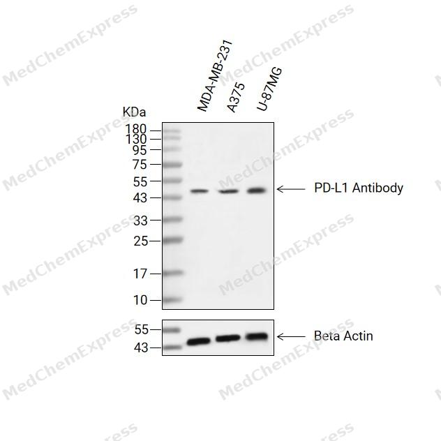 Western blot analysis of extracts from MDA-MB-231(lane 2(20ug) , A375(lane 3(20ug) and U-87MG(lane 4(20ug) using PD-L1 Antibody (HY-P80273) Rabbit mAb. Proteins were transferred to a PVDF membrane and blocked with 5% non-fat milk in TBST for 2 hour at room temperature. The primary antibody (1/1000) and Loading control antibody (Beta Actin, HY-P83730, 1/10000) was used in 5% non-fat milk in TBST at 4°C overnight. Goat Anti-Mouse/Rabbit IgG-HRP Secondary Antibody (1/10000) was used for 1 hour at room temperature.
 -
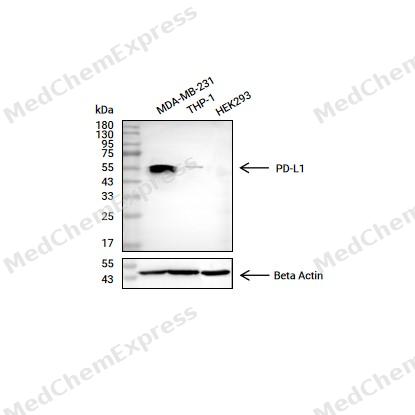 Western blot analysis of extracts from MDA-MB-231 (lane 2(20μg), THP-1(lane 3(20μg) and HEK293(lane 4(20μg) using PD-L1(HY-P80273) Rabbit mAb. Proteins were transferred to a PVDF membrane and blocked with 5% non-fat milk in TBST for 2 hour at room temperature. The primary antibody (1/1000) and Loading control antibody (Beta Actin, HY-P80438, 1/10000) was used in 5% non-fat milk in TBST at 4°C overnight. Goat Anti-Mouse IgG-HRP Secondary Antibody (1/10000) was used for 1 hour at room temperature.
 -
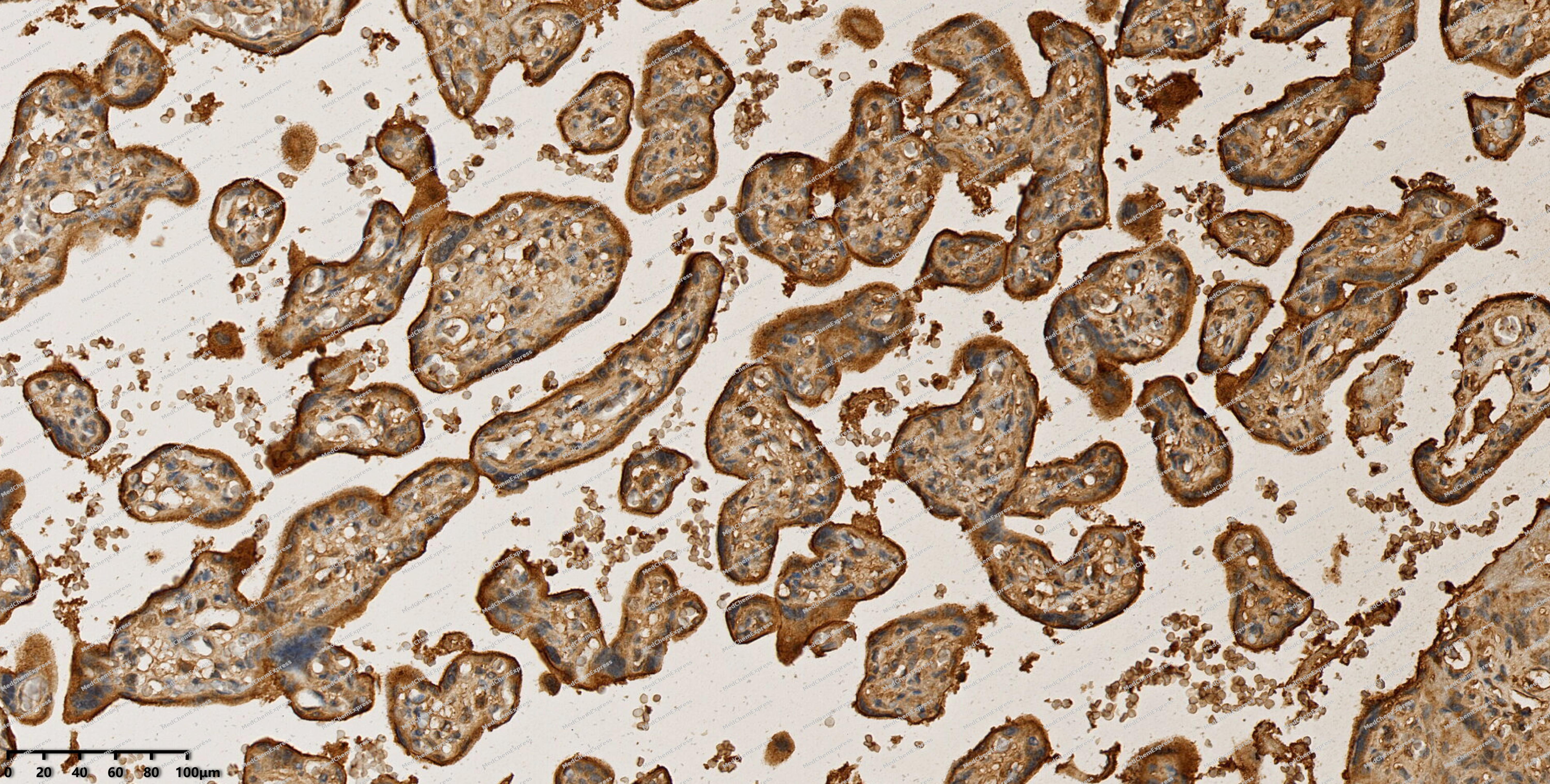 Immunohistochemical analysis of paraffin-embedded human placenta using PD-L1 antibody. The section was pre-treated using heat mediated antigen retrieval with EDTA (pH 9.0) for 14 minutes. The tissues were blocked in 5% BSA for 30 minutes at room temperature, washed with TBST, and then probed with the primary antibody (HY-P80273, 1/200) for 30 minutes at room temperature. The detection was performed using Polymer HRP-conjugated Goat Anti-Mouse/Rabbit lgG(H&L) secondary antibody (HY-P83652). DAB was used as the chromogen. Tissues were counterstained with hematoxylin and mounted with DPX.
 -
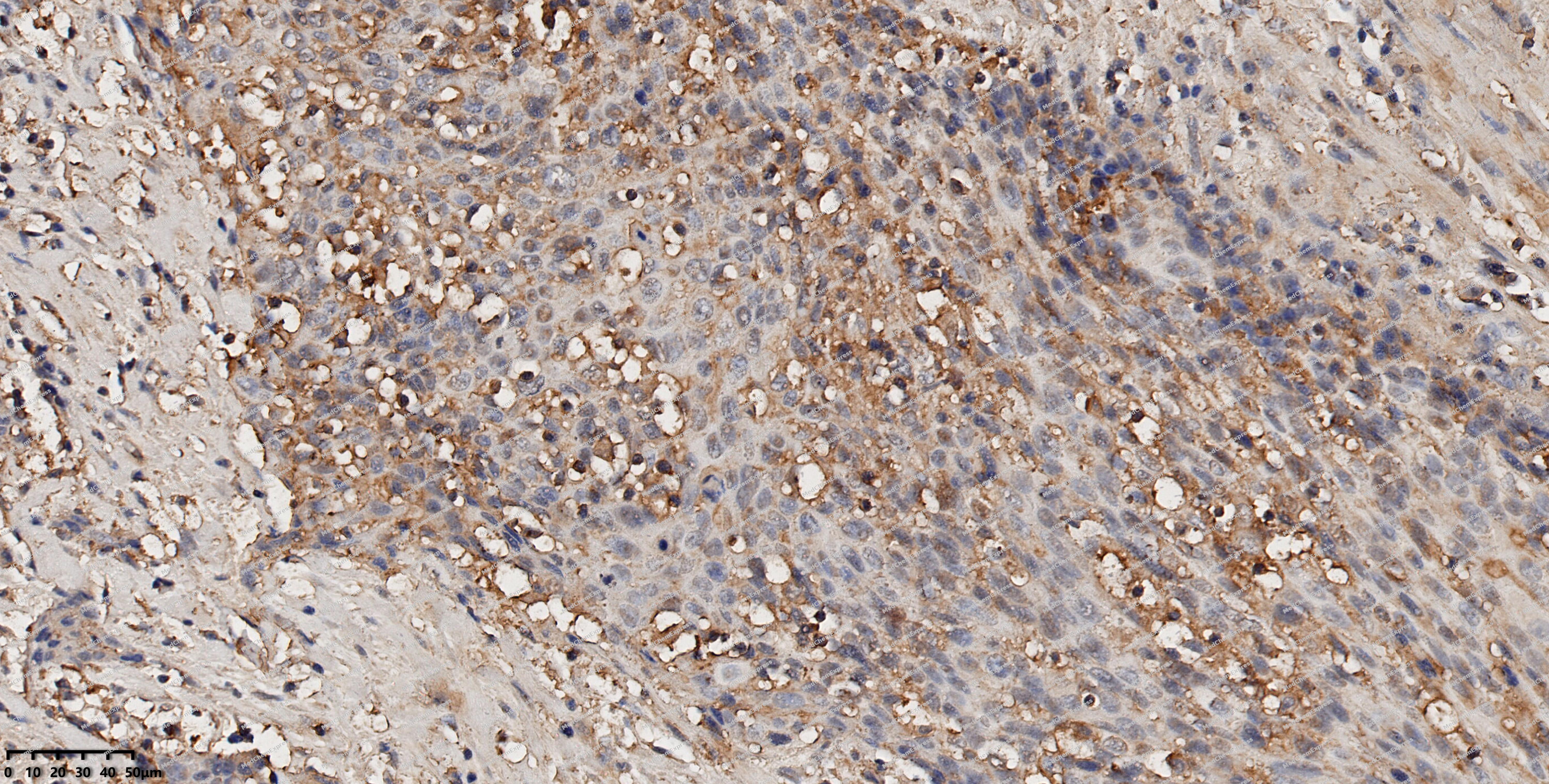 Immunohistochemical analysis of paraffin-embedded human head and neck cancer using PD-L1 antibody. The section was pre-treated using heat mediated antigen retrieval with EDTA (pH 9.0) for 14 minutes. The tissues were blocked in 5% BSA for 30 minutes at room temperature, washed with TBST, and then probed with the primary antibody (HY-P80273, 1/200) for 30 minutes at room temperature. The detection was performed using Polymer HRP-conjugated Goat Anti-Mouse/Rabbit lgG(H&L) secondary antibody (HY-P83652). DAB was used as the chromogen. Tissues were counterstained with hematoxylin and mounted with DPX.
 -
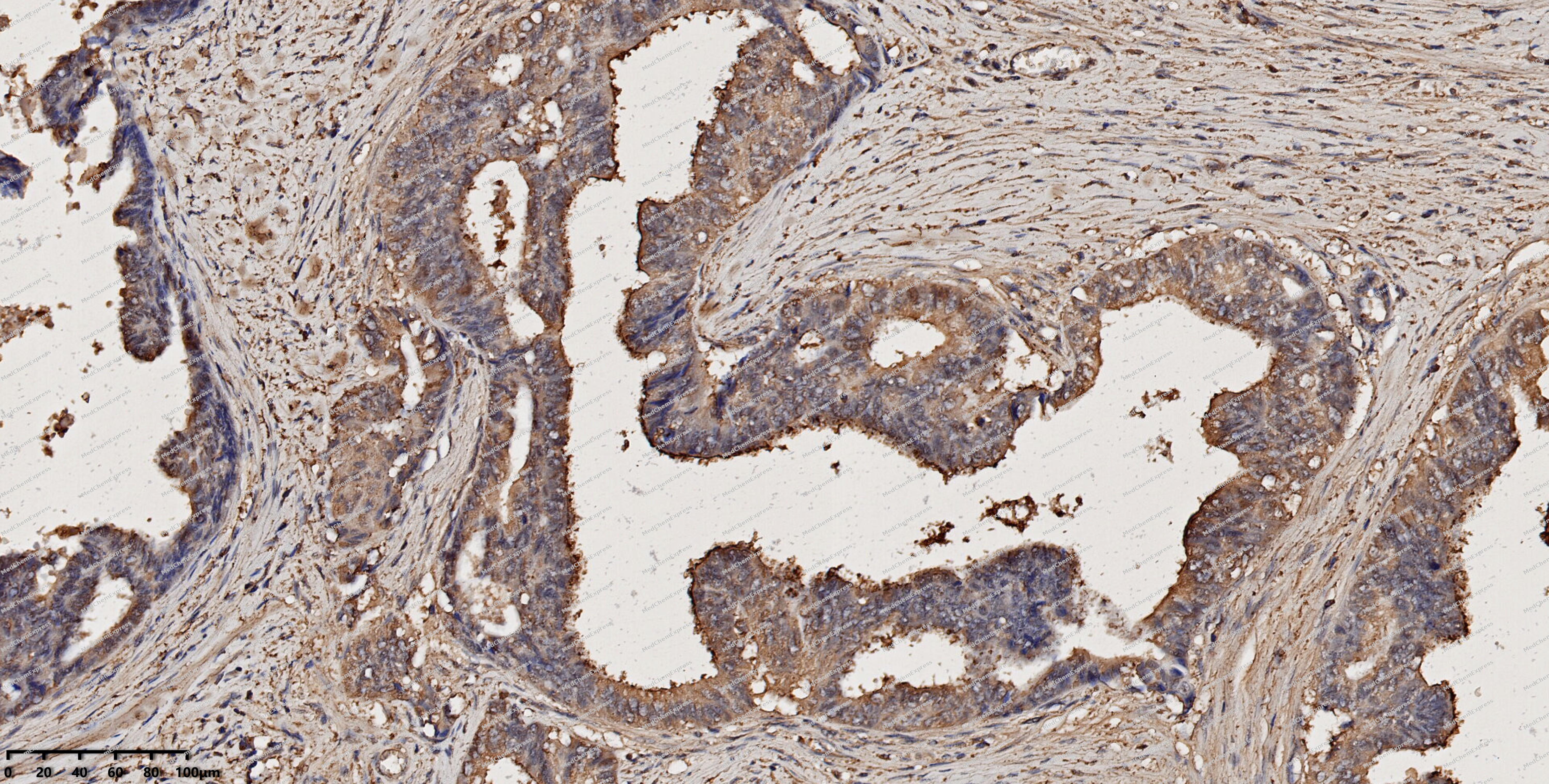 Immunohistochemical analysis of paraffin-embedded human cervical cancer using PD-L1 antibody. The section was pre-treated using heat mediated antigen retrieval with EDTA (pH 9.0) for 14 minutes. The tissues were blocked in 5% BSA for 30 minutes at room temperature, washed with TBST, and then probed with the primary antibody (HY-P80273, 1/200) for 30 minutes at room temperature. The detection was performed using Polymer HRP-conjugated Goat Anti-Mouse/Rabbit lgG(H&L) secondary antibody (HY-P83652). DAB was used as the chromogen. Tissues were counterstained with hematoxylin and mounted with DPX.


Background
-
Function
CD274 (PD-L1) is an important immune checkpoint ligand protein belonging to the B7 family of immunoregulatory molecules. By binding to PD-1 on the surface of T cells, PD-L1 inhibits T-cell activation and immune responses; it is a key mechanism of tumor immune escape and a major target for current immune checkpoint therapies.
-
Subcellular Localization
Cell membrane; Single-pass type I membrane protein; Early endosome membrane; Single-pass type I membrane protein; Recycling endosome membrane; Single-pass type I membrane protein; Nucleus; Cell membrane; Single-pass type I membrane protein; Endomembrane system; Single-pass type I membrane protein; Secreted
-
Expression
Tissue_specificity:It is highly expressed in the heart, skeletal muscle, placenta, and lungs. It is weakly expressed in the thymus, spleen, kidneys, and liver. It is expressed on activated T cells and B cells, dendritic cells, keratinocytes, and monocytes; it is widely expressed, with the highest expression in the lungs, liver, pituitary gland, and various peripheral blood cells (including neutrophils and certain subtypes of lymphocytes and myeloid cells) .
Induction:Up-regulated on T- and B-cells, dendritic cells, keratinocytes and monocytes after LPS and IFNG activation. Up-regulated in B-cells activated by surface Ig cross-linking -
Isoforms & Post-Translational Modification
CD274 (PD-L1) has 4 isomers: Q9NZQ7-1: 33275 Da (predicted); Q9NZQ7-2: 20246 Da (predicted); Q9NZQ7-3: 20454 Da (predicted); Q9NZQ7-4: 28050 Da (predicted).
Ubiquitinated; STUB1 likely mediates polyubiquitination of PD-L1/CD274 triggering its degradation. Ubiquitinated by MARCHF8; leading to degradation. Deubiquitinated by USP22; leading to stabilization -
Subunit
Interacts with PDCD1. Interacts (via transmembrane domain) with CMTM4 and CMTM6. Interacts with (phosphorylated) STAT3; promoting nuclear translocation. Interacts with CD80
-
SwissProt ID
-
Synonyms
CD274, B7H1, PDCD1L1, PDCD1LG1, PDL1, Programmed cell death 1 ligand 1, PD-L1, PDCD1 ligand 1, Programmed death ligand 1, hPD-L1, B7 homolog 1, B7-H1
-
Research Field
Immunology


Documentation
-
Data Sheet (262 KB)
-
SDS (313 KB)
- English - EN (313 KB)
- Français - FR (313 KB)
- Deutsch - DE (313 KB)
- Norwegian - NO (313 KB)
- Español - ES (313 KB)
- Swedish - SV (313 KB)
- Italian - IT (313 KB)
- Korean - KR (313 KB)
- Portuguese - PT (313 KB)
-
User Guide for Antibodies (1077 KB)
References
[1]. Freeman GJ, et al. Engagement of the PD-1 immunoinhibitory receptor by a novel B7 family member leads to negative regulation of lymphocyte activation. J Exp Med. 2000 Oct 2;192(7):1027-34. [Content Brief]
[2]. Mezzadra R, et al. Identification of CMTM6 and CMTM4 as PD-L1 protein regulators. Nature. 2017 Sep 7;549(7670):106-110. [Content Brief]
[3]. Burr ML, et al. CMTM6 maintains the expression of PD-L1 and regulates anti-tumour immunity. Nature. 2017 Sep 7;549(7670):101-105. [Content Brief]
[4]. Huang X, et al. USP22 Deubiquitinates CD274 to Suppress Anticancer Immunity. Cancer Immunol Res. 2019 Oct;7(10):1580-1590. [Content Brief]
[5]. Kennedy A, et al. The CTLA-4 immune checkpoint protein regulates PD-L1:PD-1 interaction via transendocytosis of its ligand CD80. EMBO J. 2023 Mar 1;42(5):e111556. [Content Brief]
[6]. Dong H, et al. B7-H1, a third member of the B7 family, co-stimulates T-cell proliferation and interleukin-10 secretion. Nat Med. 1999 Dec;5(12):1365-9. [Content Brief]
[7]. Hou J, et al. PD-L1-mediated gasdermin C expression switches apoptosis to pyroptosis in cancer cells and facilitates tumour necrosis. Nat Cell Biol. 2020 Oct;22(10):1264-1275. [Content Brief]

 Powered by Bioz
