



RAC1/2/3 Antibody (YA3290)
(Synonyms: Cell migration-inducing gene 5 protein; EN-7; GX; p21-Rac1; p21-Rac2; p21-Rac3; RAC1; RAC2; RAC3; TC25)
Based on 1 Customer Validation
RAC1/2/3 Antibody (YA3290) is a Rabbit-derived and non-conjugated IgG monoclonal antibody, targeting to RAC1/2/3.
-
Host:
Rabbit
-
Isotype:
IgG
-
Application:
WB
-
Reactivity :
Human, Mouse, Rat
-
Formulation:
Supplied in 10mM PBS, pH 7.4, 150mM sodium chloride, 0.05% BSA, 0.02% sodium azide and 50% glycerol.
-
Conjugation:
Non-conjugated


To place orders, for customer services and technical support, please contact: MedChemExpress USA
Tel: 609-228-6898 E-mail: [email protected] [email protected]





Applications
| Application |
WB

WB: Western Blot
|
|---|---|
| Dilution Ratio | 1:500-1:1000 |


Product Details
RAC1/2/3 Antibody (YA3290) is a Rabbit-derived and non-conjugated IgG monoclonal antibody, targeting to RAC1/2/3.
-
Host Rabbit

-
Clonality Recombinant,Monoclonal

-
Species ReactivityHuman, Mouse, Rat
-
Observed Molecular WeightObserved band size: 21 kDa
Note: Due to possible protein modifications or aggregation, the molecular weight should be confirmed by actual measurement, and the predicted value is for reference only.

-
Calculated Molecular Weight Predicted band size: 21 kDa

A synthesized peptide derived from human Rac1/2/3 aa1-192.
Endogenous
Affinity Chromatography
Non-conjugated
Unmodified
IgG


Product Properties
-
Appearance
Liquid
-
Formulation
Supplied in 10mM PBS, pH 7.4, 150mM sodium chloride, 0.05% BSA, 0.02% sodium azide and 50% glycerol.
-
Concentration
Batch-dependent, Please check the COA for the concentration of each lot. Check Lot Concentration
-
Storage & Stability
Stored at -20°C for 1 year. Avoid repeated freeze / thaw cycles.
-
Shipping
Shipping with blue ice.


Verification Images
-
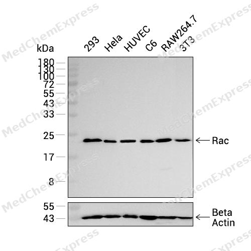
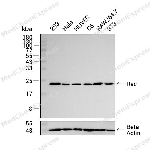 Western blot analysis was performed on extracts from 293 (lane 1, 15 μg), Hela (lane 2, 15 μg), HUVEC (lane 3, 15 μg), C6 (lane 4, 15 μg), RAW264.7 (lane 5, 15 μg), and 3T3 (lane 6, 15 μg) using Rac Rabbit mAb.Proteins were transferred to a PVDF membrane and blocked with 5% non-fat milk in TBST at 4°C overnight.The primary antibody (1:1000 dilution) and the loading control antibody (beta-Actin, HY-P83730, 1:20000 dilution) were incubated in 5% non-fat milk in TBST for 1 hour at 37°C.Goat Anti-Rabbit IgG-HRP Secondary Antibody (1:20000 dilution) was then applied for 40 minutes at 37°C.
 -
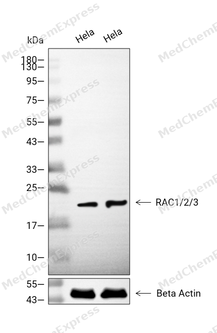
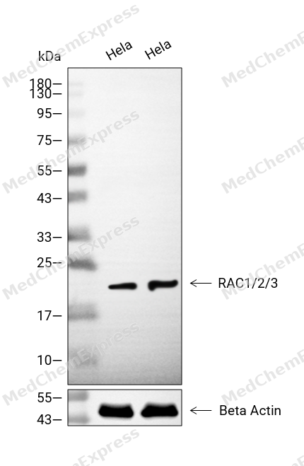 Western blot analysis of extracts from Hela (lane 2(20μg), Hela (lane 3(40μg), using RAC1/2/3 Antibody. Proteins were transferred to a PVDF membrane and blocked with 5% BSA in TBST for 2 hour at room temperature. The primary antibody and Loading control antibody (Beta Actin, HY-P80438, 1/3000) was used in 5% BSA in TBST at 4°C overnight. Goat Anti-Mouse/Rabbit IgG-HRP Secondary Antibody (HY-P8004/HY-P8001, 1/10,000) was used for 1 hour at room temperature.


Background
-
Function
RAC1/2/3 is a P63000: Plasma membrane-associated small GTPase which cycles between active GTP-bound and inactive GDP-bound states. In its active state, binds to a variety of effector proteins to regulate cellular responses such as secretory processes, phagocytosis of apoptotic cells, epithelial cell polarization, neurons adhesion, migration and differentiation, and growth-factor induced formation of membrane ruffles. Rac1 p21/rho GDI heterodimer is the active component of the cytosolic factor sigma 1, which is involved in stimulation of the NADPH oxidase activity in macrophages. Essential for the SPATA13-mediated regulation of cell migration and adhesion assembly and disassembly. Stimulates PKN2 kinase activity.
P15153: Plasma membrane-associated small GTPase which cycles between an active GTP-bound and inactive GDP-bound state. In its active state, binds to a variety of effector proteins to regulate cellular responses, such as secretory processes, phagocytose of apoptotic cells and epithelial cell polarization. Regulatory subunit of the phagocyte NADPH oxidase complex that mediates the transfer of electrons from cytosolic NADPH to O2 to produce the superoxide anion (O2- )
P60763: Plasma membrane-associated small GTPase which cycles between an active GTP-bound and inactive GDP-bound state. In active state binds to a variety of effector proteins to regulate cellular responses, such as cell spreading and the formation of actin-based protusions including lamellipodia and membrane ruffles. Promotes cell adhesion and spreading on fibrinogen in a CIB1 and alpha-IIb/beta3 integrin-mediated manner
[1][2][3][4][5][6][7]. -
Subcellular Localization
P63000: Cell membrane; Lipid-anchor; Cytoplasmic side; Melanosome; Cytoplasm; Cell projection, lamellipodium; Cell projection, dendrite; Synapse; Nucleus
P15153: Cytoplasm
P60763: Cytoplasm; Endomembrane system; Cell projection, lamellipodium; Cytoplasm, perinuclear region; Cell membrane; Cytoplasm, cytoskeleton -
Expression
Tissue_specificity: P63000: Isoform B is predominantly identified in skin and epithelial tissues from the intestinal tract. Its expression is elevated in colorectal tumors at various stages of neoplastic progression, as compared to their respective adjacent tissues
P15153: Hematopoietic specific
P60763: Highest levels in brain, also detected in heart, placenta and pancreas
Induction: P60763: Expression down-regulated in quiescent fibroblasts and clearly induced by serum stimulation -
Isoforms & Post-Translational Modification
P63000 has 2 isomers: P63000-1: 21450 Da (predicted); P63000-2: 23467 Da (predicted).
GTP-bound active form is ubiquitinated at Lys-147 by HACE1, leading to its degradation by the proteasome;Phosphorylated by AKT at Ser-71;Ubiquitinated at Lys-166 in a FBXL19-mediated manner; leading to proteasomal degradation;(Microbial infection) AMPylation at Tyr-32 and Thr-35 are mediated by bacterial enzymes in case of infection by H.somnus and V.parahaemolyticus, respectively. AMPylation occurs in the effector region and leads to inactivation of the GTPase activity by preventing the interaction with downstream effectors, thereby inhibiting actin assembly in infected cells. It is unclear whether some human enzyme mediates AMPylation; FICD has such ability in vitro but additional experiments remain to be done to confirm results in vivo;(Microbial infection) Glycosylated at Tyr-32 by Photorhabdus asymbiotica toxin PAU_02230. Mono-O-GlcNAcylation by PAU_02230 inhibits downstream signaling by an impaired interaction with diverse regulator and effector proteins of Rac and leads to actin disassembly;(Microbial infection) Glucosylated at Thr-35 by C.difficile toxins TcdA and TcdB in the colonic epithelium, and by P.sordellii toxin TcsL in the vascular endothelium (PubMed:19744486, PubMed:24905543, PubMed:7775453, PubMed:7777059, PubMed:8626575). Monoglucosylation completely prevents the recognition of the downstream effector, blocking the GTPases in their inactive form, leading to actin cytoskeleton disruption and cell death, resulting in the loss of colonic epithelial barrier function (PubMed:7775453, PubMed:7777059);(Microbial infection) Glycosylated (O-GlcNAcylated) at Thr-35 by C.novyi toxin TcdA (PubMed:8810274). O-GlcNAcylation completely prevents the recognition of the downstream effector, blocking the GTPases in their inactive form, leading to actin cytoskeleton disruption (PubMed:8810274);(Microbial infection) Palmitoylated by the N-epsilon-fatty acyltransferase F2 chain of V.cholerae toxin RtxA (PubMed:29074776). Palmitoylation inhibits activation by guanine nucleotide exchange factors (GEFs), preventing Rho GTPase signaling (PubMed:29074776) -
Subunit
P63000: Interacts with NISCH. Interacts with PIP5K1A. Interacts with the GTP-bound form of RAB7A. Interacts with SRGAP2.
P15153: Interacts with DOCK2, which may activate it. Interacts with S100A8 and calprotectin (S100A8/9) (PubMed:10559471, PubMed:15642721).
P60763: Interacts with the GEF protein DOCK7, which promotes the exchange between GDP and GTP, and therefore activates it (PubMed:16982419). Interacts with C1D (PubMed:9852280). Interacts (via C-terminal region) with CIB1; the interaction induces their association with the cytoskeleton upon alpha-IIb/beta3 integrin-mediated adhesion (PubMed:11756406). Interacts with NRBP (PubMed:11956649) -
Synonyms
Cell migration-inducing gene 5 protein; EN-7; GX; p21-Rac1; p21-Rac2; p21-Rac3; RAC1; RAC2; RAC3; TC25
-
Research Field
Signal Transduction


Documentation
-
Data Sheet (235 KB)
-
SDS (252 KB)
- English - EN (252 KB)
- Français - FR (252 KB)
- Deutsch - DE (252 KB)
- Norwegian - NO (252 KB)
- Español - ES (252 KB)
- Swedish - SV (252 KB)
- Italian - IT (252 KB)
- Korean - KR (252 KB)
- Portuguese - PT (252 KB)
-
User Guide for Antibodies (1077 KB)

 Powered by Bioz
