



RNase L Antibody (YA2314)
(Synonyms: PRCA1; Ribonuclease4; RibonucleaseL; RNase L; Rnasel; RNS4)
Based on 1 Customer Validation
RNase L Antibody (YA2314) is a Rabbit-derived and non-conjugated IgG monoclonal antibody, targeting to RNase L.
-
Host:
Rabbit
-
Isotype:
IgG
-
Application:
WB
-
Reactivity :
Human
-
Formulation:
Supplied in 10mM PBS, pH 7.4, 150mM sodium chloride, 0.05% BSA, 0.02% sodium azide and 50% glycerol.
-
Conjugation:
Non-conjugated


To place orders, for customer services and technical support, please contact: MedChemExpress USA
Tel: 609-228-6898 E-mail: [email protected] [email protected]





Applications
| Application |
WB

WB: Western Blot
|
|---|---|
| Dilution Ratio | 1:500-1:1000 |


Product Details
RNase L Antibody (YA2314) is a Rabbit-derived and non-conjugated IgG monoclonal antibody, targeting to RNase L.
-
Host Rabbit

-
Clonality Recombinant,Monoclonal

-
Species ReactivityHuman
-
Observed Molecular WeightObserved band size: 80 kDa
Note: Due to possible protein modifications or aggregation, the molecular weight should be confirmed by actual measurement, and the predicted value is for reference only.

-
Calculated Molecular Weight Predicted band size: 84 kDa

A synthesized peptide derived from human RNase L aa550-741/741.
Endogenous
Affinity Purified
Non-conjugated
Unmodified
IgG


Product Properties
-
Appearance
Solution
-
Formulation
Supplied in 10mM PBS, pH 7.4, 150mM sodium chloride, 0.05% BSA, 0.02% sodium azide and 50% glycerol.
-
Concentration
Batch-dependent, Please check the COA for the concentration of each lot. Check Lot Concentration
-
Storage & Stability
Stored at -20°C for 1 year. Avoid repeated freeze / thaw cycles.
-
Shipping
Shipping with blue ice.


Verification Images
-
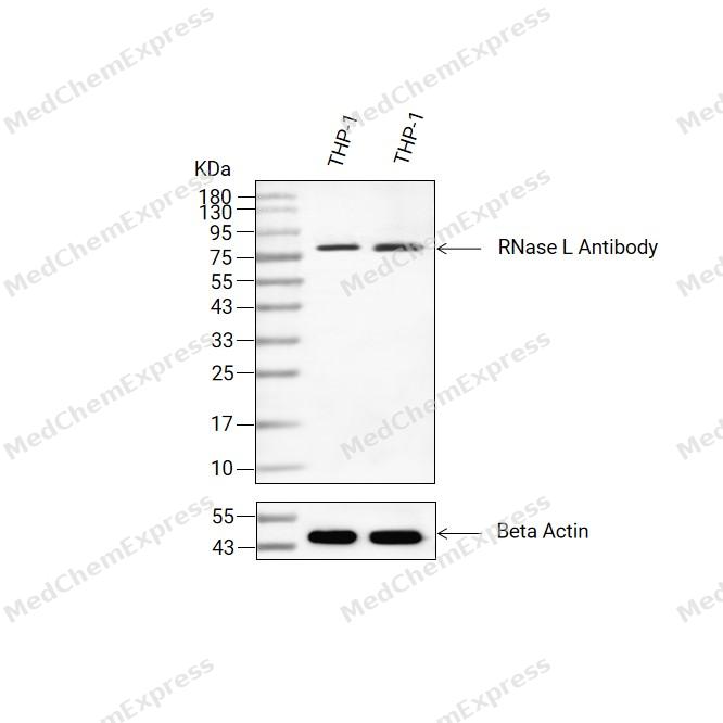 Western blot analysis of extracts from THP-1(lane 2(20ug) and THP-1(lane 3(40ug) using RNase L Antibody (HY-P82569) Rabbit mAb. Proteins were transferred to a PVDF membrane and blocked with 5% non-fat milk in TBST for 2 hour at room temperature. The primary antibody (1/1000) and Loading control antibody (Beta Actin, HY-P83730, 1/10000) was used in 5% non-fat milk in TBST at 4°C overnight. Goat Anti-Mouse/Rabbit IgG-HRP Secondary Antibody (1/10000) was used for 1 hour at room temperature.
 -
 Western blot analysis of extracts from HEK293T (lane2, 20μg), THP-1 (lane3, 20μg), K562 (lane4, 20μg) and HCT116 (lane5, 20μg) using RNase L Antibody (HY-P82569). Proteins were transferred to a PVDF membrane and blocked with 5% non-fat milk in TBST for 2 hour at room temperature. The primary antibody (1/1000) and Loading control antibody (Beta Actin, HY-P80993, 1/10,000) was used in 5% non-fat milk in TBST at 4°C overnight. Goat Anti-Rabbit IgG-HRP Secondary Antibody (HY-P8001,1/10,000) was used for 1 hour at room temperature.
 -
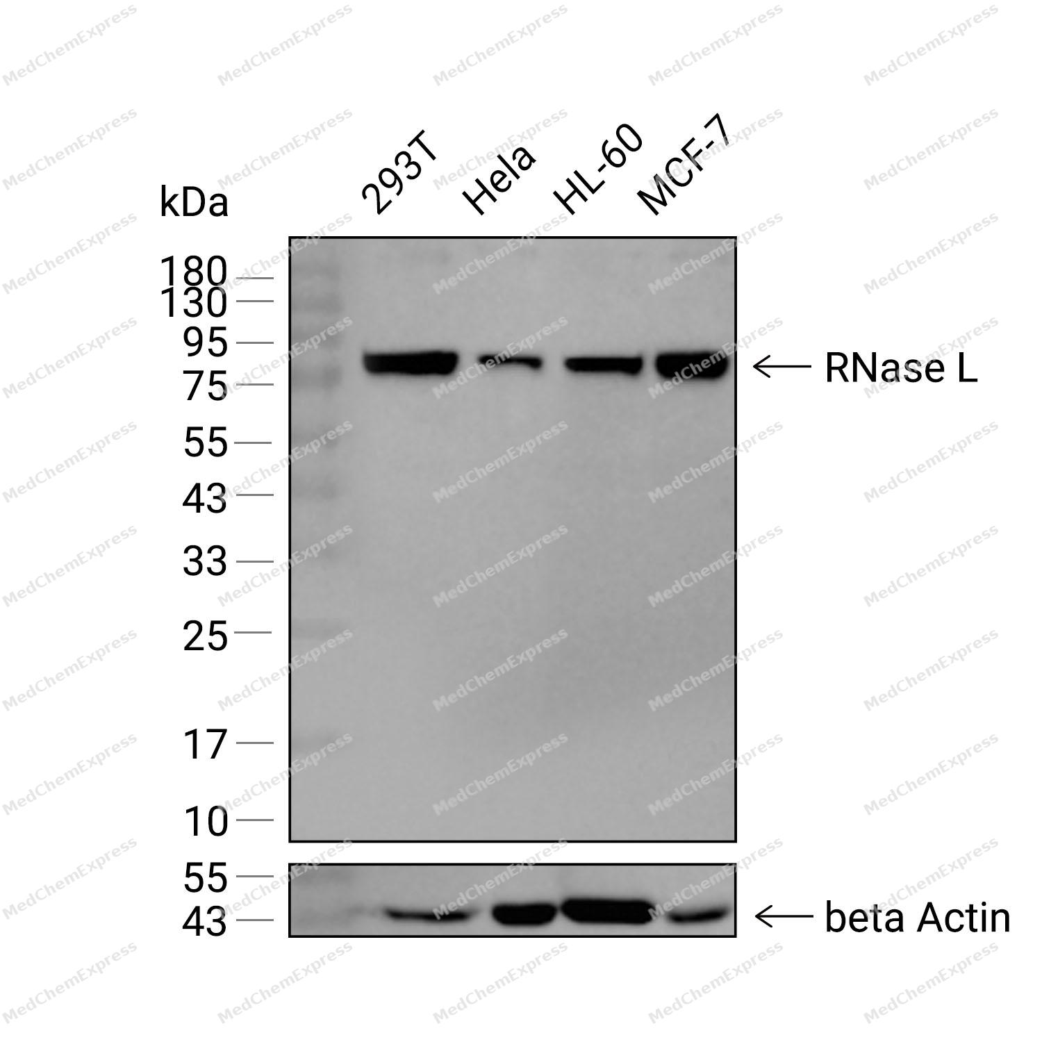 Western blot analysis was performed on extracts from 293T (lane 1, 15 μg), Hela (lane 2, 15 μg), HL-60 (lane 3, 15 μg), and MCF-7 (lane 4, 15 μg) using RNase L Rabbit mAb.Proteins were transferred to a PVDF membrane and blocked with 5% non-fat milk in TBST at 4°C overnight.The primary antibody (1:1000 dilution) and the loading control antibody (beta-Actin, HY-P80438, 1:20000 dilution) were incubated in 5% non-fat milk in TBST for 1 hour at 37°C.Goat Anti-Rabbit IgG-HRP Secondary Antibody (1:20000 dilution) was then applied for 40 minutes at 37°C.


Background
-
Function
RNASEL encodes RNase L, an interferon-inducible endoribonuclease that functions as a central effector of the 2′-5′-oligoadenylate synthetase (OAS) -RNase L pathway, a conserved arm of innate antiviral immunity[1][2]. Upon recognition of viral double-stranded RNA, OAS enzymes synthesize 2′-5′-linked oligoadenylates (2-5A), which bind and activate RNase L, leading to the cleavage of cellular and viral RNA and suppression of viral replication[1][3][4]. Mechanistically, RNase L-mediated RNA fragmentation not only restricts pathogen replication but also promotes downstream stress and immune signaling, including activation of ZAKα-dependent ribotoxic stress pathways and antiviral transcriptional programs[2]. Beyond classical antiviral defense, the OAS-RNase L axis regulates inflammatory responses, and genetic defects in this pathway have been linked to severe inflammatory manifestations associated with SARS-CoV-2 infection, highlighting its importance in immune homeostasis[5]. Disease-relevant studies further demonstrate that RNase L participates in tissue-specific biological processes, including the regulation of wound-induced hair follicle neogenesis and epithelial regeneration through modulation of innate immune signaling pathways[6]. Compared with related OAS family members that function primarily as upstream sensors and 2-5A synthetases, RNase L serves as the terminal catalytic effector that directly executes RNA degradation after pathway activation[1][3]. For experimental applications, pharmacological inhibition of RNase L has been achieved using small-molecule inhibitors that block RNase L activity and prevent excessive RNA cleavage, providing useful tools for mechanistic studies of innate immunity, inflammation, and RNA biology[7].
-
Subcellular Localization
Cytoplasm; Mitochondrion
-
Expression
Tissue_specificity:Highly expressed in spleen and thymus followed by prostate, testis, uterus, small intestine, colon and peripheral blood leukocytes
Induction:By interferons. Virus replication in higher vertebrates is restrained by IFNs that cause cells to transcribe genes encoding antiviral proteins, such as 2'-5' oligoadenylate synthetases (OASs) . oligoadenylate synthetase is stimulated by dsRNA to produce 5'-phosphorylated, 2'-5'-linked oligoadenylates (2-5A) , whose function is to activate RNASEL -
Isoforms & Post-Translational Modification
Q05823 has 2 isomers: Q05823-1: 83533 Da (predicted); Q05823-2: 73416 Da (predicted).
-
Subunit
Monomer (inactive form) or homodimer. Interacts with ABCE1; this interaction inhibits the RNASEL
-
SwissProt ID
-
Synonyms
PRCA1; Ribonuclease4; RibonucleaseL; RNase L; Rnasel; RNS4
-
Research Field
Immunology


Documentation
-
Data Sheet (261 KB)
-
SDS (251 KB)
- English - EN (251 KB)
- Français - FR (251 KB)
- Deutsch - DE (251 KB)
- Norwegian - NO (251 KB)
- Español - ES (251 KB)
- Swedish - SV (251 KB)
- Italian - IT (251 KB)
- Korean - KR (251 KB)
- Portuguese - PT (251 KB)
-
User Guide for Antibodies (1077 KB)
References
[1]. Karasik A, et al. The Unusual Role of Ribonuclease L in Innate Immunity. Wiley Interdiscip Rev RNA. 2024 Nov-Dec;15(6):e1878. [Content Brief]
[2]. Karasik A, et al. Endonucleolytic RNA cleavage drives changes in gene expression during the innate immune response. Cell Rep. 2024 Jun 25;43(6):114287. [Content Brief]
[3]. Soveg FW, et al. Endomembrane targeting of human OAS1 p46 augments antiviral activity. Elife. 2021 Aug 3;10:e71047. [Content Brief]
[4]. Ezelle HJ, et al. The Roles of RNase-L in Antimicrobial Immunity and the Cytoskeleton-Associated Innate Response. Int J Mol Sci. 2016 Jan 8;17(1):74. [Content Brief]
[5]. Lee D, et al. Inborn errors of OAS-RNase L in SARS-CoV-2-related multisystem inflammatory syndrome in children. Science. 2023 Feb 10;379(6632):eabo3627. [Content Brief]
[6]. Kirby CS, et al. RNase L represses hair follicle regeneration through altered innate immune signaling. J Clin Invest. 2025 Feb 4;135(6):e172595. [Content Brief]
[7]. Daou S, et al. A phenolic small molecule inhibitor of RNase L prevents cell death from ADAR1 deficiency. Proc Natl Acad Sci U S A. 2020 Oct 6;117(40):24802-24812. [Content Brief]

 Powered by Bioz
