



ROR gamma T Antibody (YA3559)
(Synonyms: IMD42 antibody; MGC129539 antibody; NR1F3 antibody; Nuclear receptor ROR gamma antibody; Nuclear receptor subfamily 1 group F member 3 antibody; RAR related orphan receptor C antibody; Retinoic acid binding receptor gamma antibody; Retinoid related orphan receptor gamma antibody; Rorc antibody; RORG antibody; RORG_HUMAN antibody; RZR GAMMA antibody; RZRG antibody; TOR antibody)
Based on 1 Customer Validation
ROR gamma T Antibody (YA3559) is a Rabbit-derived and non-conjugated IgG monoclonal antibody, targeting to ROR gamma T.
-
Host:
Rabbit
-
Isotype:
IgG
-
Application:
WB, IHC-P
-
Reactivity :
Human, Rat
-
Formulation:
Supplied in PBS (pH7.4), 0.1% BSA, 40% Glycerol. Preservative: 0.05% Sodium Azide.
-
Conjugation:
Non-conjugated


To place orders, for customer services and technical support, please contact: MedChemExpress USA
Tel: 609-228-6898 E-mail: [email protected] [email protected]





Applications
| Application |
WB

WB: Western Blot
|
IHC-P
IHC-P: Immunohistochemistry-Paraffin
|
|---|---|---|
| Dilution Ratio | 1:2000 | 1:500 |


Product Details
ROR gamma T Antibody (YA3559) is a Rabbit-derived and non-conjugated IgG monoclonal antibody, targeting to ROR gamma T.
-
Host Rabbit

-
Clonality Monoclonal,Recombinant

-
Species ReactivityHuman, Rat
-
Observed Molecular WeightObserved band size: 55 kDa
Note: Due to possible protein modifications or aggregation, the molecular weight should be confirmed by actual measurement, and the predicted value is for reference only.

-
Calculated Molecular Weight Predicted band size: 58 kDa;

Recombinant protein within human ROR gamma T aa 101-300 / 497 (P51449-2).
affinity purified
Non-conjugated
Unmodified
IgG


Product Properties
-
Appearance
Liquid
-
Formulation
Supplied in PBS (pH7.4), 0.1% BSA, 40% Glycerol. Preservative: 0.05% Sodium Azide.
-
Concentration
Batch-dependent, Please check the COA for the concentration of each lot. Check Lot Concentration
-
Storage & Stability
Stored at -20°C for 1 year. Avoid repeated freeze / thaw cycles.
-
Shipping
Shipping with blue ice.


Verification Images
-
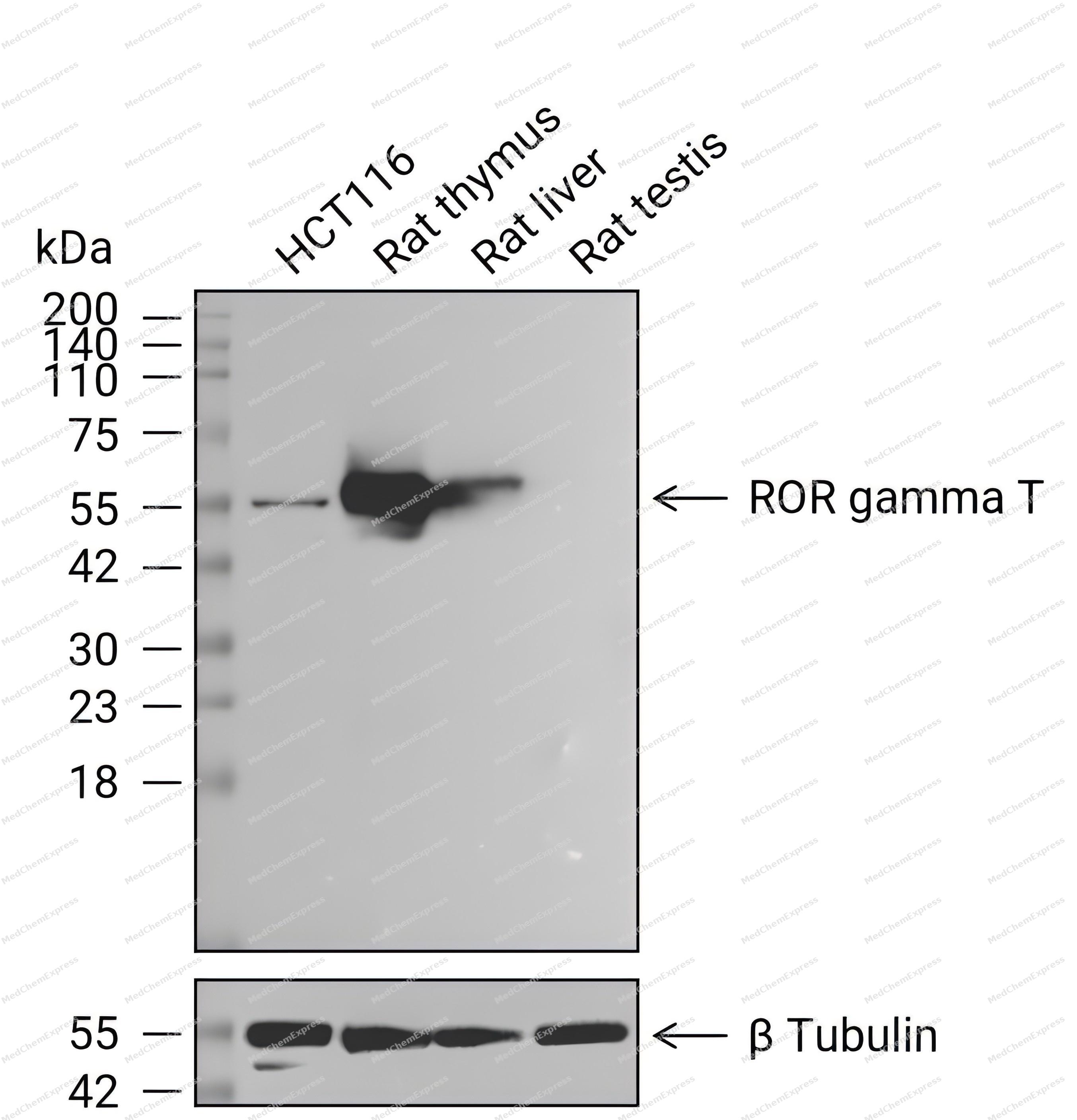 Western blot analysis of extracts from HCT116 (lane 1(20μg)) 、Rat thymus (lane 2(20μg)) 、Rat liver (lane 3(20μg)) and Rat testis (lane 4(20μg, negative)) using ROR gamma T Antibody (HY-P83758) . Proteins were transferred to a PVDF membrane and blocked with 5% nonfat dry milk in TBST for 1.5 hour at room temperature. The primary antibody (1/2000) and Loading control antibody (β Tubulin, 1/5000) was used in 5% nonfat dry milk in TBST at 4℃ overnight. Goat Anti-Rabbit IgG-HRP Secondary Antibody (1/10,000) was used for 1 hour at room temperature.
 -
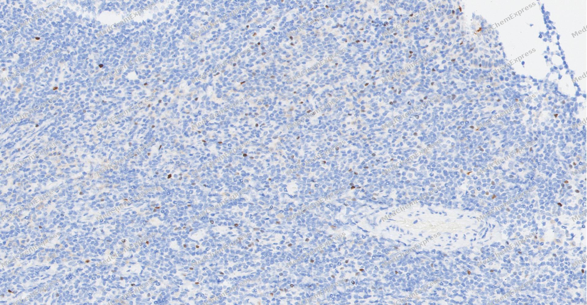 Immunohistochemical analysis of paraffin-embedded human Tonsil tissue using ROR gamma T antibody was performed. The section was pretreated using high-temperature and high-pressure mediated EDTA antigen retrieval buffer (pH 9.0), for 5 minutes. The tissues were incubated with primary antibody (HY-P83758, 1:500 dilution) at room temperature for 60 minutes. Detection was performed using an HRP conjugated compact polymer system. DAB was used as the chromogen. The tissues were counterstained with hematoxylin and mounted with neutral balsam mounting medium.
 -
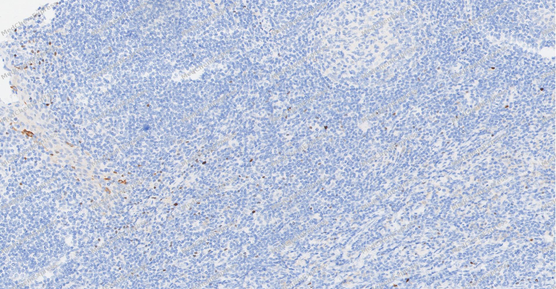 Immunohistochemical analysis of paraffin-embedded human Tonsil tissue using ROR gamma T antibody was performed. The section was pretreated using high-temperature and high-pressure mediated EDTA antigen retrieval buffer (pH 9.0), for 5 minutes. The tissues were incubated with primary antibody (HY-P83758, 1:500 dilution) at room temperature for 60 minutes. Detection was performed using an HRP conjugated compact polymer system. DAB was used as the chromogen. The tissues were counterstained with hematoxylin and mounted with neutral balsam mounting medium.
 -
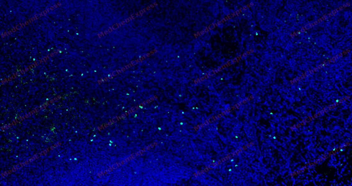 Tyramide signaling amplification based immunofluorescence was performed on paraffin-embedded human Tonsil tissue using ROR gamma T antibody. Antigen retrieval was performed in EDTA buffer pH 9.0 (95 °C, 20 min) followed by cooling to RT. Then incubated with primary antibody (HY-P83758, 1:800 dilution) at room temperature for 60 minutes and HRP conjugated secondary antibody for 10 minutes. Fluorescence was then developed with Vari Fluor 532 TSA (200×)(HY-D1832). The tissues were counterstained with DAPI and mounted with Anti-fade mounting medium.
 -
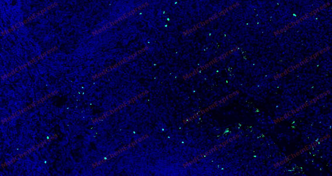 Tyramide signaling amplification based immunofluorescence was performed on paraffin-embedded human Tonsil tissue using ROR gamma T antibody. Antigen retrieval was performed in EDTA buffer pH 9.0 (95 °C, 20 min) followed by cooling to RT. Then incubated with primary antibody (HY-P83758, 1:800 dilution) at room temperature for 60 minutes and HRP conjugated secondary antibody for 10 minutes. Fluorescence was then developed with Vari Fluor 532 TSA (200×)(HY-D1832). The tissues were counterstained with DAPI and mounted with Anti-fade mounting medium.


Background
-
Function
ROR gamma T is a Nuclear receptor that binds DNA as a monomer to ROR response elements (RORE) containing a single core motif half-site 5'-AGGTCA-3' preceded by a short A-T-rich sequence. Key regulator of cellular differentiation, immunity, peripheral circadian rhythm as well as lipid, steroid, xenobiotics and glucose metabolism. Considered to have intrinsic transcriptional activity, have some natural ligands like oxysterols that act as agonists (25-hydroxycholesterol) or inverse agonists (7-oxygenated sterols), enhancing or repressing the transcriptional activity, respectively. Recruits distinct combinations of cofactors to target gene regulatory regions to modulate their transcriptional expression, depending on the tissue, time and promoter contexts. Regulates the circadian expression of clock genes such as CRY1, BMAL1 and NR1D1 in peripheral tissues and in a tissue-selective manner. Competes with NR1D1 for binding to their shared DNA response element on some clock genes such as BMAL1, CRY1 and NR1D1 itself, resulting in NR1D1-mediated repression or RORC-mediated activation of the expression, leading to the circadian pattern of clock genes expression. Therefore influences the period length and stability of the clock. Involved in the regulation of the rhythmic expression of genes involved in glucose and lipid metabolism, including PLIN2 and AVPR1A. Negative regulator of adipocyte differentiation through the regulation of early phase genes expression, such as MMP3. Controls adipogenesis as well as adipocyte size and modulates insulin sensitivity in obesity. In liver, has specific and redundant functions with RORA as positive or negative modulator of expression of genes encoding phase I and Phase II proteins involved in the metabolism of lipids, steroids and xenobiotics, such as SULT1E1. Also plays a role in the regulation of hepatocyte glucose metabolism through the regulation of G6PC1 and PCK1. Regulates the rhythmic expression of PROX1 and promotes its nuclear localization. Plays an indispensable role in the induction of IFN-gamma dependent anti-mycobacterial systemic immunity; Essential for thymopoiesis and the development of several secondary lymphoid tissues, including lymph nodes and Peyer's patches. Required for the generation of LTi (lymphoid tissue inducer) cells. Regulates thymocyte survival through DNA-binding on ROREs of target gene promoter regions and recruitment of coactivaros via the AF-2. Also plays a key role, downstream of IL6 and TGFB and synergistically with RORA, for lineage specification of uncommitted CD4(+) T-helper (T(H)) cells into T(H)17 cells, antagonizing the T(H)1 program. Probably regulates IL17 and IL17F expression on T(H) by binding to the essential enhancer conserved non-coding sequence 2 (CNS2) in the IL17-IL17F locus. May also play a role in the pre-TCR activation cascade leading to the maturation of alpha/beta T-cells and may participate in the regulation of DNA accessibility in the TCR-J(alpha) locus[1][2][3][4][5].
-
Subcellular Localization
Nucleus
-
Expression
Tissue_specificity:Isoform 1 is widely expressed in many tissues, including liver and adipose, and highly expressed in skeletal muscle. Isoform 2 is primarily expressed in immature thymocytes
Induction:Up-regulated in the state of obesity
Positive sample: HCT 116 cell lysate, rat thymus tissue lysate, human thymus tissue, rat thymus tissue. -
Isoforms & Post-Translational Modification
P51449 has 2 isomers: P51449-1: 58195 Da (predicted); P51449-2: 55813 Da (predicted).
-
Subunit
Interacts (via AF-2 motif) with the coactivator NCOA2 (via LXXLL motif) (PubMed:20211758). Interacts with the corepressor NCOR1 (By similarity). Interacts with CRY1 (PubMed:22170608). Interacts (via AF-2 motif) with the coactivators NCOA1 and PPARGC1A (via LXXLL motif) (By similarity). Interacts (via AF-2 motif) with PROX1 (By similarity). Interacts with FOXP3 (PubMed:18368049). Interacts with NR0B2 (By similarity)
-
SwissProt ID
-
Synonyms
IMD42 antibody; MGC129539 antibody; NR1F3 antibody; Nuclear receptor ROR gamma antibody; Nuclear receptor subfamily 1 group F member 3 antibody; RAR related orphan receptor C antibody; Retinoic acid binding receptor gamma antibody; Retinoid related orphan receptor gamma antibody; Rorc antibody; RORG antibody; RORG_HUMAN antibody; RZR GAMMA antibody; RZRG antibody; TOR antibody


Documentation
-
Data Sheet (235 KB)
-
SDS (315 KB)
- English - EN (315 KB)
- Français - FR (315 KB)
- Deutsch - DE (315 KB)
- Norwegian - NO (315 KB)
- Español - ES (315 KB)
- Swedish - SV (315 KB)
- Italian - IT (315 KB)
- Korean - KR (315 KB)
- Portuguese - PT (315 KB)
-
User Guide for Antibodies (1077 KB)

 Powered by Bioz
