



Smad5 Antibody (YA5909)
(Synonyms: SMAD5; MADH5; Mothers against decapentaplegic homolog 5; MAD homolog 5; Mothers against DPP homolog 5; JV5-1; SMAD family member 5; SMAD 5; Smad5; hSmad5)Based on 1 Customer Validation
Smad5 Antibody (YA5909) is a Rabbit-derived and non-conjugated IgG monoclonal antibody, targeting to Smad5.
-
Host:
Rabbit
-
Isotype:
IgG
-
Application:
WB, IHC-P, ICC/IF, IP, ELISA
-
Reactivity :
Human, Mouse, Rat
-
Formulation:
Supplied in PBS, 50% glycerol, 0.05% Proclin 300, 0.05%BSA
-
Conjugation:
Non-conjugated


To place orders, for customer services and technical support, please contact: MedChemExpress USA
Tel: 609-228-6898 E-mail: [email protected] [email protected]





Applications
| Application |
IHC-P

IHC-P: Immunohistochemistry-Paraffin
|
WB
WB: Western Blot
|
ICC/IF
ICC/IF: Immunocytochemistry/
Immunofluorescence |
ELISA
ELISA: Enzyme Linked Immunosorbent Assay
|
IP
IP: Immunoprecipitation
|
|---|---|---|---|---|---|
| Dilution Ratio | 1:200-1:1000 | 1:2000-1:10000 | 1:200-1:1000 | 1:5000-1:20000 | 1:50-1:200 |


Product Details
Smad5 Antibody (YA5909) is a Rabbit-derived and non-conjugated IgG monoclonal antibody, targeting to Smad5.
-
Host Rabbit

-
Clonality Monoclonal

-
Species ReactivityHuman, Mouse, Rat
-
Observed Molecular WeightObserved band size: 55 kDa
Note: Due to possible protein modifications or aggregation, the molecular weight should be confirmed by actual measurement, and the predicted value is for reference only.

-
Calculated Molecular Weight Predicted band size: 55 kDa

Protein A
Non-conjugated
Unmodified
IgG


Product Properties
-
Appearance
Solution
-
Formulation
Supplied in PBS, 50% glycerol, 0.05% Proclin 300, 0.05%BSA
-
Concentration
Batch-dependent, Please check the COA for the concentration of each lot. Check Lot Concentration
-
Storage & Stability
Stored at -20°C for 1 year. Avoid repeated freeze / thaw cycles.
-
Shipping
Shipping with blue ice.


Verification Images
-
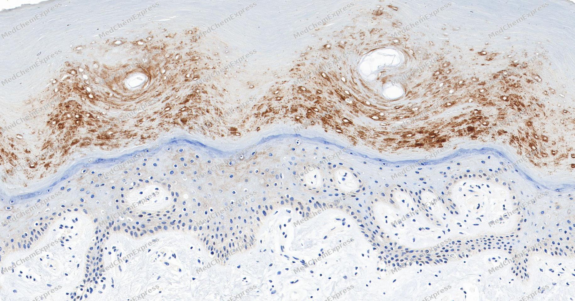 Immunohistochemical analysis of paraffin-embedded human skin tissue using Smad5 Antibody (YA5909). The section was pre-treated using heat mediated antigen retrieval with Tris/EDTA buffer (pH 9.0) for 20 minutes. The tissues were probed with the primary antibody (HY-P86217, 1/150) overnight at 4℃. The detection was performed using an HRP conjugated compact polymer system. DAB was used as the chromogen. Tissues were counterstained with hematoxylin and mounted with neutral balsam.
 -
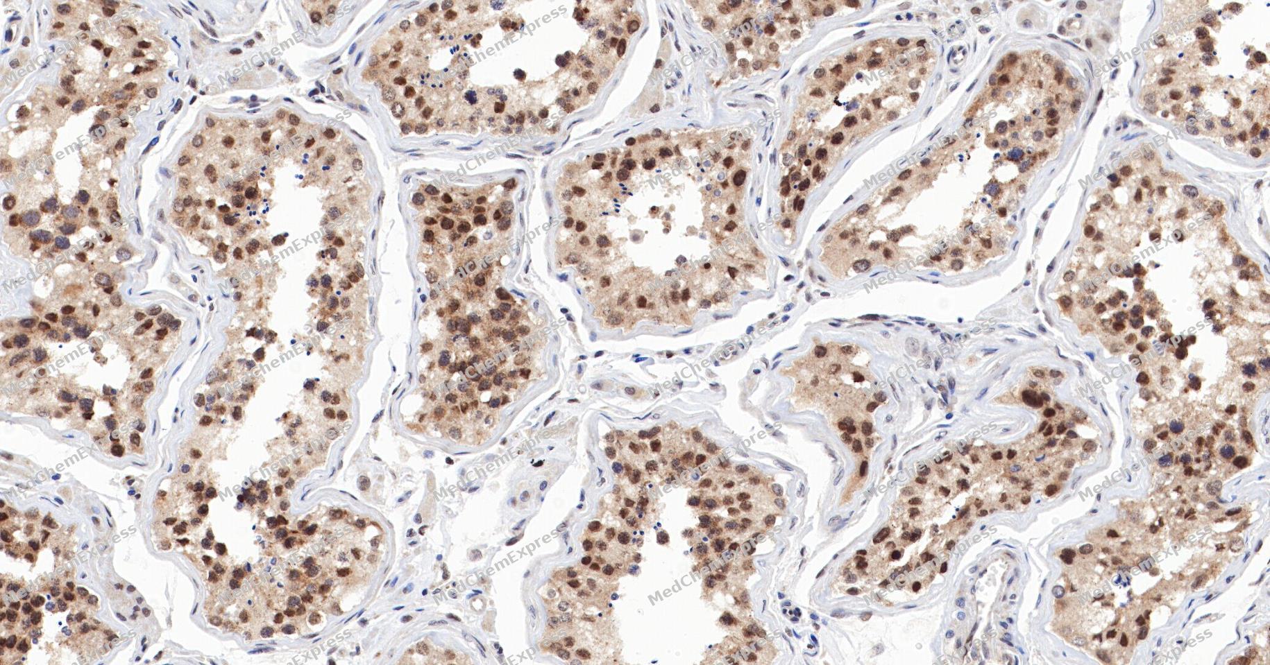 Immunohistochemical analysis of paraffin-embedded human testis tissue using Smad5 Antibody (YA5909). The section was pre-treated using heat mediated antigen retrieval with Tris/EDTA buffer (pH 9.0) for 20 minutes. The tissues were probed with the primary antibody (HY-P86217, 1/150) overnight at 4℃. The detection was performed using an HRP conjugated compact polymer system. DAB was used as the chromogen. Tissues were counterstained with hematoxylin and mounted with neutral balsam.
 -
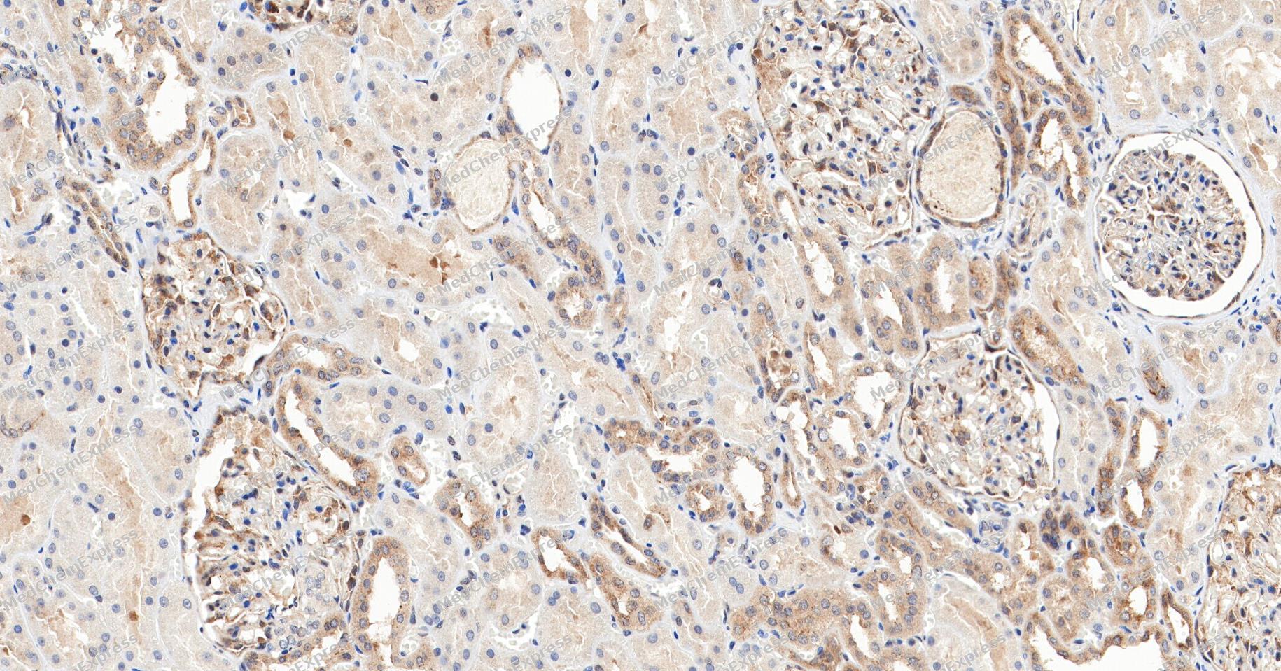 Immunohistochemical analysis of paraffin-embedded human kidney tissue using Smad5 Antibody (YA5909). The section was pre-treated using heat mediated antigen retrieval with Tris/EDTA buffer (pH 9.0) for 20 minutes. The tissues were probed with the primary antibody (HY-P86217, 1/150) overnight at 4℃. The detection was performed using an HRP conjugated compact polymer system. DAB was used as the chromogen. Tissues were counterstained with hematoxylin and mounted with neutral balsam.
 -
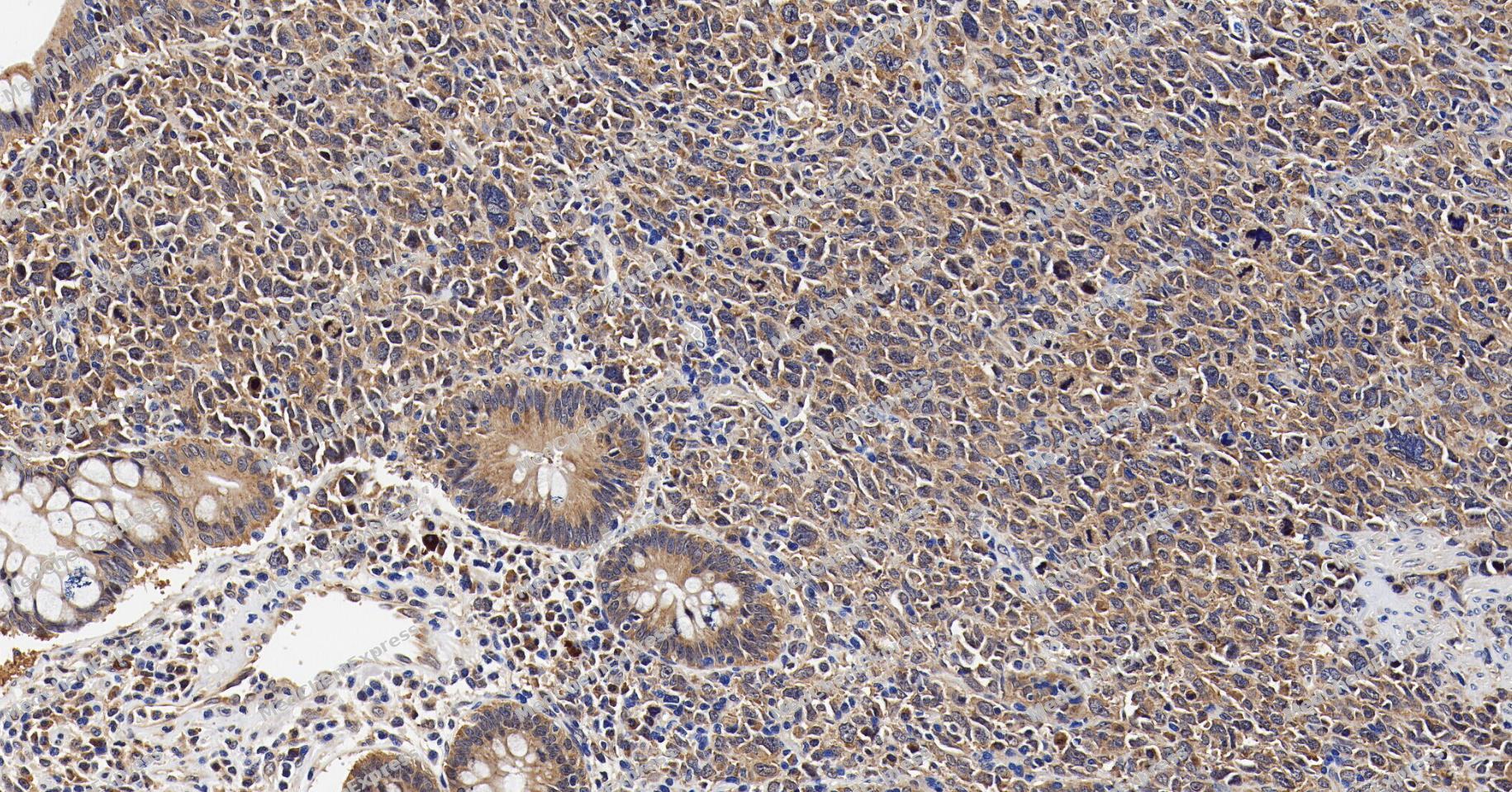 Immunohistochemical analysis of paraffin-embedded human colon cancer tissue using Smad5 Antibody (YA5909). The section was pre-treated using heat mediated antigen retrieval with Tris/EDTA buffer (pH 9.0) for 20 minutes. The tissues were probed with the primary antibody (HY-P86217, 1/150) overnight at 4℃. The detection was performed using an HRP conjugated compact polymer system. DAB was used as the chromogen. Tissues were counterstained with hematoxylin and mounted with neutral balsam.
 -
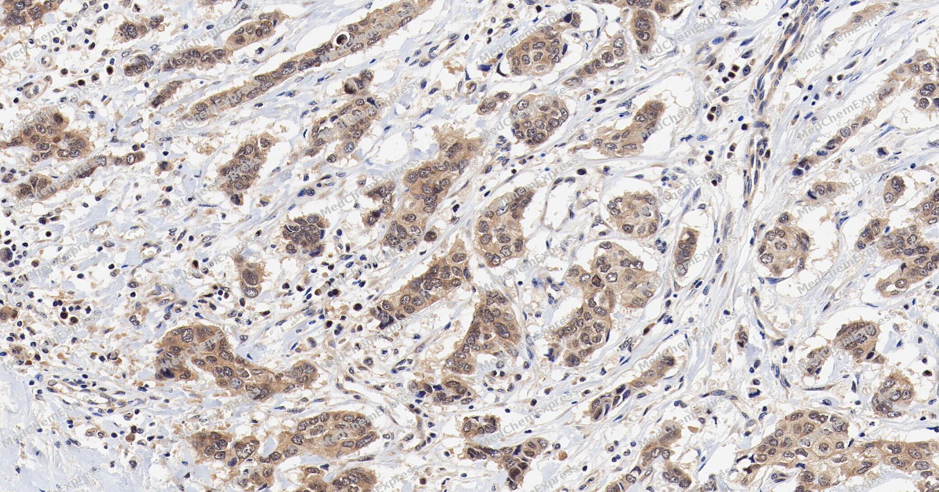 Immunohistochemical analysis of paraffin-embedded human breast cancer tissue using Smad5 Antibody (YA5909). The section was pre-treated using heat mediated antigen retrieval with Tris/EDTA buffer (pH 9.0) for 20 minutes. The tissues were probed with the primary antibody (HY-P86217, 1/150) overnight at 4℃. The detection was performed using an HRP conjugated compact polymer system. DAB was used as the chromogen. Tissues were counterstained with hematoxylin and mounted with neutral balsam.
 -
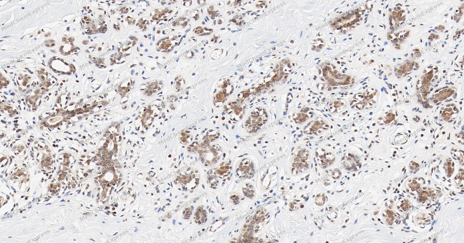 Immunohistochemical analysis of paraffin-embedded human breast tissue using Smad5 Antibody (YA5909). The section was pre-treated using heat mediated antigen retrieval with Tris/EDTA buffer (pH 9.0) for 20 minutes. The tissues were probed with the primary antibody (HY-P86217, 1/150) overnight at 4℃. The detection was performed using an HRP conjugated compact polymer system. DAB was used as the chromogen. Tissues were counterstained with hematoxylin and mounted with neutral balsam.
 -
 Immunohistochemical analysis of paraffin-embedded mouse kidney tissue using Smad5 Antibody (HY-P86217, 1/800). The section was pretreated using heat mediated antigen retrieval with sodium citrate buffer (pH 6.0) for 8 minutes. The tissues were blocked with quick block buffer for 0.5 hours at room temperature, washed with PBS and PBST, and then incubated with the primary antibody overnight at 4℃. The detection was performed using an HRP conjugated compact polymer system. DAB was used as the chromogen. Tissues were counterstained with hematoxylin and mounted with DPX.
 -
 Immunohistochemical analysis of paraffin-embedded mouse colon tissue using Smad5 Antibody (HY-P86217, 1/800). The section was pretreated using heat mediated antigen retrieval with sodium citrate buffer (pH 6.0) for 8 minutes. The tissues were blocked with quick block buffer for 0.5 hours at room temperature, washed with PBS and PBST, and then incubated with the primary antibody overnight at 4℃. The detection was performed using an HRP conjugated compact polymer system. DAB was used as the chromogen. Tissues were counterstained with hematoxylin and mounted with DPX.
 -
 Immunohistochemical analysis of paraffin-embedded mouse testis tissue using Smad5 Antibody (HY-P86217, 1/800). The section was pretreated using heat mediated antigen retrieval with sodium citrate buffer (pH 6.0) for 8 minutes. The tissues were blocked with quick block buffer for 0.5 hours at room temperature, washed with PBS and PBST, and then incubated with the primary antibody overnight at 4℃. The detection was performed using an HRP conjugated compact polymer system. DAB was used as the chromogen. Tissues were counterstained with hematoxylin and mounted with DPX.
 -
 Immunohistochemical analysis of paraffin-embedded human adrenal gland tissue using Smad5 Antibody (HY-P86217, 1/800). The section was pretreated using heat mediated antigen retrieval with sodium citrate buffer (pH 6.0) for 8 minutes. The tissues were blocked with quick block buffer for 0.5 hours at room temperature, washed with PBS and PBST, and then incubated with the primary antibody overnight at 4℃. The detection was performed using an HRP conjugated compact polymer system. DAB was used as the chromogen. Tissues were counterstained with hematoxylin and mounted with DPX.
 -
 Immunohistochemical analysis of paraffin-embedded human tonsil tissue using Smad5 Antibody (HY-P86217, 1/800). The section was pretreated using heat mediated antigen retrieval with sodium citrate buffer (pH 6.0) for 8 minutes. The tissues were blocked with quick block buffer for 0.5 hours at room temperature, washed with PBS and PBST, and then incubated with the primary antibody overnight at 4℃. The detection was performed using an HRP conjugated compact polymer system. DAB was used as the chromogen. Tissues were counterstained with hematoxylin and mounted with DPX.
 -
 Immunohistochemical analysis of paraffin-embedded human gallbladder tissue using Smad5 Antibody (HY-P86217, 1/800). The section was pretreated using heat mediated antigen retrieval with sodium citrate buffer (pH 6.0) for 8 minutes. The tissues were blocked with quick block buffer for 0.5 hours at room temperature, washed with PBS and PBST, and then incubated with the primary antibody overnight at 4℃. The detection was performed using an HRP conjugated compact polymer system. DAB was used as the chromogen. Tissues were counterstained with hematoxylin and mounted with DPX.
 -
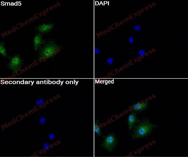 Immunocytochemistry analysis of A549 cells labeling Smad5 with Smad5 Antibody (HY-P86217) at 1:200 dilution. Cells were fixed in 4% paraformaldehyde for 15 minutes at room temperature, permeabilized with 0.1% Triton X-100 in PBS for 15 minutes at room temperature, then blocked with quick block buffer for 10 minutes at room temperature. Cells were then incubated with Smad5 Antibody (HY-P86217) at 1:200 dilution in quick block buffer overnight at 4 ℃. AF488-conjugated Goat Anti-Rabbit IgG H&L(HY-P8002, Green) was used as the secondary antibody at 1/1,000 dilution. PBS instead of the primary antibody was used as the secondary antibody only control. The Nuclear counterstain was DAPI (Blue).
 -
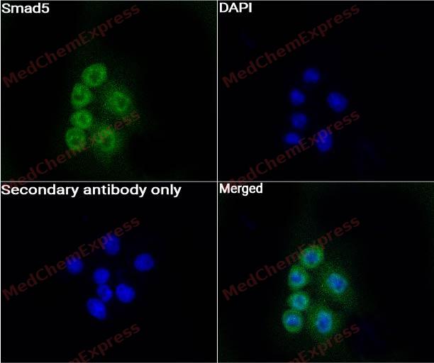 Immunocytochemistry analysis of Hela cells labeling Smad5 with Smad5 Antibody (HY-P86217) at 1:200 dilution. Cells were fixed in 4% paraformaldehyde for 15 minutes at room temperature, permeabilized with 0.1% Triton X-100 in PBS for 15 minutes at room temperature, then blocked with quick block buffer for 10 minutes at room temperature. Cells were then incubated with Smad5 Antibody (HY-P86217) at 1:200 dilution in quick block buffer overnight at 4 ℃. AF488-conjugated Goat Anti-Rabbit IgG H&L(HY-P8002, Green) was used as the secondary antibody at 1/1,000 dilution. PBS instead of the primary antibody was used as the secondary antibody only control. The Nuclear counterstain was DAPI (Blue).


Background
-
Function
SMAD5 is a receptor activated Smad (R-Smad), also known as dwarfin C, JV5-1 and MAD5, which belong to the dwarfin/SMAD family. The SMAD5 protein consists of an N-terminal domain responsible for DNA binding, a C-terminal domain primarily used for protein-protein interactions, and a junction region containing moths necessary for ubiquitination degradation. SMAD5 is activated by an activated type I TGF-β receptor, and phosphorylated SMAD5 binds to SMAD4 (common SMAD) to form heterotrimer complexes that enter the nucleus as transcription factors. At the same time, SMAD4 is also bound by inhibitory SMAD (SMAD6 and SMAD7), making R-Smad compete for receptors or interact with Smad4, and target degradation and negative regulation of TGF-β signaling. SMAD5 mainly responds to BMP signaling pathways. During embryonic development, SMAD5 is widely expressed in mice from gastrum formation. During this period, the loss of SMAD5 leads to mutations in the embryo, or defects in the yolk sac outside the embryo, and death in the second trimester. Furthermore, the BMP/SMAD5 pathway can regulate osteogenesis by up-regulating transcription of the transcription factor Runx2, or by targeting multiple cis-acting promoter elements in osteogenic specific genes, including osteocalcin and alkaline phosphatase (ALP) . Alps bind to Smad interacting protein 1 (SIP1) and inhibit Smad5-induced SIP1 activation. A significant expansion of primitive multipotent progenitor cells showing normal terminal maturation was reported in embryonic stem cells from Smad5-deficient mice, suggesting that conditional activation or deactivation of Smad5-specific signaling pathways may contribute to hematopoietic and osteogenic tissue engineering[1].
-
Subcellular Localization
Cytoplasm; Nucleus; Mitochondrion
-
Expression
Tissue_specificity:general expression -
Subunit
Homodimer (PubMed:33510867). Forms trimers with the co-SMAD SMAD4 (PubMed:9442019). Interacts with PEBP2-alpha subunit and SMURF1. Interacts with SUV39H1 and SUV39H2. Interacts (via MH2 domain) with LEMD3. Interacts with WWP1. Interacts with TMEM119 (By similarity). Interacts with ZNF8 (PubMed:12370310). Interacts with RANBP3L (PubMed:25755279). Interacts with HK1 (PubMed:28675158). Interacts with HGS; this interaction attenuates BMP signaling (PubMed:16516194)
-
SwissProt ID
-
Synonyms
SMAD5; MADH5; Mothers against decapentaplegic homolog 5; MAD homolog 5; Mothers against DPP homolog 5; JV5-1; SMAD family member 5; SMAD 5; Smad5; hSmad5


Documentation
-
Data Sheet (260 KB)
-
SDS (252 KB)
- English - EN (252 KB)
- Français - FR (252 KB)
- Deutsch - DE (252 KB)
- Norwegian - NO (252 KB)
- Español - ES (252 KB)
- Swedish - SV (252 KB)
- Italian - IT (252 KB)
- Korean - KR (252 KB)
- Portuguese - PT (252 KB)
-
User Guide for Antibodies (1077 KB)
References

 Powered by Bioz
