



SOCS2 Antibody (YA072)
(Synonyms: CIS2, SSI2, STATI2, SOCS2, Suppressor of cytokine signaling 2, SOCS-2, Cytokine-inducible SH2 protein 2, STAT-induced STAT inhibitor 2, CIS-2, SSI-2)Based on 1 Customer Validation
SOCS2 Antibody (YA072) is a Rabbit-derived and non-conjugated IgG monoclonal antibody, targeting to SOCS2.
-
Host:
Rabbit
-
Isotype:
IgG
-
Application:
WB, ICC/IF, IP, IHC-P
-
Reactivity :
Human, Mouse, Rat
-
Formulation:
Supplied in 1*TBS (pH7.4), 0.05% BSA and 40% Glycerol. Preservative: 0.05% Sodium Azide.
-
Conjugation:
Non-conjugated


To place orders, for customer services and technical support, please contact: MedChemExpress USA
Tel: 609-228-6898 E-mail: [email protected] [email protected]





Applications
| Application |
WB

WB: Western Blot
|
ICC/IF
ICC/IF: Immunocytochemistry/
Immunofluorescence |
IP
IP: Immunoprecipitation
|
IHC-P
IHC-P: Immunohistochemistry-Paraffin
|
|---|---|---|---|---|
| Dilution Ratio | 1:500-1:2000 | 1:50-1:200 | 1:10-1:50 | 1:1000 |


Product Details
SOCS2 Antibody (YA072) is a Rabbit-derived and non-conjugated IgG monoclonal antibody, targeting to SOCS2.
-
Host Rabbit

-
Clonality Recombinant,Monoclonal

-
Species ReactivityHuman, Mouse, Rat
-
Observed Molecular WeightObserved band size: 22 kDa
Note: Due to possible protein modifications or aggregation, the molecular weight should be confirmed by actual measurement, and the predicted value is for reference only.

-
Calculated Molecular Weight Predicted band size: 22 kDa

Synthetic peptide corresponding to Human SOCS2.AA range:1-198.
Endogenous
Protein A affinity purified.
Non-conjugated
Unmodified
IgG


Product Properties
-
Appearance
Solution
-
Formulation
Supplied in 1*TBS (pH7.4), 0.05% BSA and 40% Glycerol. Preservative: 0.05% Sodium Azide.
-
Concentration
Batch-dependent, Please check the COA for the concentration of each lot. Check Lot Concentration
-
Storage & Stability
Stored at -20°C for 1 year. Avoid repeated freeze / thaw cycles.
-
Shipping
Shipping with blue ice.


Verification Images
-
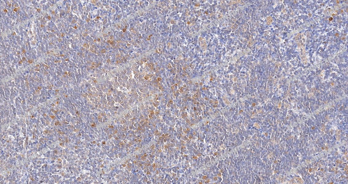 Immunohistochemical analysis of paraffin-embedded human tonsil tissue using SOCS2 Antibody (HY-P80328, 1/1000). The section was pretreated using heat mediated antigen retrieval with sodium citrate buffer (pH 6.0) for 8 minutes. The tissues were blocked with quick block buffer for 0.5 hours at room temperature, washed with PBS and PBST, and then incubated with the primary antibody overnight at 4℃. The detection was performed using an HRP conjugated compact polymer system. DAB was used as the chromogen. Tissues were counterstained with hematoxylin and mounted with DPX.
 -
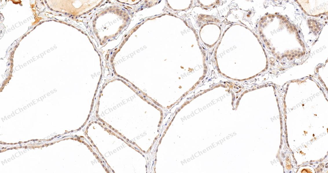 Immunohistochemical analysis of paraffin-embedded human thyroid gland tissue using SOCS2 Antibody (HY-P80328, 1/1000). The section was pretreated using heat mediated antigen retrieval with sodium citrate buffer (pH 6.0) for 8 minutes. The tissues were blocked with quick block buffer for 0.5 hours at room temperature, washed with PBS and PBST, and then incubated with the primary antibody overnight at 4℃. The detection was performed using an HRP conjugated compact polymer system. DAB was used as the chromogen. Tissues were counterstained with hematoxylin and mounted with DPX.
 -
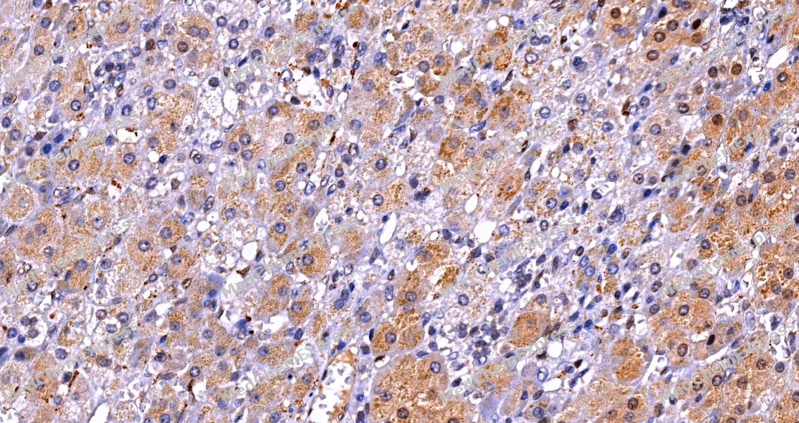 Immunohistochemical analysis of paraffin-embedded human adrenal gland tissue using SOCS2 Antibody (HY-P80328, 1/1000). The section was pretreated using heat mediated antigen retrieval with sodium citrate buffer (pH 6.0) for 8 minutes. The tissues were blocked with quick block buffer for 0.5 hours at room temperature, washed with PBS and PBST, and then incubated with the primary antibody overnight at 4℃. The detection was performed using an HRP conjugated compact polymer system. DAB was used as the chromogen. Tissues were counterstained with hematoxylin and mounted with DPX.
 -
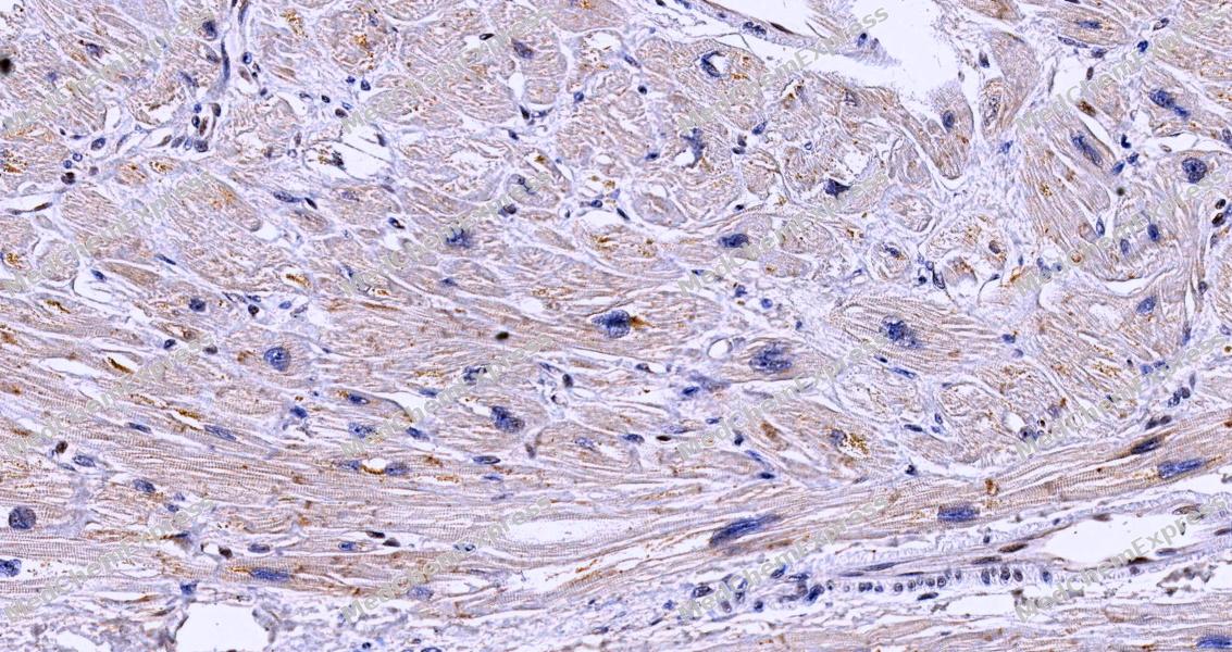 Immunohistochemical analysis of paraffin-embedded human heart muscle tissue using SOCS2 Antibody (HY-P80328, 1/1000). The section was pretreated using heat mediated antigen retrieval with sodium citrate buffer (pH 6.0) for 8 minutes. The tissues were blocked with quick block buffer for 0.5 hours at room temperature, washed with PBS and PBST, and then incubated with the primary antibody overnight at 4℃. The detection was performed using an HRP conjugated compact polymer system. DAB was used as the chromogen. Tissues were counterstained with hematoxylin and mounted with DPX.
 -
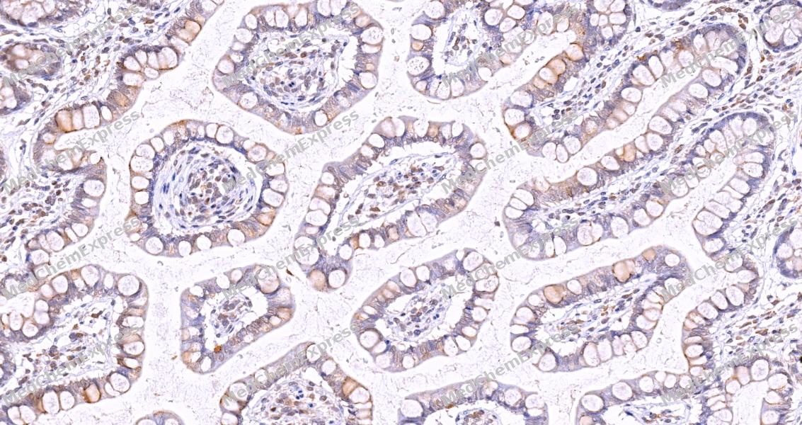 Immunohistochemical analysis of paraffin-embedded human small ntestine tissue using SOCS2 Antibody (HY-P80328, 1/1000). The section was pretreated using heat mediated antigen retrieval with sodium citrate buffer (pH 6.0) for 8 minutes. The tissues were blocked with quick block buffer for 0.5 hours at room temperature, washed with PBS and PBST, and then incubated with the primary antibody overnight at 4℃. The detection was performed using an HRP conjugated compact polymer system. DAB was used as the chromogen. Tissues were counterstained with hematoxylin and mounted with DPX.
 -
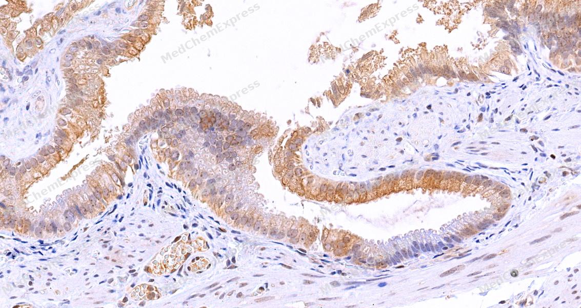 Immunohistochemical analysis of paraffin-embedded human gallbladder tissue using SOCS2 Antibody (HY-P80328, 1/1000). The section was pretreated using heat mediated antigen retrieval with sodium citrate buffer (pH 6.0) for 8 minutes. The tissues were blocked with quick block buffer for 0.5 hours at room temperature, washed with PBS and PBST, and then incubated with the primary antibody overnight at 4℃. The detection was performed using an HRP conjugated compact polymer system. DAB was used as the chromogen. Tissues were counterstained with hematoxylin and mounted with DPX.
 -
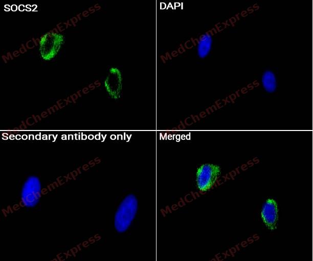 Immunocytochemistry analysis of PC-3 cells labeling SOCS2 Antibody (HY-P80328) at 1/50 dilution. Cells were fixed in 4% paraformaldehyde for 15 minutes at room temperature, permeabilized with 0.1% Triton X-100 for 10 minutes at room temperature, then blocked with BSA for Immunol Staining for 10 min at room temperature. Cells were then incubated with SOCS2 Antibody (HY-P80328) at 1/50 dilution in BSA for Immunol Staining at 4 ℃ overnight. AF488-conjugated AffiniPure Goat Anti-Rabbit /Mouse IgG H&L(HY-P8002/HY-P8005, Green) was used as the secondary antibody at 1/1,000 dilution. PBS instead of the primary antibody was used as the secondary antibody only control. The Nuclear counterstain was DAPI (Blue).
 -
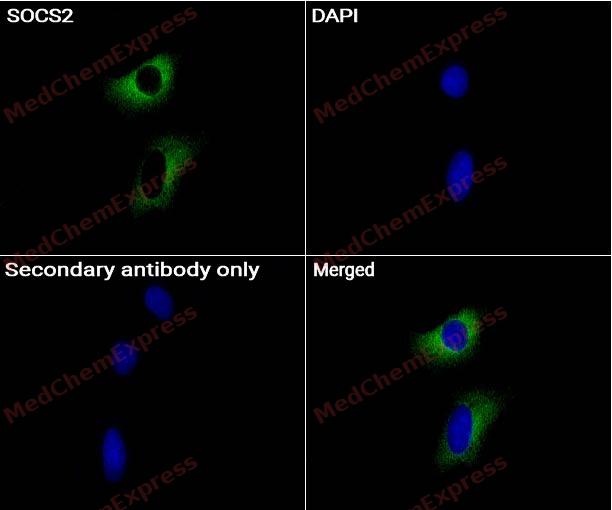 Immunocytochemistry analysis of SH-SY-5Y cells labeling SOCS2 Antibody (HY-P80328) at 1/50 dilution. Cells were fixed in 4% paraformaldehyde for 15 minutes at room temperature, permeabilized with 0.1% Triton X-100 for 10 minutes at room temperature, then blocked with BSA for Immunol Staining for 10 min at room temperature. Cells were then incubated with SOCS2 Antibody (HY-P80328) at 1/50 dilution in BSA for Immunol Staining at 4 ℃ overnight. AF488-conjugated AffiniPure Goat Anti-Rabbit /Mouse IgG H&L(HY-P8002/HY-P8005, Green) was used as the secondary antibody at 1/1,000 dilution. PBS instead of the primary antibody was used as the secondary antibody only control. The Nuclear counterstain was DAPI (Blue).


Background
-
Function
SOCS2 is a Substrate-recognition component of a cullin-5-RING E3 ubiquitin-protein ligase complex (ECS complex, also named CRL5 complex), which mediates the ubiquitination and subsequent proteasomal degradation of target proteins, such as EPOR and GHR. Specifically recognizes and binds phosphorylated proteins via its SH2 domain, promoting their ubiquitination. The ECS(SOCS2) complex acts as a key regulator of growth hormone receptor (GHR) levels by mediating ubiquitination and degradation of GHR, following GHR phosphorylation by JAK2. The ECS(SOCS2) also catalyzes ubiquitination and degradation of JAK2-phosphorylated EPOR[1][2][3][4][5][6].
-
Subcellular Localization
Cytoplasm
-
Expression
Tissue_specificity:High expression in heart, placenta, lung, kidney and prostate. Predominantly expressed in pulmonary epithelia cells, specifically type II pneumocytes
Induction:By a subset of cytokines, including EPO/erythropoietin and CSF2/GM-CSF -
Subunit
Substrate-recognition component of the ECS(SOCS2) complex, composed of SOCS2, CUL5, ELOB, ELOC and RNF7/RBX2 (PubMed:16675548, PubMed:21980433, PubMed:23897481, PubMed:25505247, PubMed:31182716, PubMed:34857742, PubMed:37816714). Interacts with IGF1R (PubMed:9727029). Interacts with DCUN1D1 (PubMed:23401859)
-
SwissProt ID
-
Synonyms
CIS2, SSI2, STATI2, SOCS2, Suppressor of cytokine signaling 2, SOCS-2, Cytokine-inducible SH2 protein 2, STAT-induced STAT inhibitor 2, CIS-2, SSI-2
-
Research Field
Cell Biology


Documentation
-
Data Sheet (261 KB)
-
SDS (252 KB)
- English - EN (252 KB)
- Français - FR (252 KB)
- Deutsch - DE (252 KB)
- Norwegian - NO (252 KB)
- Español - ES (252 KB)
- Swedish - SV (252 KB)
- Italian - IT (252 KB)
- Korean - KR (252 KB)
- Portuguese - PT (252 KB)
-
User Guide for Antibodies (1077 KB)
References
[1]. Eyckerman S, et al. Design and application of a cytokine-receptor-based interaction trap. Nat Cell Biol. 2001 Dec;3(12):1114-9. [Content Brief]
[2]. Vesterlund M, et al. The SOCS2 ubiquitin ligase complex regulates growth hormone receptor levels. PLoS One. 2011;6(9):e25358. [Content Brief]
[3]. Bulatov E, et al. Biophysical studies on interactions and assembly of full-size E3 ubiquitin ligase: suppressor of cytokine signaling 2 (SOCS2)-elongin BC-cullin 5-ring box protein 2 (RBX2). J Biol Chem. 2015 Feb 13;290(7):4178-91. [Content Brief]
[4]. Kung WW, et al. Structural insights into substrate recognition by the SOCS2 E3 ubiquitin ligase. Nat Commun. 2019 Jun 10;10(1):2534. [Content Brief]
[5]. Linossi EM, et al. Discovery of an exosite on the SOCS2-SH2 domain that enhances SH2 binding to phosphorylated ligands. Nat Commun. 2021 Dec 2;12(1):7032. [Content Brief]
[6]. Ramachandran S, et al. Structure-based design of a phosphotyrosine-masked covalent ligand targeting the E3 ligase SOCS2. Nat Commun. 2023 Oct 10;14(1):6345. [Content Brief]

 Powered by Bioz
