



Transcription Factor EB Antibody (YA5545)

Based on 1 Customer Validation
Transcription Factor EB Antibody (YA5545) is a Mouse-derived and non-conjugated IgG1 monoclonal antibody, targeting to Transcription Factor EB.
-
Host:
Mouse
-
Isotype:
IgG1
-
Application:
IHC-P, WB, ICC/IF, ELISA
-
Reactivity :
Human, Mouse,
-
Formulation:
Supplied in PBS, 50% glycerol, 0.05% Proclin 300, 0.05%BSA
-
Conjugation:
Non-conjugated


To place orders, for customer services and technical support, please contact: MedChemExpress USA
Tel: 609-228-6898 E-mail: [email protected] [email protected]





Applications
| Application |
IHC-P

IHC-P: Immunohistochemistry-Paraffin
|
WB
WB: Western Blot
|
ICC/IF
ICC/IF: Immunocytochemistry/
Immunofluorescence |
ELISA
ELISA: Enzyme Linked Immunosorbent Assay
|
|---|---|---|---|---|
| Dilution Ratio | 1:200-1000 | 1:500-2000 | 1:100-500 | 1:1000-5000 |


Product Details
Transcription Factor EB Antibody (YA5545) is a Mouse-derived and non-conjugated IgG1 monoclonal antibody, targeting to Transcription Factor EB.
-
Host Mouse

-
Clonality Monoclonal

-
Species ReactivityHuman, Mouse,
-
Observed Molecular WeightObserved band size: 53 kDa
Note: Due to possible protein modifications or aggregation, the molecular weight should be confirmed by actual measurement, and the predicted value is for reference only.

-
Calculated Molecular Weight Predicted band size: 53 kDa

Synthesized peptide derived from human TFEB AA range: 300-440
Protein G
Non-conjugated
Unmodified
IgG1


Product Properties
-
Appearance
Liquid
-
Formulation
Supplied in PBS, 50% glycerol, 0.05% Proclin 300, 0.05%BSA
-
Concentration
Batch-dependent, Please check the COA for the concentration of each lot. Check Lot Concentration
-
Storage & Stability
Stored at -20°C for 1 year. Avoid repeated freeze / thaw cycles.
-
Shipping
Shipping with blue ice.


Verification Images
-
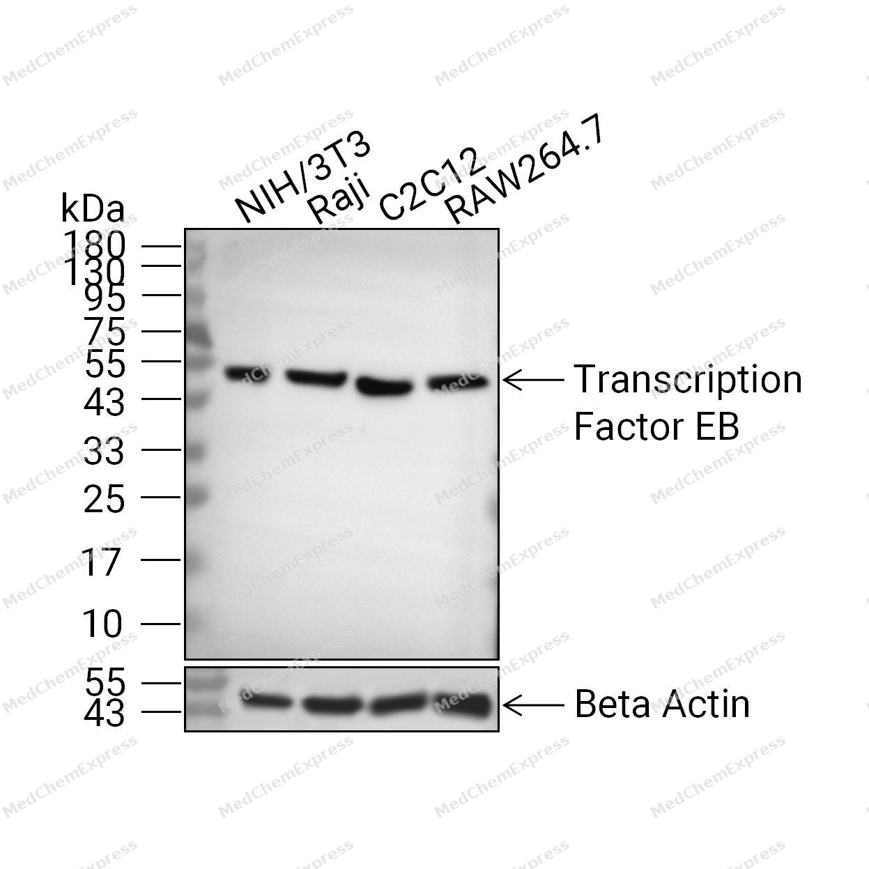 Western blot analysis of extracts from NIH/3T3 lane2(20μg),Raji(lane3(20μg), C2C12(lane4(20μg) and RAW264.7(lane5(20μg) using Transcription Factor EB Antibody (HY-P85853). Proteins were transferred to a PVDF membrane and blocked with 5% non-fat milk in TBST at 4°C overnight. The primary antibody (1/1000) and Loading control antibody (Beta Actin, HY-P80993, 1/10,000) was used in 5% non-fat milk in TBST for 2 hour at room temperature. Goat Anti-Mouse IgG-HRP Secondary Antibody (HY-P8004, 1/10,000) was used for 1 hour at room temperature.
 -
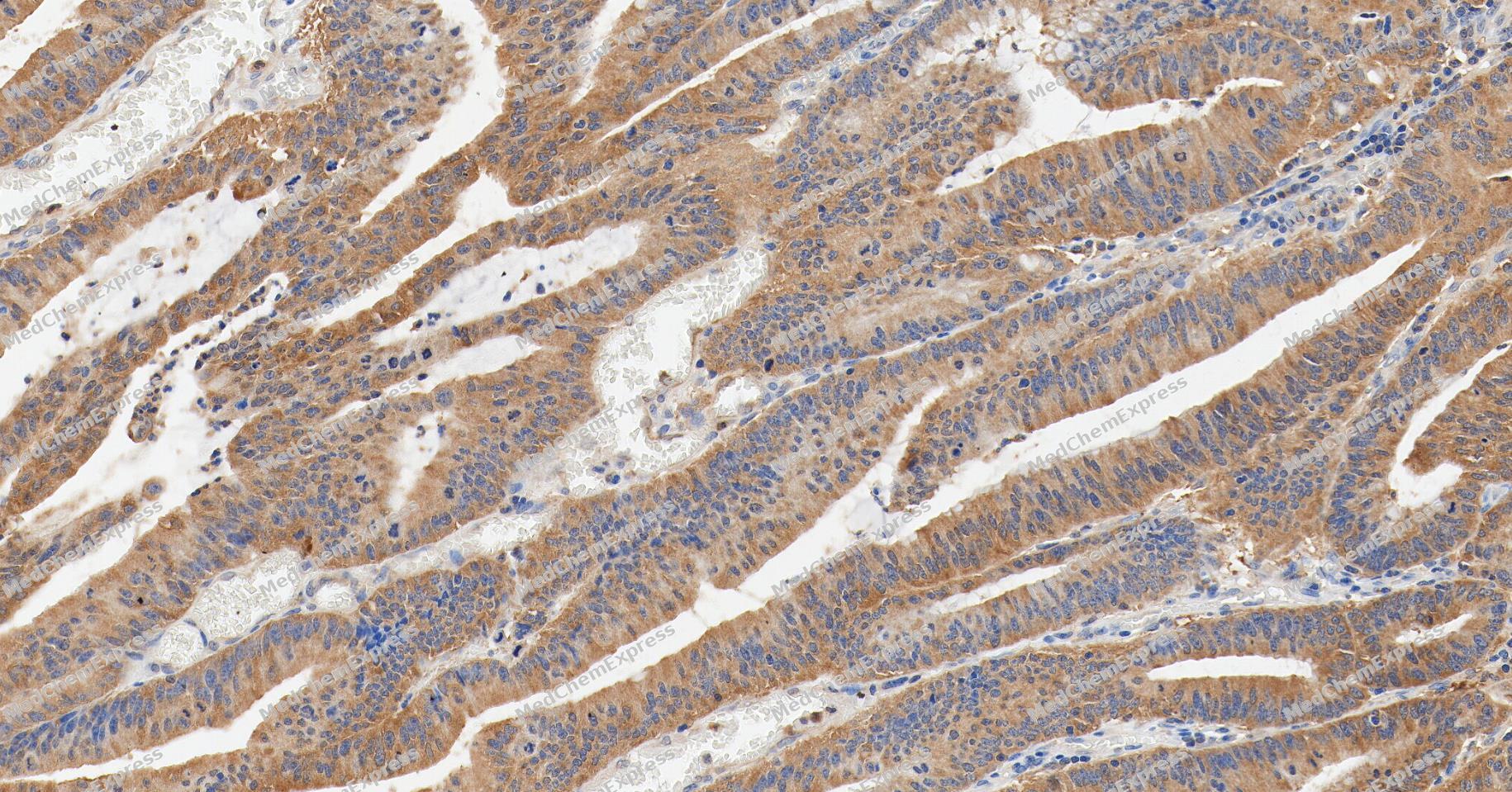 Immunohistochemical analysis of paraffin-embedded human colon cancer tissue using Transcription Factor EB Antibody (YA5545). The section was pre-treated using heat mediated antigen retrieval with Tris/EDTA buffer (pH 9.0) for 20 minutes. The tissues were probed with the primary antibody (HY-P85853, 1/200) overnight at 4℃. The detection was performed using an HRP conjugated compact polymer system. DAB was used as the chromogen. Tissues were counterstained with hematoxylin and mounted with neutral balsam.
 -
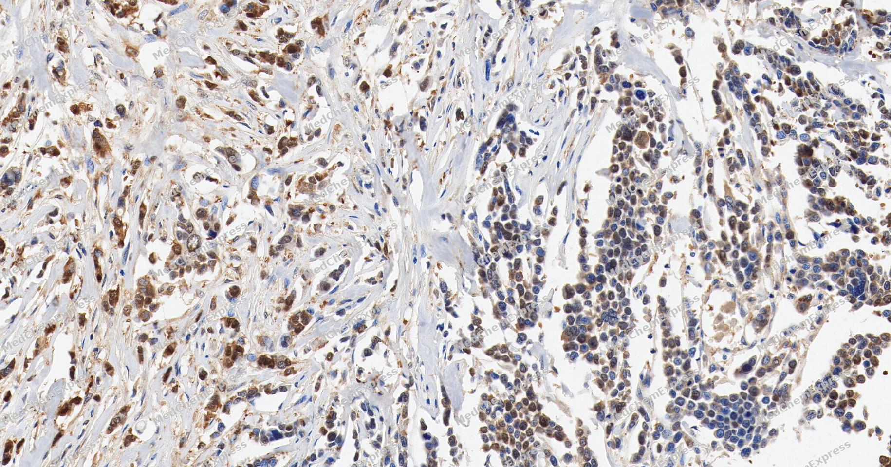 Immunohistochemical analysis of paraffin-embedded human breast cancer tissue using Transcription Factor EB Antibody (YA5545). The section was pre-treated using heat mediated antigen retrieval with Tris/EDTA buffer (pH 9.0) for 20 minutes. The tissues were probed with the primary antibody (HY-P85853, 1/200) overnight at 4℃. The detection was performed using an HRP conjugated compact polymer system. DAB was used as the chromogen. Tissues were counterstained with hematoxylin and mounted with neutral balsam.
 -
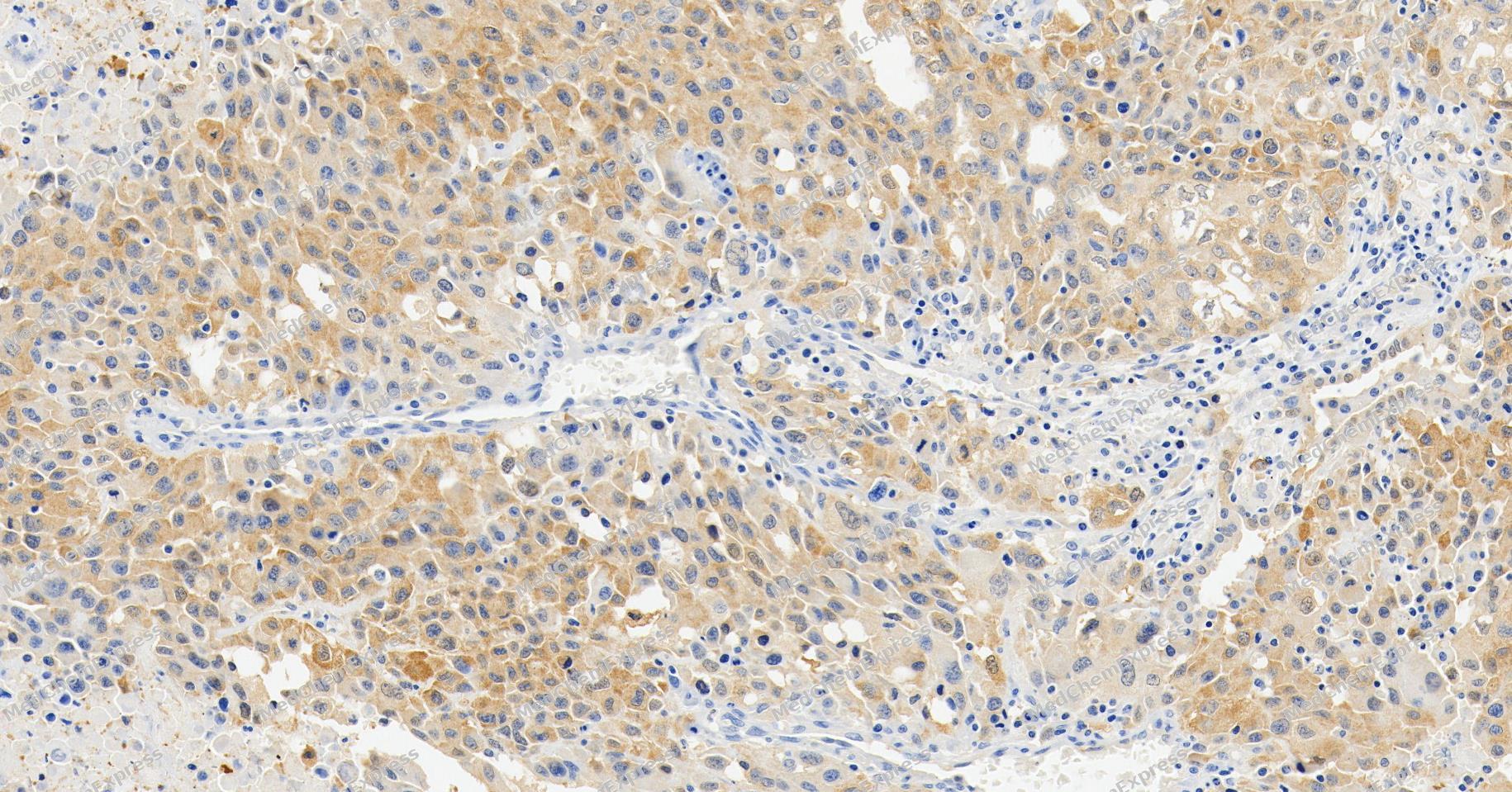 Immunohistochemical analysis of paraffin-embedded human lung cancer tissue using Transcription Factor EB Antibody (YA5545). The section was pre-treated using heat mediated antigen retrieval with Tris/EDTA buffer (pH 9.0) for 20 minutes. The tissues were probed with the primary antibody (HY-P85853, 1/200) overnight at 4℃. The detection was performed using an HRP conjugated compact polymer system. DAB was used as the chromogen. Tissues were counterstained with hematoxylin and mounted with neutral balsam.
 -
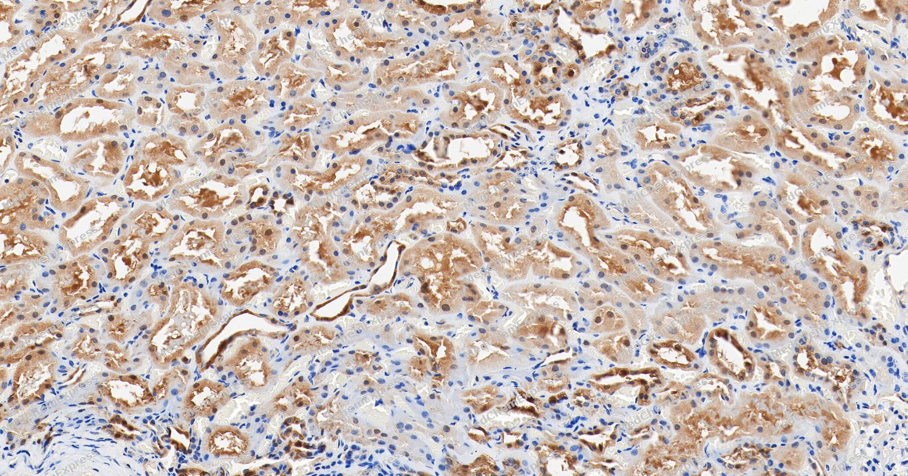 Immunohistochemical analysis of paraffin-embedded human kidney tissue using Transcription Factor EB Antibody (YA5545). The section was pre-treated using heat mediated antigen retrieval with Tris/EDTA buffer (pH 9.0) for 20 minutes. The tissues were probed with the primary antibody (HY-P85853, 1/200) overnight at 4℃. The detection was performed using an HRP conjugated compact polymer system. DAB was used as the chromogen. Tissues were counterstained with hematoxylin and mounted with neutral balsam.
 -
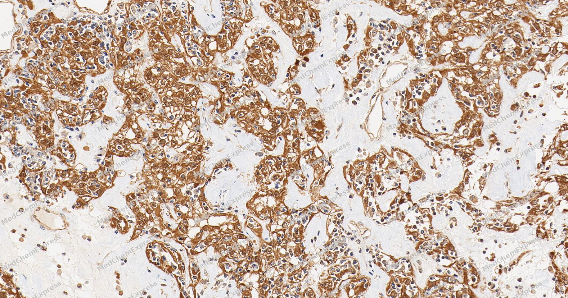 Immunohistochemical analysis of paraffin-embedded human renal cell carcinoma tissue using Transcription Factor EB Antibody (YA5545). The section was pre-treated using heat mediated antigen retrieval with Tris/EDTA buffer (pH 9.0) for 20 minutes. The tissues were probed with the primary antibody (HY-P85853, 1/200) overnight at 4℃. The detection was performed using an HRP conjugated compact polymer system. DAB was used as the chromogen. Tissues were counterstained with hematoxylin and mounted with neutral balsam.
 -
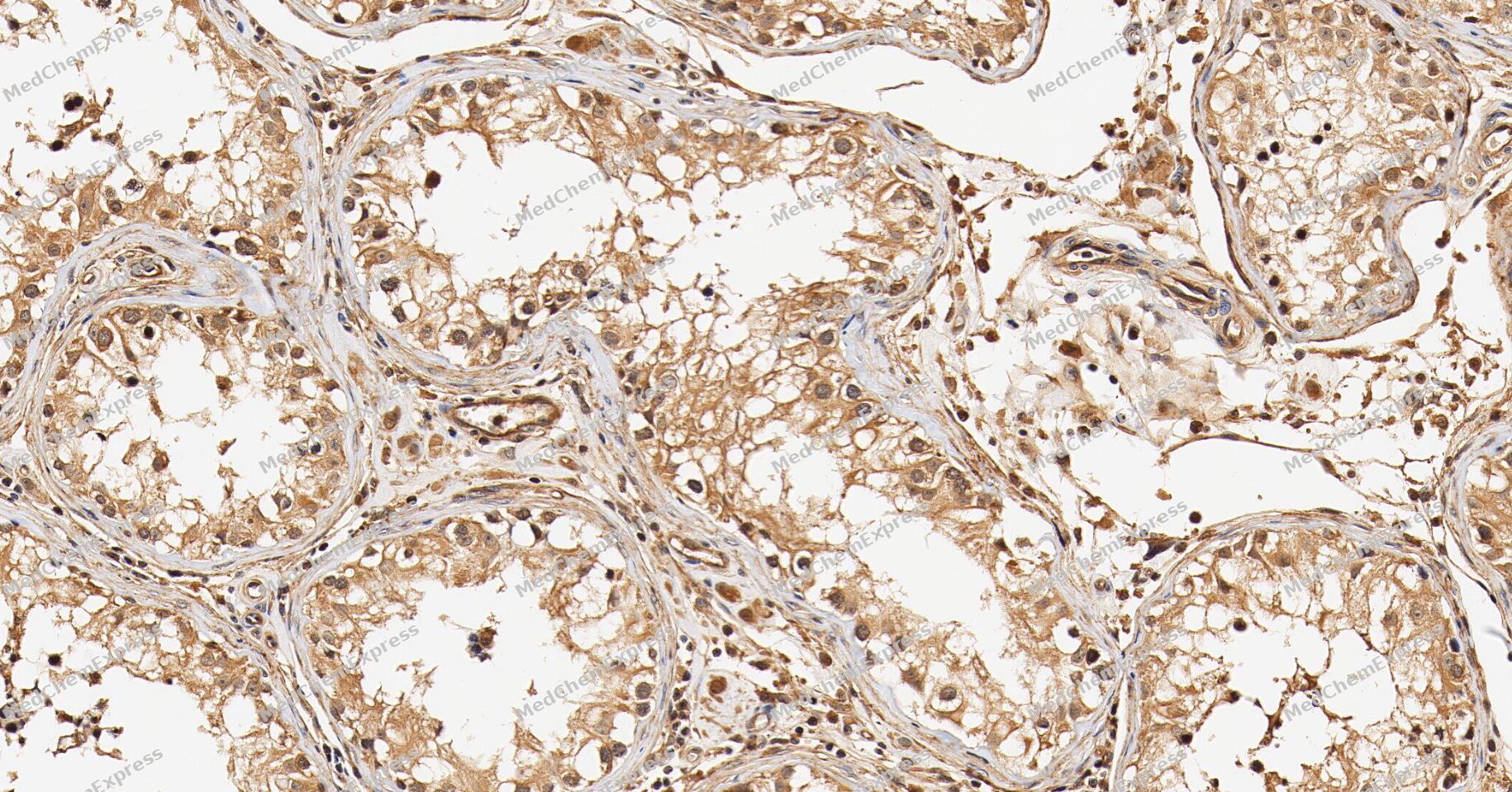 Immunohistochemical analysis of paraffin-embedded human testis tissue using Transcription Factor EB Antibody (YA5545). The section was pre-treated using heat mediated antigen retrieval with Tris/EDTA buffer (pH 9.0) for 20 minutes. The tissues were probed with the primary antibody (HY-P85853, 1/200) overnight at 4℃. The detection was performed using an HRP conjugated compact polymer system. DAB was used as the chromogen. Tissues were counterstained with hematoxylin and mounted with neutral balsam.
 -
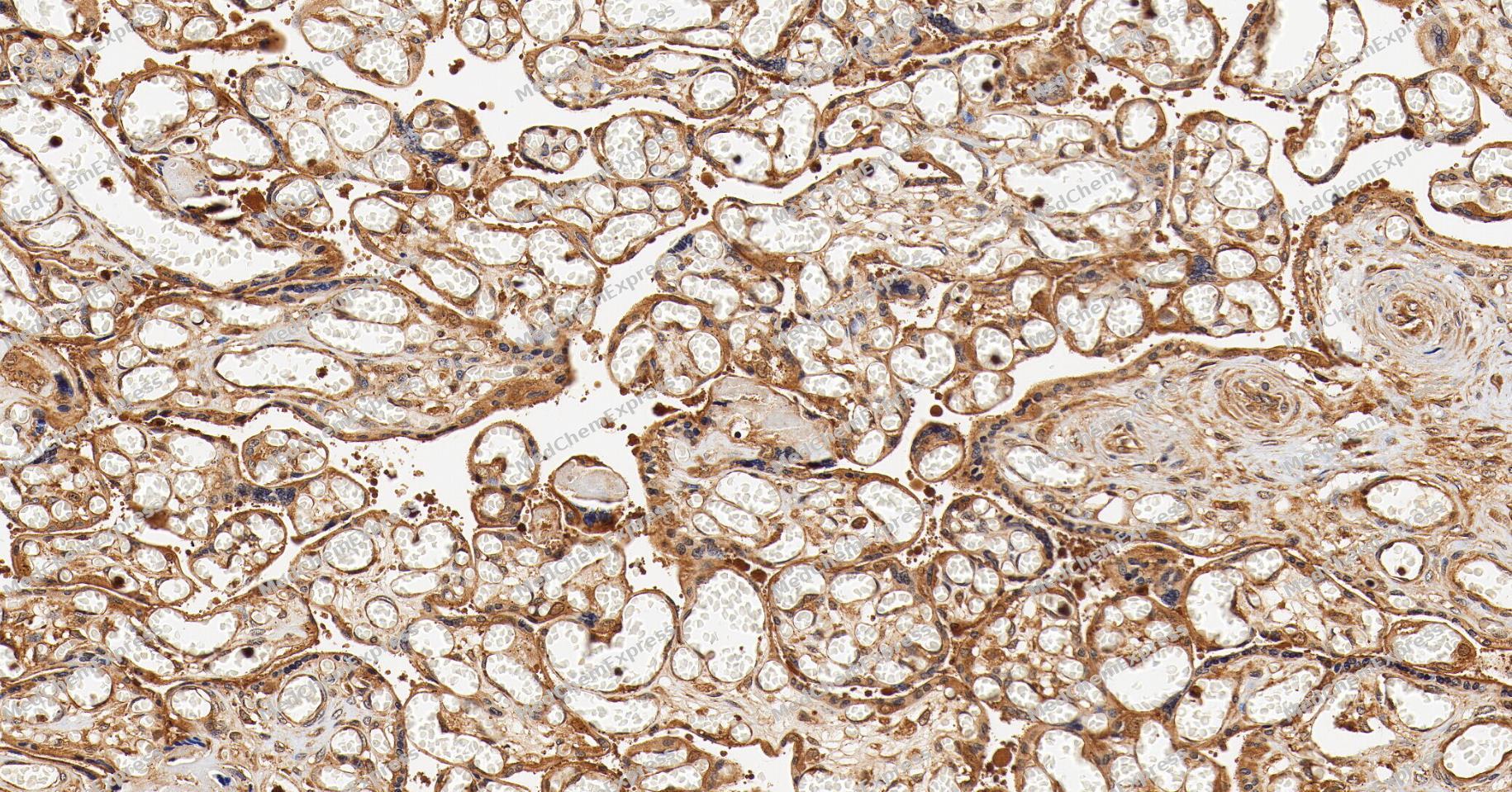 Immunohistochemical analysis of paraffin-embedded human placenta tissue using Transcription Factor EB Antibody (YA5545). The section was pre-treated using heat mediated antigen retrieval with Tris/EDTA buffer (pH 9.0) for 20 minutes. The tissues were probed with the primary antibody (HY-P85853, 1/200) overnight at 4℃. The detection was performed using an HRP conjugated compact polymer system. DAB was used as the chromogen. Tissues were counterstained with hematoxylin and mounted with neutral balsam.
 -
 Immunocytochemistry analysis of Hela cells labeling Transcription Factor EB with Transcription Factor EB Antibody (HY-P85853) at 1/100 dilution. Cells were fixed in 4% paraformaldehyde for 15 minutes at room temperature, permeabilized with 0.1% Triton X-100 in PBS for 15 minutes at room temperature, then blocked with quick block buffer for 10 minutes at room temperature. Cells were then incubated with Transcription Factor EB Antibody (HY-P85853) at 1/100 dilution in quick block buffer overnight at 4 ℃.AF488-conjugated Goat Anti-Mouse IgG H&L (HY-P8005, Green) was used as the secondary antibody at 1/1,000 dilution. PBS instead of the primary antibody was used as the secondary antibody only control. The Nuclear counterstain was DAPI (Blue).
 -
 Immunocytochemistry analysis of Hela cells labeling Transcription Factor EB with Transcription Factor EB Antibody (HY-P85853) at 1/300 dilution. Cells were fixed in 4% paraformaldehyde for 15 minutes at room temperature, permeabilized with 0.1% Triton X-100 in PBS for 15 minutes at room temperature, then blocked with quick block buffer for 10 minutes at room temperature. Cells were then incubated with Transcription Factor EB Antibody (HY-P85853) at 1/300 dilution in quick block buffer overnight at 4 ℃.AF488-conjugated Goat Anti-Mouse IgG H&L (HY-P8005, Green) was used as the secondary antibody at 1/1,000 dilution. PBS instead of the primary antibody was used as the secondary antibody only control. The Nuclear counterstain was DAPI (Blue).


Background
-
Function
Transcription Factor EB transcription factor that acts as a master regulator of lysosomal biogenesis, autophagy, lysosomal exocytosis, lipid catabolism, energy metabolism and immune response. Specifically recognizes and binds E-box sequences (5'-CANNTG-3'); efficient DNA-binding requires dimerization with itself or with another MiT/TFE family member such as TFE3 or MITF. Involved in the cellular response to amino acid availability by acting downstream of MTOR: in the presence of nutrients, TFEB phosphorylation by MTOR promotes its cytosolic retention and subsequent inactivation. Upon starvation or lysosomal stress, inhibition of MTOR induces TFEB dephosphorylation, resulting in nuclear localization and transcription factor activity. Specifically recognizes and binds the CLEAR-box sequence (5'-GTCACGTGAC-3') present in the regulatory region of many lysosomal genes, leading to activate their expression, thereby playing a central role in expression of lysosomal genes. Regulates lysosomal positioning in response to nutrient deprivation by promoting the expression of PIP4P1. Acts as a positive regulator of autophagy by promoting expression of genes involved in autophagy. In association with TFE3, activates the expression of CD40L in T-cells, thereby playing a role in T-cell-dependent antibody responses in activated CD4(+) T-cells and thymus-dependent humoral immunity. Specifically recognizes the gamma-E3 box, a subset of E-boxes, present in the heavy-chain immunoglobulin enhancer. Plays a role in the signal transduction processes required for normal vascularization of the placenta. Involved in the immune response to infection by the bacteria S.aureus, S.typhimurium or S.enterica: infection promotes itaconate production, leading to alkylation, resulting in nuclear localization and transcription factor activity. Itaconate-mediated alkylation activates TFEB-dependent lysosomal biogenesis, facilitating the bacteria clearance during the antibacterial innate immune response. In association with ACSS2, promotes the expression of genes involved in lysosome biogenesis and both autophagy upon glucose deprivation[1][2][3][4][5][6][7][8][9][10][11][12][13][14][15][16][17][18][19][20].
-
Subcellular Localization
Nucleus; Cytoplasm, cytosol; Lysosome membrane; Nucleus
-
Isoforms & Post-Translational Modification
P19484 has 2 isomers: P19484-1: 52865 Da (predicted); P19484-2: 43803 Da (predicted).
Phosphorylation at Ser-211 by MTOR via non-canonical mTORC1 pathway regulates its subcellular location and activity (PubMed:21617040, PubMed:22343943, PubMed:22576015, PubMed:22692423, PubMed:23401004, PubMed:24081491, PubMed:25720963, PubMed:30120233, PubMed:32612235, PubMed:32753672, PubMed:35662396, PubMed:36697823). When nutrients are present, phosphorylation by MTOR promotes association with 14-3-3/YWHA adapters and retention in the cytosol (PubMed:22343943, PubMed:22576015, PubMed:22692423, PubMed:23401004, PubMed:25720963, PubMed:32612235, PubMed:32753672, PubMed:35662396). Inhibition of mTORC1, starvation and lysosomal disruption, promotes dephosphorylation by calcineurin PPP3CB and translocation to the nucleus (PubMed:22343943, PubMed:22576015, PubMed:22692423, PubMed:25720963, PubMed:32753672). Dephosphorylated by calcineurin PPP3CB in response to lysosomal Ca(2+) release (PubMed:25720963). IRGM promotes dephosphorylation by calcineurin PPP3CB, resulting in TFEB nuclear translocation and stimulation of lysosomal biogenesis (PubMed:32753672). Dephosphorylated by phosphatase PPP3CA following Coxsackievirus B3 infection, leading to nuclear translocation (PubMed:33691586). Exported from the nucleus in a mTORC1-dependent manner in response to nutrient availability (PubMed:30120233);Alkylated via a non-enzymatic covalent modification (PubMed:35662396). Itaconate, an anti-inflammatory metabolite generated in response to lipopolysaccharide, alkylates Cys-212, preventing association with 14-3-3/YWHA adapters, thereby promoting nuclear translocation and activity (PubMed:35662396);Sumoylated; does not affect dimerization with MITF;(Microbial infection) Cleavage by Coxsackievirus B3 protease 3C after site Gln-60. This non-phosphorylated cleavage product retains its ability to interact with TFEB, TFE3 or MITF and presents impaired transcriptional activity, resulting in disruption of lysosomal functions and increased viral infection -
Subunit
Homodimer and heterodimer; with TFE3 or MITF (PubMed:15507434, PubMed:1748288). Interacts (when phosphorylated by MTOR) with YWHAZ; promoting retention in the cytosol (PubMed:35662396). Interacts with IRGM; promoting association between TFEB and PPP3CB and dephosphorylation (PubMed:32753672). Interacts with small GTPases Rag (RagA/RRAGA, RagB/RRAGB, RagC/RRAGC and/or RagD/RRAGD); promoting its recruitment to lysosomal membrane in the presence of nutrients (PubMed:23401004). Interacts with ACSS2 (PubMed:28552616)
-
SwissProt ID


Documentation
-
Data Sheet (234 KB)
-
SDS (252 KB)
- English - EN (252 KB)
- Français - FR (252 KB)
- Deutsch - DE (252 KB)
- Norwegian - NO (252 KB)
- Español - ES (252 KB)
- Swedish - SV (252 KB)
- Italian - IT (252 KB)
- Korean - KR (252 KB)
- Portuguese - PT (252 KB)
-
User Guide for Antibodies (1077 KB)

 Powered by Bioz
