



TGF beta Receptor II Antibody
(Synonyms: TGF-beta receptor type-2; TGF beta R2; TGFBR2; AAT 3; AAT3; FAA 3; FAA3; HNPCC6; MFS 2; MFS2; RIIC; TAAD 2; TAAD2; TbetaR II; TGF beta receptor type 2; TGF beta receptor type II; TGF beta type II receptor; TGFB R2; TGFbeta RII; TGFBR 2; TGFR 2; TGFR2; Transforming growth factor beta receptor II; Transforming growth factor beta receptor type II. TGF-βRⅡ; TGFβRⅡ; TGF βRⅡ; TGFβ RⅡ.)
Based on 1 Customer Validation
TGF beta Receptor II Antibody is a Rabbit-derived and non-conjugated IgG polyclonal antibody, targeting to TGF beta Receptor II.
-
Host:
Rabbit
-
Isotype:
IgG
-
Application:
WB, ELISA, IHC-P, ICC/IF
-
Reactivity :
Human, Mouse, Rat
-
Formulation:
Supplied in PBS containing 50% glycerol, 0.5% BSA and 0.02% sodium azide, pH 7.3.
-
Conjugation:
Non-conjugated




To place orders, for customer services and technical support, please contact: MedChemExpress USA
Tel: 609-228-6898 E-mail: [email protected] [email protected]





Applications
| Application |
WB

WB: Western Blot
|
IHC-P
IHC-P: Immunohistochemistry-Paraffin
|
ICC/IF
ICC/IF: Immunocytochemistry/
Immunofluorescence |
ELISA
ELISA: Enzyme Linked Immunosorbent Assay
|
|---|---|---|---|---|
| Dilution Ratio | 1:500-2000 | 1:100-1:300 | 1:50-200 | 1:20000 |


Product Details
TGF beta Receptor II Antibody is a Rabbit-derived and non-conjugated IgG polyclonal antibody, targeting to TGF beta Receptor II.
-
Host Rabbit

-
Clonality Polyclonal

-
Species ReactivityHuman, Mouse, Rat
-
Observed Molecular WeightObserved band size: 65 kDa
Note: Due to possible protein modifications or aggregation, the molecular weight should be confirmed by actual measurement, and the predicted value is for reference only.

-
Calculated Molecular Weight Predicted band size: 65kDa;

Entrez Gene: 7048 Human ; 21813 Mouse ; 81810 Rat
SwissProt: P37173 Human ; Q62312 Mouse ; P38438 Rat
OMIM: 614331 Human
KLH conjugated synthetic peptide derived from human TGF beta Receptor II: 241-330/567
Endogenous
affinity purified
Non-conjugated
Unmodified
IgG


Product Properties
-
Appearance
Liquid
-
Formulation
Supplied in PBS containing 50% glycerol, 0.5% BSA and 0.02% sodium azide, pH 7.3.
-
Concentration
Batch-dependent, Please check the COA for the concentration of each lot. Check Lot Concentration
-
Storage & Stability
Stored at -20°C for 1 year. Avoid repeated freeze / thaw cycles.
-
Shipping
Shipping with blue ice.


Verification Images
-
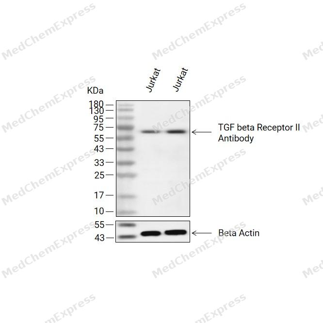 Western blot analysis of extracts from Jurkat(lane 2(20ug) and Jurkat(lane 3(40ug) using TGF beta Receptor II Antibody (HY-P81131) Rabbit pAb. Proteins were transferred to a PVDF membrane and blocked with 5% non-fat milk in TBST for 2 hour at room temperature. The primary antibody (1/1000) and Loading control antibody (Beta Actin, HY-P83730, 1/10000) was used in 5% non-fat milk in TBST at 4°C overnight. Goat Anti-Mouse/Rabbit IgG-HRP Secondary Antibody (1/10000) was used for 1 hour at room temperature.
 -
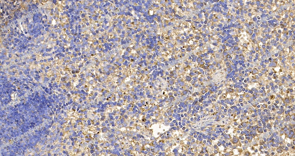
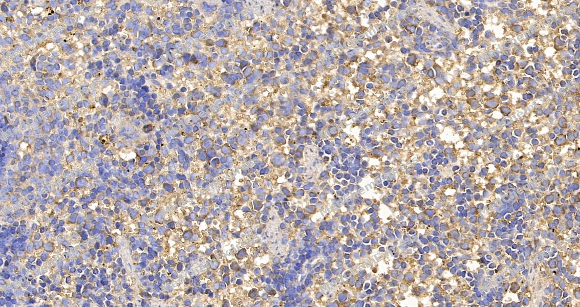 Immunohistochemical analysis of paraffin-embedded rat spleen tissue using TGF beta Receptor II Antibody. The section was pre-treated using heat mediated antigen retrieval with sodium citrate buffer (pH 6.0) for 8 minutes. The tissues were blocked in QuickBlock for 20 minutes at room temperature, washed with ddH2O and PBS, and then probed with the primary antibody at 1/100 dilution in 4℃ overnight. The detection was performed using an HRP conjugated compact polymer system. DAB was used as the chromogen. Tissues were counterstained with hematoxylin and mounted with DPX.
 -
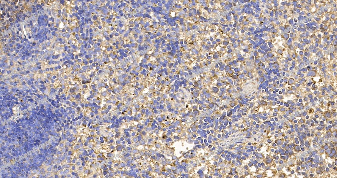 Immunohistochemical analysis of paraffin-embedded rat spleen tissue using TGF beta Receptor II Antibody. The section was pre-treated using heat mediated antigen retrieval with sodium citrate buffer (pH 6.0) for 8 minutes. The tissues were blocked in QuickBlock for 20 minutes at room temperature, washed with ddH2O and PBS, and then probed with the primary antibody at 1/100 dilution in 4℃ overnight. The detection was performed using an HRP conjugated compact polymer system. DAB was used as the chromogen. Tissues were counterstained with hematoxylin and mounted with DPX.
 -
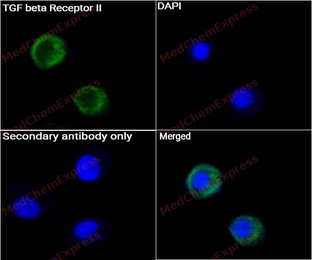
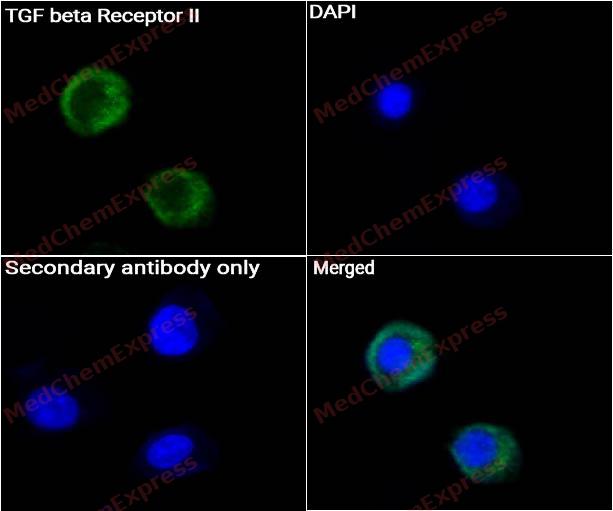 Immunocytochemistry analysis of A549 cells labeling TGF beta Receptor II Antibody (HY-P81131) at 1/50 dilution. Cells were fixed in 4% paraformaldehyde for 15 minutes at room temperature, permeabilized with 0.1% Triton X-100 for 10 minutes at room temperature, then blocked with QuickBlock™ Blocking Buffer for Immunol Staining for 10 min at room temperature. Cells were then incubated with TGF beta Receptor II Antibody (HY-P81131) at 1/50 dilution in QuickBlock™ Blocking Buffer for Immunol Staining at 4 ℃. Alexa Fluor® 488-conjugated AffiniPure Goat Anti-Rabbit IgG H&L(HY-P8002, Green) was used as the secondary antibody at 1/1,000 dilution. PBS instead of the primary antibody was used as the secondary antibody only control. The Nuclear counterstain was DAPI (Blue).
 -
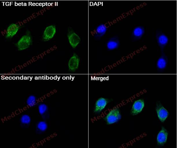 Immunocytochemistry analysis of A549 cells labeling TGF beta Receptor II Antibody (HY-P81131) at 1/150 dilution. Cells were fixed in 4% paraformaldehyde for 15 minutes at room temperature, permeabilized with 0.1% Triton X-100 for 10 minutes at room temperature, then blocked with QuickBlock™ Blocking Buffer for Immunol Staining for 10 min at room temperature. Cells were then incubated with TGF beta Receptor II Antibody (HY-P81131) at 1/150 dilution in QuickBlock™ Blocking Buffer for Immunol Staining at 4 ℃. Alexa Fluor® 488-conjugated AffiniPure Goat Anti-Rabbit IgG H&L(HY-P8002, Green) was used as the secondary antibody at 1/1,000 dilution. PBS instead of the primary antibody was used as the secondary antibody only control. The Nuclear counterstain was DAPI (Blue).


Background
-
Function
TGF beta Receptor II is a Transmembrane serine/threonine kinase forming with the TGF-beta type I serine/threonine kinase receptor, TGFBR1, the non-promiscuous receptor for the TGF-beta cytokines TGFB1, TGFB2 and TGFB3. Transduces the TGFB1, TGFB2 and TGFB3 signal from the cell surface to the cytoplasm and thus regulates a plethora of physiological and pathological processes including cell cycle arrest in epithelial and hematopoietic cells, control of mesenchymal cell proliferation and differentiation, wound healing, extracellular matrix production, immunosuppression and carcinogenesis. The formation of the receptor complex composed of 2 TGFBR1 and 2 TGFBR2 molecules symmetrically bound to the cytokine dimer results in the phosphorylation and activation of TGFBR1 by the constitutively active TGFBR2. Activated TGFBR1 phosphorylates SMAD2 which dissociates from the receptor and interacts with SMAD4. The SMAD2-SMAD4 complex is subsequently translocated to the nucleus where it modulates the transcription of the TGF-beta-regulated genes. This constitutes the canonical SMAD-dependent TGF-beta signaling cascade. Also involved in non-canonical, SMAD-independent TGF-beta signaling pathways; Has transforming growth factor beta-activated receptor activity; Has transforming growth factor beta-activated receptor activity; Binds TGFB1, TGFB2 and TGFB3 in the picomolar affinity range without the participation of additional receptors. Blocks activation of SMAD2 and SMAD3 by TGFB1
-
Subcellular Localization
Cell membrane; Single-pass type I membrane protein; Membrane raft; Secreted
-
Isoforms & Post-Translational Modification
P37173 has 3 isomers: P37173-1: 64568 Da (predicted); P37173-2: 67457 Da (predicted); P37173-3: 9162 Da (predicted).
Phosphorylated on a Ser/Thr residue in the cytoplasmic domain -
Subunit
Homodimer. Heterohexamer; TGFB1, TGFB2 and TGFB3 homodimeric ligands assemble a functional receptor composed of two TGFBR1 and TGFBR2 heterodimers to form a ligand-receptor heterohexamer. The respective affinity of TGFRB1 and TGFRB2 for the ligands may modulate the kinetics of assembly of the receptor and may explain the different biological activities of TGFB1, TGFB2 and TGFB3. Component of a complex composed of TSC22D1 (via N-terminus), TGFBR1 and TGFBR2; the interaction between TSC22D1 and TGFBR1 is inhibited by SMAD7 and promoted by TGFB1 (PubMed:21791611). Interacts with DAXX. Interacts with DYNLT4. Interacts with ZFYVE9; ZFYVE9 recruits SMAD2 and SMAD3 to the TGF-beta receptor. Interacts with and is activated by SCUBE3; this interaction does not affect TGFB1-binding to TGFBR2. Interacts with VPS39; this interaction is independent of the receptor kinase activity and of the presence of TGF-beta. Interacts with CLU (PubMed:8555189)
-
SwissProt ID
-
Synonyms
TGF-beta receptor type-2; TGF beta R2; TGFBR2; AAT 3; AAT3; FAA 3; FAA3; HNPCC6; MFS 2; MFS2; RIIC; TAAD 2; TAAD2; TbetaR II; TGF beta receptor type 2; TGF beta receptor type II; TGF beta type II receptor; TGFB R2; TGFbeta RII; TGFBR 2; TGFR 2; TGFR2; Transforming growth factor beta receptor II; Transforming growth factor beta receptor type II. TGF-βRⅡ; TGFβRⅡ; TGF βRⅡ; TGFβ RⅡ.


Documentation
-
Data Sheet (234 KB)
-
SDS (316 KB)
- English - EN (316 KB)
- Français - FR (316 KB)
- Deutsch - DE (316 KB)
- Norwegian - NO (316 KB)
- Español - ES (316 KB)
- Swedish - SV (316 KB)
- Italian - IT (316 KB)
- Korean - KR (316 KB)
- Portuguese - PT (316 KB)
-
User Guide for Antibodies (1077 KB)

 Powered by Bioz
