



TNFRSF1A Antibody (YA3855)
(Synonyms: FPF; p55; p60; TBP1; TNF-R; TNFAR; TNFR1; p55-R; CD120a; TNFR55; TNFR60; TNF-R-I; TNF-R55)Based on 1 Customer Validation
TNFRSF1A Antibody (YA3855) is a Mouse-derived and non-conjugated IgG1 monoclonal antibody, targeting to TNFRSF1A.
-
Host:
Mouse
-
Isotype:
IgG1
-
Application:
ICC/IF, FC, ELISA
-
Reactivity :
Human
-
Formulation:
Supplied in PBS with 0.05% sodium azide
-
Conjugation:
Non-conjugated


To place orders, for customer services and technical support, please contact: MedChemExpress USA
Tel: 609-228-6898 E-mail: [email protected] [email protected]





Applications
| Application |
ICC/IF

ICC/IF: Immunocytochemistry/
Immunofluorescence |
FC
FC: Flow Cytometry
|
ELISA
ELISA: Enzyme Linked Immunosorbent Assay
|
|---|---|---|---|
| Dilution Ratio | 1:200-1:1000 | 1:200-1:400 | 1:10000 |


Product Details
TNFRSF1A Antibody (YA3855) is a Mouse-derived and non-conjugated IgG1 monoclonal antibody, targeting to TNFRSF1A.
-
Host Mouse

-
Clonality Monoclonal

-
Species ReactivityHuman
-
Observed Molecular WeightObserved band size: 55 kDa
Note: Due to possible protein modifications or aggregation, the molecular weight should be confirmed by actual measurement, and the predicted value is for reference only.

-
Calculated Molecular Weight Predicted band size: 50 kDa

Purified recombinant fragment of human TNFRSF1A (AA: extra 30-211) expressed in E. Coli.
affinity purified.
Non-conjugated
Unmodified
IgG1


Product Properties
-
Appearance
Solution
-
Formulation
Supplied in PBS with 0.05% sodium azide
-
Concentration
Batch-dependent, Please check the COA for the concentration of each lot. Check Lot Concentration
-
Storage & Stability
Stored at -20°C for 1 year. Avoid repeated freeze / thaw cycles.
-
Shipping
Shipping with blue ice.


Verification Images
-
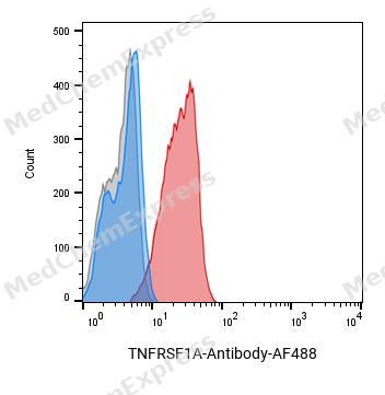 Flow cytometric analysis of 1X10^6 HepG2 cells labeling TNFRSF1A Antibody(red). Cells were fixed with 4% paraformaldehyde and permeabilised with 0.2% Triton X-100. Then stained with the primary antibody at 1/400 dilution overnight at 4℃. AF 488 Goat Anti-mouse IgG H&L was used as the secondary antibody at 1/1,000 dilution for 45 minutes at room temperature. Mouse IgG Isotype Control (blue) was used as the isotype control, cells without incubation with primary antibody were used as the unlabeled control (black).
 -
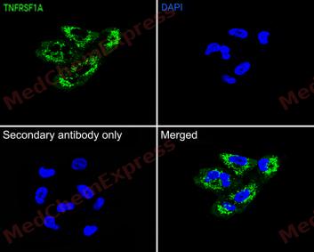 Immunofluorescence analysis of A549 cells labeling TNFRSF1A antibody at 1/50 dilution. Cells were fixed in 4% paraformaldehyde for 15 minutes at room temperature, permeabilized with 0.1% Triton X-100 in PBS for 15 minutes at room temperature, then blocked with 1% BSA in 10% negative goat serum for 1 hour at room temperature.Cells were then incubated with TNFRSF1A antibody at 1/50 dilution in 1% BSA in PBST overnight at 4 ℃. AF 488 Goat Anti-mouse IgG H&L (green) was used as the secondary antibody at 1/500 dilution. PBS instead of the primary antibody was used as the secondary antibody only control. The Nuclear counterstain was DAPI (Blue)


Background
-
Function
TNFRSF1A receptor for TNFSF2/TNF-alpha and homotrimeric TNFSF1/lymphotoxin-alpha. The adapter molecule FADD recruits caspase-8 to the activated receptor. The resulting death-inducing signaling complex (DISC) performs caspase-8 proteolytic activation which initiates the subsequent cascade of caspases (aspartate-specific cysteine proteases) mediating apoptosis. Contributes to the induction of non-cytocidal TNF effects including anti-viral state and activation of the acid sphingomyelinase
-
Subcellular Localization
Cell membrane; Single-pass type I membrane protein; Golgi apparatus membrane; Single-pass type I membrane protein; Secreted; Secreted
-
Isoforms & Post-Translational Modification
P19438 has 5 isomers: P19438-1: 50495 Da (predicted); P19438-2: 38651 Da (predicted); P19438-4: 25577 Da (predicted); P19438-3: 24794 Da (predicted); P19438-5: 24194 Da (predicted).
The soluble form is produced from the membrane form by proteolytic processing;(Microbial infection) Glycosylated at Arg-376 by enteropathogenic E.coli protein NleB1 and S.typhimurium protein Ssek3: arginine GlcNAcylation prevents homotypic/heterotypic death domain interactions -
Subunit
Binding of TNF to the extracellular domain leads to homotrimerization. The aggregated death domains provide a novel molecular interface that interacts specifically with the death domain of TRADD. Various TRADD-interacting proteins such as TRAFS, RIPK1 and possibly FADD, are recruited to the complex by their association with TRADD. This complex activates at least two distinct signaling cascades, apoptosis and NF-kappa-B signaling. Interacts with BAG4, BABAM2, FEM1B, GRB2, SQSTM1 and TRPC4AP (PubMed:10356400, PubMed:10359574, PubMed:10542291, PubMed:15465831, PubMed:8387891, PubMed:9915703). Interacts directly with NOL3 (via CARD domain); inhibits TNF-signaling pathway (By similarity). Interacts with SH3RF2, TRADD and RIPK1. SH3RF2 facilitates the recruitment of RIPK1 and TRADD to TNFRSF1A in a TNF-alpha-dependent process (PubMed:24130170). Interacts with PGLYRP1; this interaction is important for cell death induction (PubMed:26183779). Interacts (via death domain) with MADD (via death domain) (PubMed:9115275)
-
SwissProt ID
-
Synonyms
FPF; p55; p60; TBP1; TNF-R; TNFAR; TNFR1; p55-R; CD120a; TNFR55; TNFR60; TNF-R-I; TNF-R55


Documentation
-
Data Sheet (231 KB)
-
SDS (252 KB)
- English - EN (252 KB)
- Français - FR (252 KB)
- Deutsch - DE (252 KB)
- Norwegian - NO (252 KB)
- Español - ES (252 KB)
- Swedish - SV (252 KB)
- Italian - IT (252 KB)
- Korean - KR (252 KB)
- Portuguese - PT (252 KB)
-
User Guide for Antibodies (1077 KB)

 Powered by Bioz
