



TrkB Antibody (YA026)
(Synonyms: TRKB, NTRK2, BDNF/NT-3 growth factors receptor, GP145-TrkB, Neurotrophic tyrosine kinase receptor type 2, TrkB tyrosine kinase, Tropomyosin-related kinase B, Trk-B)
Based on 1 Customer Validation
TrkB Antibody (YA026) is a Rabbit-derived and non-conjugated IgG monoclonal antibody, targeting to TrkB.
-
Host:
Rabbit
-
Isotype:
IgG
-
Application:
WB, IHC-P
-
Reactivity :
Mouse, Rat
-
Formulation:
Supplied in 1*TBS (pH7.4), 0.05% BSA and 40% Glycerol. Preservative: 0.05% Sodium Azide.
-
Conjugation:
Non-conjugated


To place orders, for customer services and technical support, please contact: MedChemExpress USA
Tel: 609-228-6898 E-mail: [email protected] [email protected]





Applications
| Application |
WB

WB: Western Blot
|
IHC-P
IHC-P: Immunohistochemistry-Paraffin
|
|---|---|---|
| Dilution Ratio | 1:500-1:2000 | 1:200 |


Product Details
TrkB Antibody (YA026) is a Rabbit-derived and non-conjugated IgG monoclonal antibody, targeting to TrkB.
-
Host Rabbit

-
Clonality Recombinant,Monoclonal

-
Species ReactivityMouse, Rat
-
Observed Molecular WeightObserved band size: 92 kDa
Note: Due to possible protein modifications or aggregation, the molecular weight should be confirmed by actual measurement, and the predicted value is for reference only.

-
Calculated Molecular Weight Predicted band size: 92 kDa

Synthetic peptide corresponding to Human TrkB.AA range:100-450.
Endogenous
Protein A affinity purified.
Non-conjugated
Unmodified
IgG


Product Properties
-
Appearance
Solution
-
Formulation
Supplied in 1*TBS (pH7.4), 0.05% BSA and 40% Glycerol. Preservative: 0.05% Sodium Azide.
-
Concentration
Batch-dependent, Please check the COA for the concentration of each lot. Check Lot Concentration
-
Storage & Stability
Stored at -20°C for 1 year. Avoid repeated freeze / thaw cycles.
-
Shipping
Shipping with blue ice.


Verification Images
-
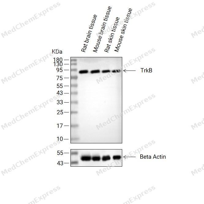 Western blot analysis of extracts from Rat brain tissue (lane2(20μg), Mouse brain tissue (lane3(20μg), Rat skin tissue (lane4(20μg) and Mouse skin tissue (lane5(20μg) using TrkB Antibody (HY-P80360). Proteins were transferred to a PVDF membrane and blocked with 5% non-fat milk in TBST at 4°C overnight. The primary antibody (1/1000) and Loading control antibody (Beta Actin, HY-P80993, 1/10,000) was used in 5% non-fat milk in TBST for 2 hour at room temperature. Goat Anti-Rabbit IgG-HRP Secondary Antibody (HY-P8001, 1/10,000) was used for 1 hour at room temperature.
 -
 Immunohistochemical analysis of paraffin-embedded mouse hippocampus tissue using TrkB Antibody (HY-P80360, 1/200). The section was pretreated using heat mediated antigen retrieval with sodium citrate buffer (pH 6.0) for 8 minutes. The tissues were blocked with quick block buffer for 0.5 hours at room temperature, washed with PBS and PBST, and then incubated with the primary antibody overnight at 4℃. The detection was performed using an HRP conjugated compact polymer system. DAB was used as the chromogen. Tissues were counterstained with hematoxylin and mounted with DPX.
 -
 Immunohistochemical analysis of paraffin-embedded mouse cerebral cortex tissue using TrkB Antibody (HY-P80360, 1/200). The section was pretreated using heat mediated antigen retrieval with sodium citrate buffer (pH 6.0) for 8 minutes. The tissues were blocked with quick block buffer for 0.5 hours at room temperature, washed with PBS and PBST, and then incubated with the primary antibody overnight at 4℃. The detection was performed using an HRP conjugated compact polymer system. DAB was used as the chromogen. Tissues were counterstained with hematoxylin and mounted with DPX.
 -
 Immunohistochemical analysis of paraffin-embedded mouse cerebellum tissue using TrkB Antibody (HY-P80360, 1/200). The section was pretreated using heat mediated antigen retrieval with sodium citrate buffer (pH 6.0) for 8 minutes. The tissues were blocked with quick block buffer for 0.5 hours at room temperature, washed with PBS and PBST, and then incubated with the primary antibody overnight at 4℃. The detection was performed using an HRP conjugated compact polymer system. DAB was used as the chromogen. Tissues were counterstained with hematoxylin and mounted with DPX.
 -
 Immunohistochemical analysis of paraffin-embedded mouse stomach tissue using TrkB Antibody (HY-P80360, 1/200). The section was pretreated using heat mediated antigen retrieval with sodium citrate buffer (pH 6.0) for 8 minutes. The tissues were blocked with quick block buffer for 0.5 hours at room temperature, washed with PBS and PBST, and then incubated with the primary antibody overnight at 4℃. The detection was performed using an HRP conjugated compact polymer system. DAB was used as the chromogen. Tissues were counterstained with hematoxylin and mounted with DPX.
 -
 Immunohistochemical analysis of paraffin-embedded rat cerebral cortex tissue using TrkB Antibody (HY-P80360, 1/200). The section was pretreated using heat mediated antigen retrieval with sodium citrate buffer (pH 6.0) for 8 minutes. The tissues were blocked with quick block buffer for 0.5 hours at room temperature, washed with PBS and PBST, and then incubated with the primary antibody overnight at 4℃. The detection was performed using an HRP conjugated compact polymer system. DAB was used as the chromogen. Tissues were counterstained with hematoxylin and mounted with DPX.
 -
 Immunohistochemical analysis of paraffin-embedded rat hippocampus tissue using TrkB Antibody (HY-P80360, 1/200). The section was pretreated using heat mediated antigen retrieval with sodium citrate buffer (pH 6.0) for 8 minutes. The tissues were blocked with quick block buffer for 0.5 hours at room temperature, washed with PBS and PBST, and then incubated with the primary antibody overnight at 4℃. The detection was performed using an HRP conjugated compact polymer system. DAB was used as the chromogen. Tissues were counterstained with hematoxylin and mounted with DPX.
 -
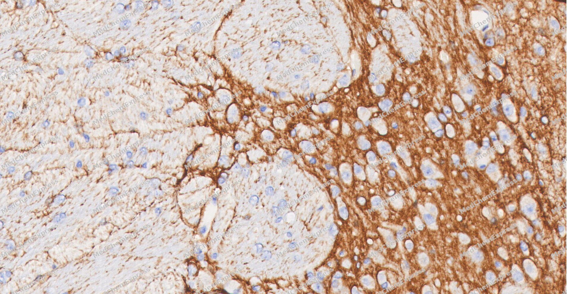 Immunohistochemical analysis of paraffin-embedded Rat Brain tissue using TrkB antibody was performed. The section was pretreated using high-temperature and high-pressure mediated EDTA antigen retrieval buffer (pH 9.0), for 5 minutes. The tissues were incubated with primary antibody (HY-P80360, 1:200 dilution) at room temperature for 60 minutes. Detection was performed using an HRP conjugated compact polymer system. DAB was used as the chromogen. The tissues were counterstained with hematoxylin and mounted with neutral balsam mounting medium.
 -
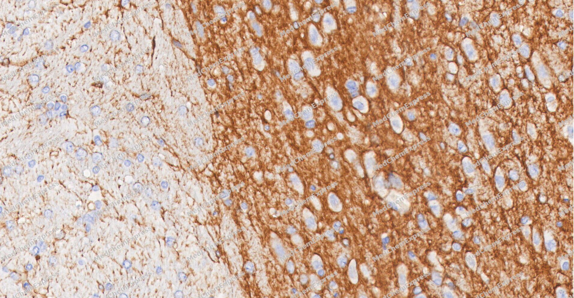 Immunohistochemical analysis of paraffin-embedded Rat Brain tissue using TrkB antibody was performed. The section was pretreated using high-temperature and high-pressure mediated EDTA antigen retrieval buffer (pH 9.0), for 5 minutes. The tissues were incubated with primary antibody (HY-P80360, 1:200 dilution) at room temperature for 60 minutes. Detection was performed using an HRP conjugated compact polymer system. DAB was used as the chromogen. The tissues were counterstained with hematoxylin and mounted with neutral balsam mounting medium.
 -
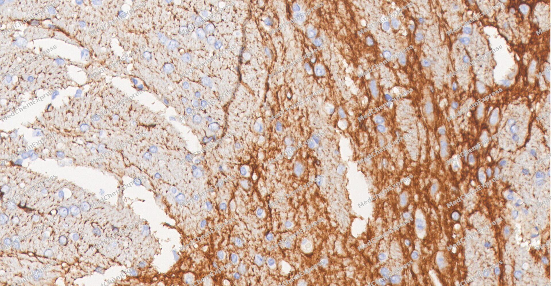 Immunohistochemical analysis of paraffin-embedded Rat Brain tissue using TrkB antibody was performed. The section was pretreated using high-temperature and high-pressure mediated EDTA antigen retrieval buffer (pH 9.0), for 5 minutes. The tissues were incubated with primary antibody (HY-P80360, 1:200 dilution) at room temperature for 60 minutes. Detection was performed using an HRP conjugated compact polymer system. DAB was used as the chromogen. The tissues were counterstained with hematoxylin and mounted with neutral balsam mounting medium.
 -
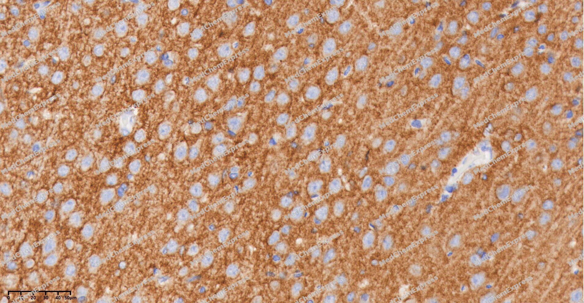 Immunohistochemical analysis of paraffin-embedded Rat Brain tissue using TrkB antibody was performed. The section was pretreated using high-temperature and high-pressure mediated EDTA antigen retrieval buffer (pH 9.0), for 5 minutes. The tissues were incubated with primary antibody (HY-P80360, 1:200 dilution) at room temperature for 60 minutes. Detection was performed using an HRP conjugated compact polymer system. DAB was used as the chromogen. The tissues were counterstained with hematoxylin and mounted with neutral balsam mounting medium.
 -
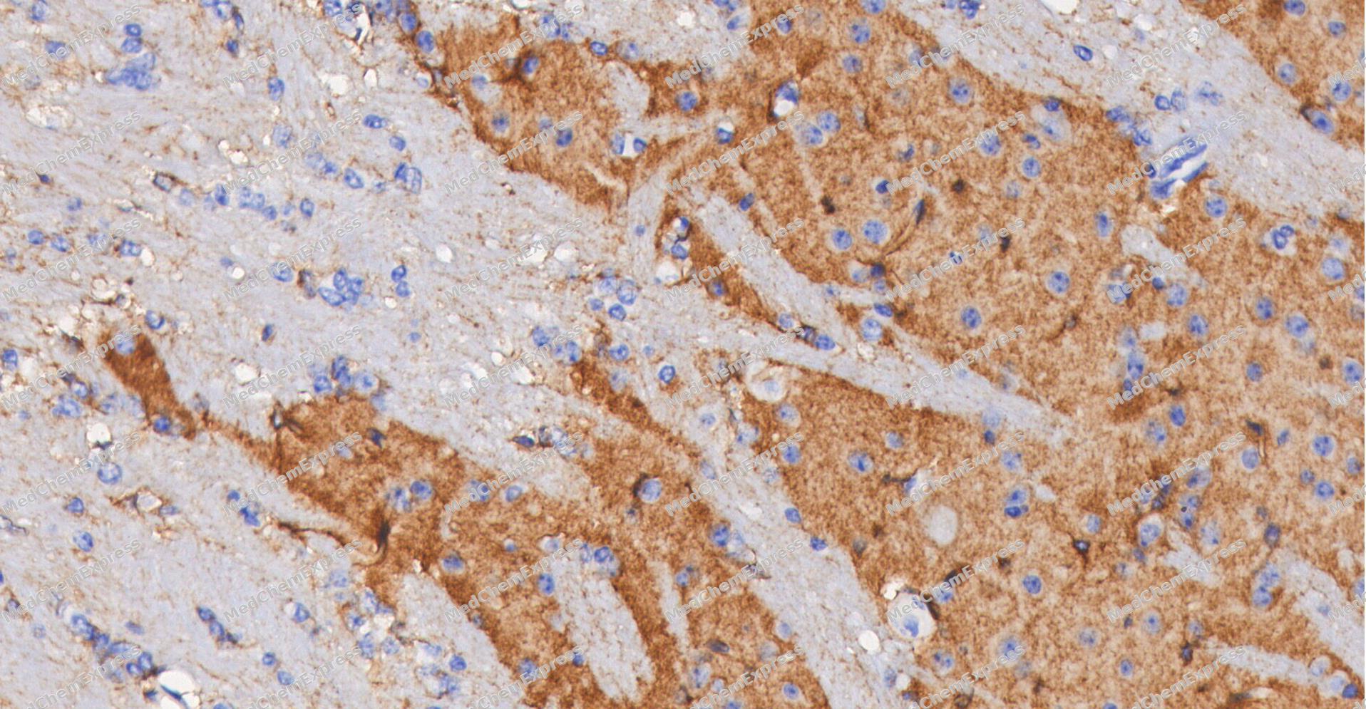 Immunohistochemical analysis of paraffin-embedded Mouse Brain tissue using TrkB antibody was performed. The section was pretreated using high-temperature and high-pressure mediated EDTA antigen retrieval buffer (pH 9.0), for 5 minutes. The tissues were incubated with primary antibody (HY-P80360, 1:200 dilution) at room temperature for 60 minutes. Detection was performed using an HRP conjugated compact polymer system. DAB was used as the chromogen. The tissues were counterstained with hematoxylin and mounted with neutral balsam mounting medium.
 -
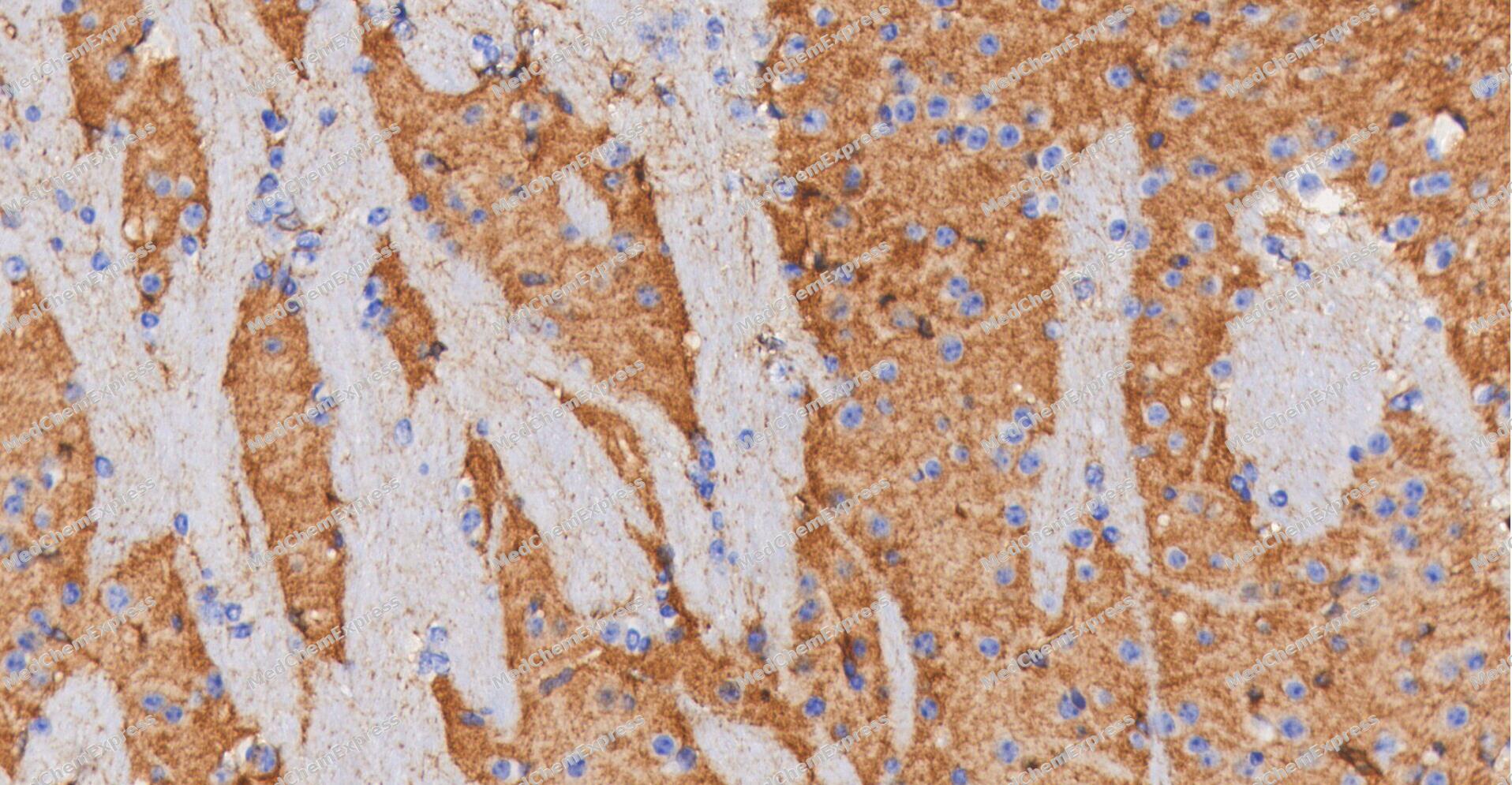 Immunohistochemical analysis of paraffin-embedded Mouse Brain tissue using TrkB antibody was performed. The section was pretreated using high-temperature and high-pressure mediated EDTA antigen retrieval buffer (pH 9.0), for 5 minutes. The tissues were incubated with primary antibody (HY-P80360, 1:200 dilution) at room temperature for 60 minutes. Detection was performed using an HRP conjugated compact polymer system. DAB was used as the chromogen. The tissues were counterstained with hematoxylin and mounted with neutral balsam mounting medium.


Background
-
Function
TrkB (tropomyosin receptor kinase B), encoded by NTRK2, is a neurotrophic tyrosine kinase receptor that mediates the cellular actions of brain-derived neurotrophic factor (BDNF) and contributes to neuronal differentiation, neuroplasticity, and homeostasis[1][2]. Mechanistically, ligand-induced activation of full-length TrkB (TrkB.FL) triggers receptor autophosphorylation and engages intracellular signaling pathways including PI3K, MAPK/ERK, and PLCγ, thereby supporting neurotrophic responses and synaptic function[3][4]. Alternative splicing of NTRK2 generates distinct receptor isoforms, including TrkB.FL and truncated variants such as TrkB.T1, TrkB.T2, and TrkB-T-ShC, which lack the tyrosine kinase domain and therefore differ fundamentally in signaling capacity[2][5]. Compared with TrkB.FL, TrkB.T1 is the major truncated isoform in the adult brain and can negatively regulate full-length receptor signaling through dominant-inhibitory mechanisms while also exhibiting independent biological activities[5][4]. Altered TrkB isoform balance has been associated with neurodegeneration, ischemic injury, amyotrophic lateral sclerosis, spinal muscular atrophy, psychiatric disorders, and cognitive dysfunction, highlighting the importance of isoform-specific regulation in disease biology[2][6][4][7]. In experimental systems, modulation of BDNF-TrkB signaling and manipulation of TrkB isoform expression are widely used to investigate neuronal survival, differentiation, synaptic plasticity, and disease mechanisms, making TrkB a valuable target for mechanistic and translational neuroscience research[3][5][4].
-
Subcellular Localization
Cell membrane; Single-pass type I membrane protein; Endosome membrane; Single-pass type I membrane protein; Early endosome membrane; Cell projection, axon; Cell projection, dendrite; Cytoplasm, perinuclear region; Postsynaptic density
-
Expression
Tissue_specificity:The TrkB subtype is expressed in both the central and peripheral nervous systems. In the central nervous system (CNS) , it is expressed in the cerebral cortex, hippocampus, thalamus, choroid plexus, cerebellar granular layer, brainstem, and spinal cord. In the peripheral nervous system, it is expressed in multiple cranial ganglia, the ophthalmic nerve, the vestibular system, various facial structures, the submandibular gland, and the dorsal root ganglion. The TrkB-T1 subtype is primarily expressed in the brain, but is also found in other tissues, including the pancreas, kidneys, and heart. The TrkB-T-Shc subtype is primarily expressed in the brain. -
Subunit
Exists in a dynamic equilibrium between monomeric (low affinity) and dimeric (high affinity) structures. Interacts (phosphorylated upon activation by BDNF) with SHC1; mediates SHC1 phosphorylation and activation. Interacts (phosphorylated upon activation by BDNF) with PLCG1 and/or PLCG2; mediates PLCG1 phosphorylation and activation. Interacts with SH2B1 and SH2B2. Interacts with NGFR; may regulate the ligand specificity of the receptor (By similarity). Interacts with SORCS2; this interaction is important for normal targeting to post-synaptic densities in response to high-frequency stimulation (By similarity). Interacts (phosphorylated upon ligand-binding) with SH2D1A; regulates NTRK2. Interacts with SQSTM1 and KIDINS220 (By similarity). Interacts (phosphorylated upon ligand-binding) with FRS2; activates the MAPK signaling pathway (PubMed:10092678). Interacts with APPL1 (By similarity). Interacts with MAPK8IP3/JIP3 and KLC1; interaction with KLC1 is mediated by MAPK8IP3/JIP3 (By similarity). Interacts with SORL1; this interaction facilitates NTRK2 trafficking between synaptic plasma membranes, postsynaptic densities and cell soma, hence positively regulates BDNF signaling (By similarity). Interacts with SLITRK2 (PubMed:35840571)
-
SwissProt ID
-
Synonyms
TRKB, NTRK2, BDNF/NT-3 growth factors receptor, GP145-TrkB, Neurotrophic tyrosine kinase receptor type 2, TrkB tyrosine kinase, Tropomyosin-related kinase B, Trk-B
-
Research Field
Neuroscience


Documentation
-
Data Sheet (262 KB)
-
SDS (252 KB)
- English - EN (252 KB)
- Français - FR (252 KB)
- Deutsch - DE (252 KB)
- Norwegian - NO (252 KB)
- Español - ES (252 KB)
- Swedish - SV (252 KB)
- Italian - IT (252 KB)
- Korean - KR (252 KB)
- Portuguese - PT (252 KB)
-
User Guide for Antibodies (1077 KB)
References
[1]. Duarte-Ruiz M, et al. Regulation of NTRK2 alternative splicing by PRPF40B controls neural differentiation and synaptic plasticity. Cell Death Dis. 2025 Dec 8;17(1):73. [Content Brief]
[2]. Li Y, et al. Tropomyosin receptor kinase B (TrkB) signalling: targeted therapy in neurogenic tumours. J Pathol Clin Res. 2023 Mar;9(2):89-99. [Content Brief]
[3]. TrkB gene information from NCBI.
[4]. Rabezanahary H, et al. Live virus neutralizing antibodies against pre and post Omicron strains in food and retail workers in Québec, Canada. Heliyon. 2024 May 21;10(10):e31026. [Content Brief]
[5]. Tessarollo L, et al. TrkB Truncated Isoform Receptors as Transducers and Determinants of BDNF Functions. Front Neurosci. 2022 Mar 7;16:847572. [Content Brief]
[6]. Vidaurre OG, et al. Imbalance of neurotrophin receptor isoforms TrkB-FL/TrkB-T1 induces neuronal death in excitotoxicity. Cell Death Dis. 2012 Jan 19;3(1):e256. [Content Brief]
[7]. Chang C, et al. Recent advances in deciphering hippocampus complexity using single-cell transcriptomics. Neurobiol Dis. 2023 Apr;179:106062. [Content Brief]

 Powered by Bioz
