



Tyrosinase Antibody (YA2522)
(Synonyms: ATN; CMM8; LB24 AB; SHEP3; SK29 AB; Tumor rejection antigen AB; TYR)
Based on 1 Customer Validation
Tyrosinase Antibody (YA2522) is a Rabbit-derived and non-conjugated IgG monoclonal antibody, targeting to Tyrosinase.
-
Host:
Rabbit
-
Isotype:
IgG
-
Application:
WB, IHC-P, ICC/IF
-
Reactivity :
Human
-
Formulation:
Supplied in 50mM Tris-Glycine(pH 7.4), 0.15M NaCl, 40%Glycerol, 0.01% sodium azide and 0.05% BSA.
-
Conjugation:
Non-conjugated


To place orders, for customer services and technical support, please contact: MedChemExpress USA
Tel: 609-228-6898 E-mail: [email protected] [email protected]





Applications
| Application |
WB

WB: Western Blot
|
IHC-P
IHC-P: Immunohistochemistry-Paraffin
|
ICC/IF
ICC/IF: Immunocytochemistry/
Immunofluorescence |
|---|---|---|---|
| Dilution Ratio | 1:500-1:1000 | 1:50-1:100 | 1:50-1:200 |


Product Details
Tyrosinase Antibody (YA2522) is a Rabbit-derived and non-conjugated IgG monoclonal antibody, targeting to Tyrosinase.
-
Host Rabbit

-
Clonality Recombinant,Monoclonal

-
Species ReactivityHuman
-
Observed Molecular WeightObserved band size: 70-80 kDa
Note: Due to possible protein modifications or aggregation, the molecular weight should be confirmed by actual measurement, and the predicted value is for reference only.

-
Calculated Molecular Weight Predicted band size: 60 kDa

A synthesized peptide derived from human Tyrosinase aa1-300/529.
Endogenous
Affinity Chromatography
Non-conjugated
Unmodified
IgG


Product Properties
-
Appearance
Solution
-
Formulation
Supplied in 50mM Tris-Glycine(pH 7.4), 0.15M NaCl, 40%Glycerol, 0.01% sodium azide and 0.05% BSA.
-
Concentration
Batch-dependent, Please check the COA for the concentration of each lot. Check Lot Concentration
-
Storage & Stability
Stored at -20°C for 1 year. Avoid repeated freeze / thaw cycles.
-
Shipping
Shipping with blue ice.


Verification Images
-
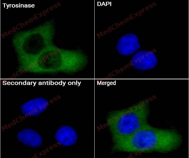 Immunocytochemistry analysis of Hela cells labeling Tyrosinase Antibody (HY-P82777) at 1/50 dilution. Cells were fixed in 4% paraformaldehyde for 15 minutes at room temperature, permeabilized with 0.1% Triton X-100 for 10 minutes at room temperature, then blocked with BSA for Immunol Staining for 10 min at room temperature. Cells were then incubated with Tyrosinase Antibody (HY-P82777) at 1/50 dilution in BSA for Immunol Staining at 4 ℃ Overnight. AF488-conjugated AffiniPure Goat Anti-Rabbit IgG H&L(HY-P8002, Green) was used as the secondary antibody at 1/1,000 dilution. PBS instead of the primary antibody was used as the secondary antibody only control. The Nuclear counterstain was DAPI (Blue).
 -
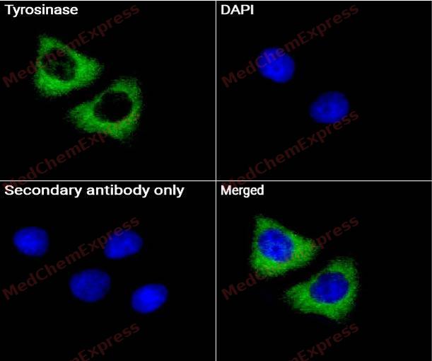 Immunocytochemistry analysis of Hela cells labeling Tyrosinase Antibody (HY-P82777) at 1/200 dilution. Cells were fixed in 4% paraformaldehyde for 15 minutes at room temperature, permeabilized with 0.1% Triton X-100 for 10 minutes at room temperature, then blocked with BSA for Immunol Staining for 10 min at room temperature. Cells were then incubated with Tyrosinase Antibody (HY-P82777) at 1/200dilution in BSA for Immunol Staining at 4 ℃ Overnight. AF488-conjugated AffiniPure Goat Anti-Rabbit IgG H&L(HY-P8002, Green) was used as the secondary antibody at 1/1,000 dilution. PBS instead of the primary antibody was used as the secondary antibody only control. The Nuclear counterstain was DAPI (Blue).


Background
-
Function
Tyrosinase is a copper-containing metalloprotein belonging to the type-3 copper protein family, together with haemocyanins and catechol oxidases. Tyrosinases are the catalysts in mammals responsible for the formation of melanin in skin and hair color, as well as browning in fruit and vegetables following cell damage.
Tyrosinases are found in various prokaryotes as well as in plants, fungi, arthropods, and mammals and are responsible for pigmentation, wound healing, radiation protection, and primary immune response. Tyrosinases perform two sequential enzymatic reactions: hydroxylation of monophenols and oxidation of diphenols to form quinones which polymerize spontaneously to melanin. In plants, sponges, and many invertebrates, tyrosinases are important for wound healing and primary immune responses; in arthropods, they play a role in sclerotization, and in bacteria, tyrosinases protect DNA from UV damage.
-
Subcellular Localization
Melanosome membrane; Single-pass type I membrane protein; Melanosome
-
Expression
Induction:Increased expression after UVB irradiation -
Isoforms & Post-Translational Modification
P14679 has 2 isomers: P14679-1: 60393 Da (predicted); P14679-2: 42914 Da (predicted).
Glycosylated -
Subunit
Forms an OPN3-dependent complex with DCT in response to blue light in melanocytes
-
SwissProt ID
-
Synonyms
ATN; CMM8; LB24 AB; SHEP3; SK29 AB; Tumor rejection antigen AB; TYR
-
Research Field
Tags & Cell Markers


Documentation
-
Data Sheet (234 KB)
-
SDS (252 KB)
- English - EN (252 KB)
- Français - FR (252 KB)
- Deutsch - DE (252 KB)
- Norwegian - NO (252 KB)
- Español - ES (252 KB)
- Swedish - SV (252 KB)
- Italian - IT (252 KB)
- Korean - KR (252 KB)
- Portuguese - PT (252 KB)
-
User Guide for Antibodies (1077 KB)

 Powered by Bioz
