



PH11
PH11 is a novel focal adhesion kinase (FAK) inhibitor that rapidly induces apoptosis in TRAIL-resistant PANC-1 cells when combined with TRAIL, but has no effect on normal human fibroblasts. The study found that PH11 downregulates c-FLIP through inhibition of FAK and phosphatidylinositol-3-kinase (PI3K)/AKT pathways, thereby restoring the TRAIL apoptotic pathway, suggesting that this combination therapy may provide an attractive therapeutic strategy for the safe and effective treatment of pancreatic cancer. PH11 selectively inhibits c-FLIP expression by modulating upstream signaling pathways and may represent an innovative therapeutic strategy. Although further work is needed to fully elucidate the mechanism of PH11-induced TRAIL sensitization, we believe that our results will provide a new approach to target c-FLIP without the risk of interfering with caspase-8 processing, which could potentially lead to TRAIL resistance. This study also suggests a role for the FAK/AKT signaling pathway in regulating c-FLIP expression in TRAIL-induced apoptosis, and this understanding will provide important clues to control the resistance mechanism to optimize the potential of TRAIL-based pancreatic cancer treatment.
Nos produits utilisent uniquement pour la recherche. Nous ne vendons pas aux patients.
- CAS No.: 1627843-95-1
- Formule: C22H22N6O5
- Masse moléculaire:450.45
-
Stockage:
Please store the product under the recommended conditions in the Certificate of Analysis.


To place orders, for customer services and technical support, please contact: MedChemExpress USA
Tel: 609-228-6898 E-mail: [email protected] [email protected]


-
 Activité biologique
-
Chemical Information
- Protocole
- Pureté et documentation
- Références
-
 Help & FAQs


-
HY-12289
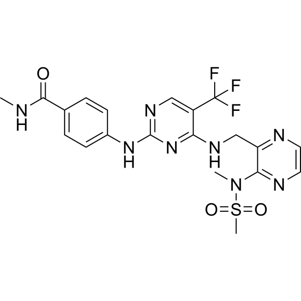
DefactinibFAK Inhibitor -
HY-12444
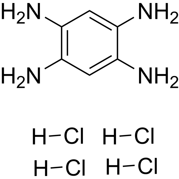
Y15FAK Inhibitor -
HY-12289A
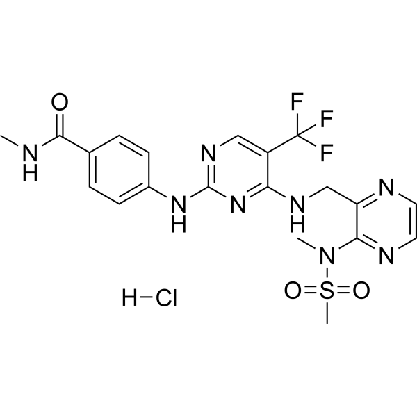
Defactinib hydrochlorideFAK Inhibitor -
HY-145652
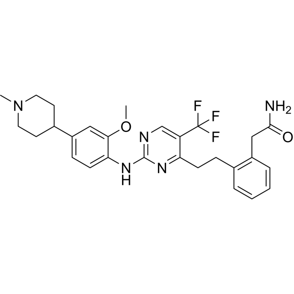
NarmafotinibFAK Inhibitor -
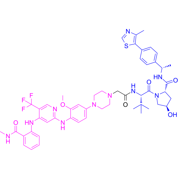
HY-132296
GSK215PROTAC FAK Degrader -
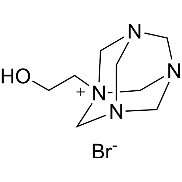
HY-103471
Y11FAK1 Inhibitor -
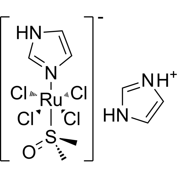
HY-19376
NAMI-ARuthenium Anticancer Agent -
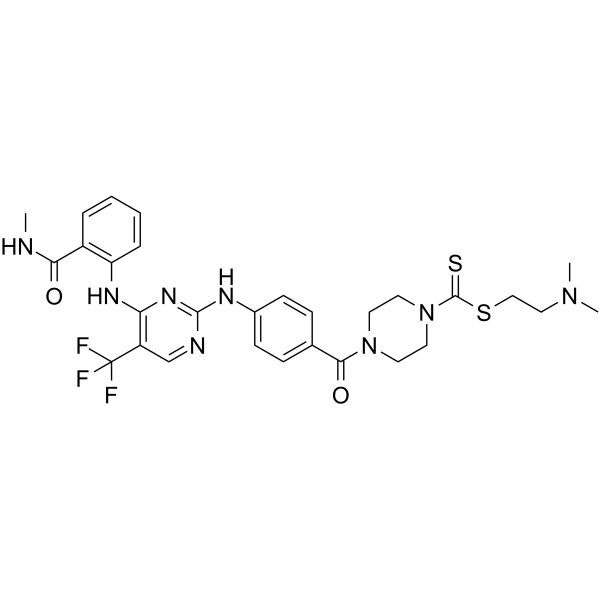
HY-128580
FAK inhibitor 2FAK Inhibitor -
HY-153697
FAK-IN-10FAK Inhibitor -

HY-12289R
Defactinib (Standard)FAK Inhibitor -

HY-12289AR
Defactinib hydrochloride (Standard)FAK Inhibitor -
HY-18928
FAK inhibitor 5FAK Inhibitor


Activité biologique
Description


Chemical Information
-
CAS No. 1627843-95-1
-
Masse moléculaire 450.45
-
Formule C22H22N6O5
-
SMILES
O=C(C1=C(NC2=NC3=NC=CN3C(NC4=CC(OC)=C(C(OC)=C4)OC)=N2)C=CC=C1)OC
-
Livraison
Room temperature in continental US; may vary elsewhere.
-
Stockage
Please store the product under the recommended conditions in the Certificate of Analysis.


Pureté et documentation
Références

Calculators
Concentration (start) × Volume (start) = Concentration (final) × Volume (final)


PH11 Related Classifications
HY-116497 Related Classifications
 Powered by Bioz