



Carteolol
Based on 2 publication(s) in Google Scholar
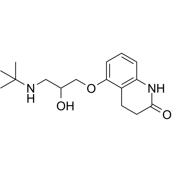
Carteolol is a non-selective β-adrenoceptor antagonist. Carteolol induces apoptosis via a caspase activated and mitochondrial-dependent pathway. Carteolol can be used for glaucoma research.
Nos produits utilisent uniquement pour la recherche. Nous ne vendons pas aux patients.
- CAS No.: 51781-06-7
- Formule: C16H24N2O3
- Masse moléculaire:292.37
-
Stockage:
Please store the product under the recommended conditions in the Certificate of Analysis.

To place orders, for customer services and technical support, please contact: MedChemExpress USA
Tel: 609-228-6898 E-mail: [email protected] [email protected]


-
 Activité biologique
-
Chemical Information
- Protocole
- Pureté et documentation
- Références
-
 Help & FAQs


Publications Citing Use of MedChemExpress (MCE) Carteolol
MoreVoir tous les produits spécifiques à Isoform Adrenergic Receptor
MoreVoir tous les produits spécifiques à Isoform Caspase
More

Activité biologique
Description
In Vitro
Carteolol (0-2%; 0-28 hours; HCECs) has cytotoxicity and decreases cell viability in a dose- and time-dependent manner[1].
Carteolol (0.25%; 4-12 hours; HCECs) induces apoptosis and necroptotic protein expression in HCECs[1].
MedChemExpress (MCE) has not independently confirmed the accuracy of these methods. They are for reference only.
-
Cell Line:HCECs
-
Concentration:0.00390625-2%
-
Incubation Time:0, 2, 4, 8, 16, 20,24 and 28 hours
-
Result:Decreased cell viability with the concentrations above 0.0015625% in a dose- and time-dependent manner.
-
Cell Line:HCECs
-
Concentration:0.25%
-
Incubation Time:4, 8 and 12 hours
-
Result:Dampened expression of the anti-apoptotic protein Bcl-2 and Bcl-xL, enhanced expression of the pro-apoptotic proteins Bax and Bad, and mitochondrial-released pro-apoptotic proteins Cyt.c and AIF.
-
Cell Line:HCECs
-
Concentration:0.25%
-
Incubation Time:4, 8 and 12 hours
-
Result:Increased the number of G1 phase of the cell cycle, whereas decreased S phase.
Essai clinique
| NCT Number | Sponsor | Condition | Start Date |
Phase

|
|---|---|---|---|---|
| NCT01329991 | 2011-05 | PHASE1 |


Chemical Information
-
CAS No. 51781-06-7
-
Masse moléculaire 292.37
-
Formule C16H24N2O3
-
SMILES
O=C1NC2=C(C(OCC(O)CNC(C)(C)C)=CC=C2)CC1
-
Synonyms
OPC-1085
-
Livraison
Room temperature in continental US; may vary elsewhere.
-
Stockage
Please store the product under the recommended conditions in the Certificate of Analysis.


Publications (2)
-
Journal Impact Factor
-
Most Recent
-
ACS Omega
Computational and Experimental Approaches Identify Beta-Blockers as Potential SARS-CoV-2 Spike Inhibitors. [Abstract]2022 Aug 8;7(32):27950-27958. PMID: 35983371 -
J Pharm Biomed Anal
Screening method of mildronate and over 300 doping agents by reversed-phase liquid chromatography-high resolution mass spectrometry. [Abstract]2021 Feb 20:195:113870. PMID: 33453569


Pureté et documentation
Références

Calculators
Concentration (start) × Volume (start) = Concentration (final) × Volume (final)


Carteolol Related Classifications
HY-17495 Related Classifications
 Powered by Bioz