



SBI-581

Based on 1 Customer Validation
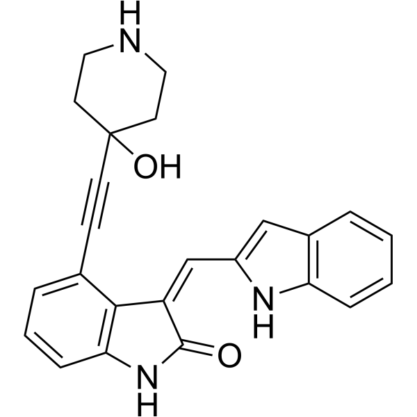
SBI-581 is an orally active and potent selective serine-threonine kinase TAO3 inhibitor, with an IC50 of 42 nM. SBI-581 promotes TKS5α accumulation at RAB11-positive vesicles. SBI-581 inhibits invadopodia formation. SBI-581 shows reasonable pharmacokinetics in mice using IP injection. SBI-581 shows antitumor activity. SBI-581 is a click chemistry reagent, it contains an Alkyne group and can undergo copper-catalyzed azide-alkyne cycloaddition (CuAAc) with molecules containing Azide groups.
Nur für Forschungszwecke. Wir verkaufen nicht an Patienten.
- Reinheit : 98.17%
- Formel: C24H21N3O2
- Molecular Weight:383.44
-
Speicherung:Powder -20°C, 3 years , 4°C, 2 years ; In solvent -80°C, 6 months , -20°C, 1 month

To place orders, for customer services and technical support, please contact: MedChemExpress USA
Tel: 609-228-6898 E-mail: [email protected] [email protected]


-
 Biologische Aktivität
-
Chemical Information
-
Lösungsmittel & Löslichkeit
- Protokoll
- Reinheit & Dokumentation
- Verweise
-
 Help & FAQs




Biologische Aktivität
Beschreibung
IC50 & Target
IC50: 42 nM (TAO3), 237 nM (MEKK3)[1]
In Vitro
In Vivo


Chemical Information
-
Appearance Solid
-
Molecular Weight 383.44
-
Formel C24H21N3O2
-
Color Yellow to orange
-
SMILES
O=C1/C(C2=C(C=CC=C2C#CC3(O)CCNCC3)N1)=C\C4=CC5=C(N4)C=CC=C5
-
Versand
Room temperature in continental US; may vary elsewhere.
-
Speicherung
Powder -20°C 3 years 4°C 2 years In solvent -80°C 6 months -20°C 1 month


Lösungsmittel & Löslichkeit
In Vitro:
DMSO : 50 mg/mL (130.40 mM; ultrasonic and warming and heat to 60°C; Hygroscopic DMSO has a significant impact on the solubility of product, please use newly opened DMSO)
Please refer to the solubility information to select the appropriate solvent. Once prepared, please aliquot and store the solution to prevent product inactivation from repeated freeze-thaw cycles.
Storage method and period of stock solution: -80°C, 6 months; -20°C, 1 month. When stored at -80°C, please use it within 6 months. When stored at -20°C, please use it within 1 month.
Please refer to the solubility information to select the appropriate solvent. Once prepared, please aliquot and store the solution to prevent product inactivation from repeated freeze-thaw cycles.
Storage method and period of stock solution: -80°C, 6 months; -20°C, 1 month. When stored at -80°C, please use it within 6 months. When stored at -20°C, please use it within 1 month.
Konzentration (Stammlösung) × Volumen (Stammlösung) = Konzentration (Ziellösung) × Volumen (Ziellösung)


Reinheit & Dokumentation
-
Data Sheet (279 KB)
-
SDS (251 KB)
- English - EN (251 KB)
- Français - FR (251 KB)
- Deutsch - DE (251 KB)
- Norwegian - NO (251 KB)
- Español - ES (251 KB)
- Swedish - SV (251 KB)
- Italian - IT (251 KB)
- Korean - KR (251 KB)
- Portuguese - PT (251 KB)
-
Handling Instructions (2659 KB)
Verweise


Complete Stock Solution Preparation Table
Please refer to the solubility information to select the appropriate solvent. Once prepared, please aliquot and store the solution to prevent product inactivation from repeated freeze-thaw cycles.
Storage method and period of stock solution: -80°C, 6 months; -20°C, 1 month. When stored at -80°C, please use it within 6 months. When stored at -20°C, please use it within 1 month.
| Optional Solvent | Concentration Solvent Mass | 1 mg | 5 mg | 10 mg | 25 mg |
|---|---|---|---|---|---|
| DMSO | 1 mM | 2.6080 mL | 13.0398 mL | 26.0797 mL | 65.1992 mL |
| 5 mM | 0.5216 mL | 2.6080 mL | 5.2159 mL | 13.0398 mL | |
| 10 mM | 0.2608 mL | 1.3040 mL | 2.6080 mL | 6.5199 mL | |
| 15 mM | 0.1739 mL | 0.8693 mL | 1.7386 mL | 4.3466 mL | |
| 20 mM | 0.1304 mL | 0.6520 mL | 1.3040 mL | 3.2600 mL | |
| 25 mM | 0.1043 mL | 0.5216 mL | 1.0432 mL | 2.6080 mL | |
| 30 mM | 0.0869 mL | 0.4347 mL | 0.8693 mL | 2.1733 mL | |
| 40 mM | 0.0652 mL | 0.3260 mL | 0.6520 mL | 1.6300 mL | |
| 50 mM | 0.0522 mL | 0.2608 mL | 0.5216 mL | 1.3040 mL | |
| 60 mM | 0.0435 mL | 0.2173 mL | 0.4347 mL | 1.0867 mL | |
| 80 mM | 0.0326 mL | 0.1630 mL | 0.3260 mL | 0.8150 mL | |
| 100 mM | 0.0261 mL | 0.1304 mL | 0.2608 mL | 0.6520 mL |


SBI-581 Related Classifications
HY-139439 Related Classifications
 Powered by Bioz