



Anticancer agent 104
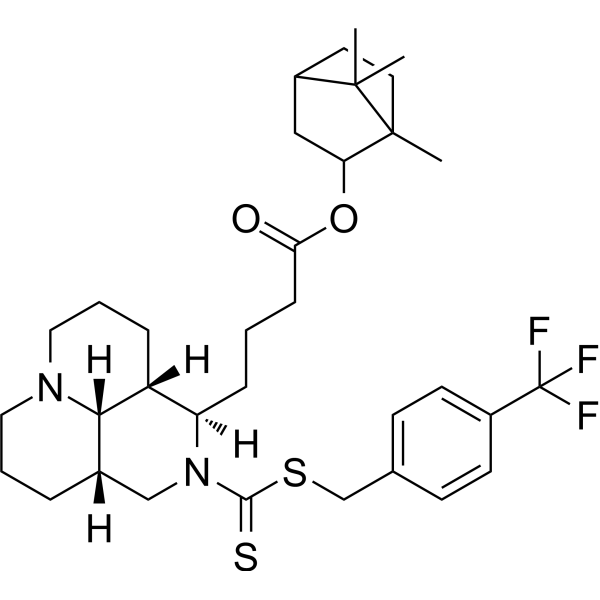
Anticancer agent 104 has anticancer activity, and induces cancer cell apoptosis.
For research use only. We do not sell to patients.
- CAS No.: 3047612-51-8
- Formula: C34H47F3N2O2S2
- Molecular Weight:636.87
-
Storage:
Please store the product under the recommended conditions in the Certificate of Analysis.

To place orders, for customer services and technical support, please contact: MedChemExpress USA
Tel: 609-228-6898 E-mail: [email protected] [email protected]


-
 Biological Activity
-
Chemical Information
- Protocols
- Purity & Documentation
- References
-
 Help & FAQs




Biological Activity
Description
Cellular Effect
|
Cell Line

|
Type | Value | Description | References |
|---|---|---|---|---|
| 786-0 | IC50 |
91.67 μM
Compound: 4l
|
Anticancer activity against human 786-0 cells assessed as reduction in cell viability incubated for 48 hrs by MTT assay
Anticancer activity against human 786-0 cells assessed as reduction in cell viability incubated for 48 hrs by MTT assay
|
[PMID: 37084600] |
| Calu-1 | IC50 |
78.34 μM
Compound: 4l
|
Anticancer activity against human Calu-1 cells assessed as reduction in cell viability incubated for 48 hrs by MTT assay
Anticancer activity against human Calu-1 cells assessed as reduction in cell viability incubated for 48 hrs by MTT assay
|
[PMID: 37084600] |
| HEK-293T | IC50 |
195.17 μM
Compound: 4l
|
Cytotoxicity against human HEK293T cells assessed as reduction in cell viability incubated for 48 hrs by MTT assay
Cytotoxicity against human HEK293T cells assessed as reduction in cell viability incubated for 48 hrs by MTT assay
|
[PMID: 37084600] |
| HepG2 | IC50 |
31.39 μM
Compound: 4l
|
Anticancer activity against human HepG2 cells assessed as reduction in cell viability incubated for 48 hrs by MTT assay
Anticancer activity against human HepG2 cells assessed as reduction in cell viability incubated for 48 hrs by MTT assay
|
[PMID: 37084600] |
| Huh-7 | IC50 |
75.18 μM
Compound: 4l
|
Anticancer activity against human Huh-7 cells assessed as reduction in cell viability incubated for 48 hrs by MTT assay
Anticancer activity against human Huh-7 cells assessed as reduction in cell viability incubated for 48 hrs by MTT assay
|
[PMID: 37084600] |
| KGN | IC50 |
176.38 μM
Compound: 4l
|
Cytotoxicity against human KGN cells assessed as reduction in cell viability incubated for 48 hrs by MTT assay
Cytotoxicity against human KGN cells assessed as reduction in cell viability incubated for 48 hrs by MTT assay
|
[PMID: 37084600] |
| LX-2 | IC50 |
96.33 μM
Compound: 4l
|
Cytotoxicity against human LX2 cells assessed as reduction in cell viability incubated for 48 hrs by MTT assay
Cytotoxicity against human LX2 cells assessed as reduction in cell viability incubated for 48 hrs by MTT assay
|
[PMID: 37084600] |
| MCF-10A | IC50 |
21.06 μM
Compound: 4l
|
Cytotoxicity against human MCF-10A cells assessed as reduction in cell viability incubated for 48 hrs by MTT assay
Cytotoxicity against human MCF-10A cells assessed as reduction in cell viability incubated for 48 hrs by MTT assay
|
[PMID: 37084600] |
| SK-BR-3 | IC50 |
44.18 μM
Compound: 4l
|
Anticancer activity against human SK-BR-3 cells assessed as reduction in cell viability incubated for 48 hrs by MTT assay
Anticancer activity against human SK-BR-3 cells assessed as reduction in cell viability incubated for 48 hrs by MTT assay
|
[PMID: 37084600] |
| SK-OV-3 | IC50 |
112.19 μM
Compound: 4l
|
Anticancer activity against human SK-OV-3 cells assessed as reduction in cell viability incubated for 48 hrs by MTT assay
Anticancer activity against human SK-OV-3 cells assessed as reduction in cell viability incubated for 48 hrs by MTT assay
|
[PMID: 37084600] |
| WI-38 | IC50 |
81.48 μM
Compound: 4l
|
Cytotoxicity against human WI-38 cells assessed as reduction in cell viability incubated for 48 hrs by MTT assay
Cytotoxicity against human WI-38 cells assessed as reduction in cell viability incubated for 48 hrs by MTT assay
|
[PMID: 37084600] |
In Vitro
Anticancer agent 104 (Compound 4l) (48 h) is toxic to HepG2 cells (IC50: 31.39 μM)[1].
Anticancer agent 104 (10-200 μg/mL, 48 h) exhibits volume reduction, chromatin margination, and eventually formed apoptotic vesicles[1].
Anticancer agent 104 (10-100 μg/mL, 48 h) induces HepG2 cell apoptosis[1].
MedChemExpress (MCE) has not independently confirmed the accuracy of these methods. They are for reference only.
-
Cell Line:Calu-1, SK-BR-3, HUH-7, 786-O, SK-OV-3 cells
-
Concentration:0-200 μM approximately
-
Incubation Time:48 h
-
Result:Shows cytotoxic activities with IC50s of 78.34, 44.18, 75.18, 91.67, 112.19 μM.
-
Cell Line:HepG2 cells
-
Concentration:10, 50 and 100 μg/mL
-
Incubation Time:48 h
-
Result:Showed apoptotic rate of 23.67%, 41.21% and 61.16% respectively.


Chemical Information
-
CAS No. 3047612-51-8
-
Molecular Weight 636.87
-
Formula C34H47F3N2O2S2
-
SMILES
C[C@@]1(C(C)([C@@](CC1)([H])C2)C)[C@@H]2OC(CCC[C@@H]3[C@]4([H])[C@]5([H])[C@](CCCN5CCC4)([H])CN3C(SCC6=CC=C(C=C6)C(F)(F)F)=S)=O
-
Shipping
Room temperature in continental US; may vary elsewhere.
-
Storage
Please store the product under the recommended conditions in the Certificate of Analysis.


Purity & Documentation
References

Calculators
Concentration (start) × Volume (start) = Concentration (final) × Volume (final)


Anticancer agent 104 Related Classifications
HY-149231 Related Classifications
 Powered by Bioz