



Cotadutide
Based on 1 publication(s) in Google Scholar
Cotadutide (MEDI0382) is a potent dual agonist of glucagon-like peptide-1 (GLP-1) and GCGR with EC50 values of 6.9 pM and 10.2 pM, respectively. Cotadutide exhibits ability to facilitate both weight loss and glycaemic control, and alleviate fibrosis. Cotadutide can be used in the research of obesity and type 2 diabetes (T2D).
For research use only. We do not sell to patients.
- Purity : 99.80%
- CAS No.: 1686108-82-6
- Formula: C167H252N42O55
- Molecular Weight:3728.09
-
Storage:
Sealed storage, away from moisture.
Powder -80°C, 2 years , -20°C, 1 year* In solvent : -80°C, 6 months; -20°C, 1 month (sealed storage, away from moisture)

To place orders, for customer services and technical support, please contact: MedChemExpress USA
Tel: 609-228-6898 E-mail: [email protected] [email protected]


-
 Biological Activity
-
Chemical Information
-
Solvent & Solubility
- Protocols
- Purity & Documentation
- References
-
 Help & FAQs


-

HY-P3375
MazdutideGLP-1R/GCGR Agonist -

HY-P4146
SurvodutideGCGR/GLP-1R Agonist -
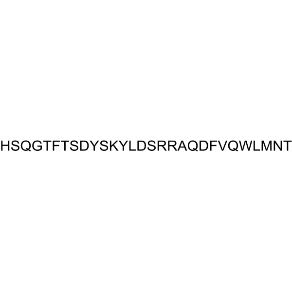
HY-P0082
Glucagon (1-29), bovine, humanPeptide Hormone -

HY-114118
SemaglutideGLP-1R Agonist -
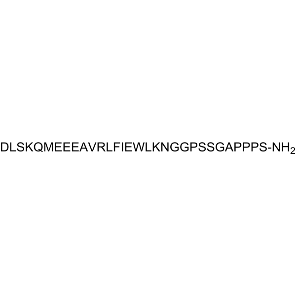
HY-P0264
Exendin(9-39) amideGLP-1R Antagonist -

HY-145632
PemvidutideGLP-1R/GCGR Agonist -
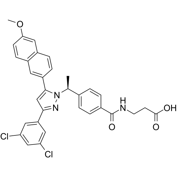
HY-50663
MK 0893GCGR Antagonist -
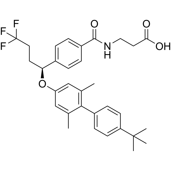
HY-19904
AdomeglivantGluR Antagonist -
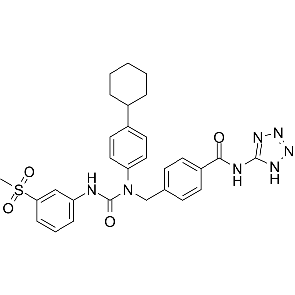
HY-124622
NNC-0640GCGR Modulator -
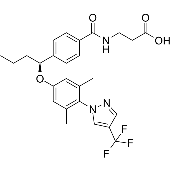
HY-19947
PF-06291874Glucagon Receptor Antagonist -
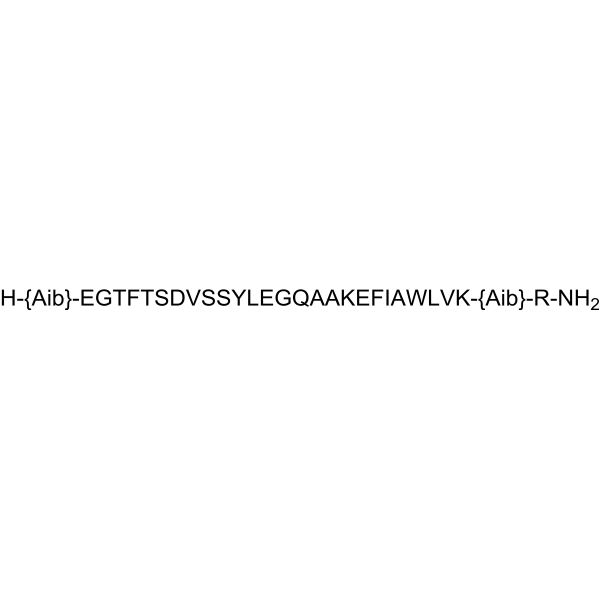
HY-P0165
TaspoglutideGCGR Agonist -
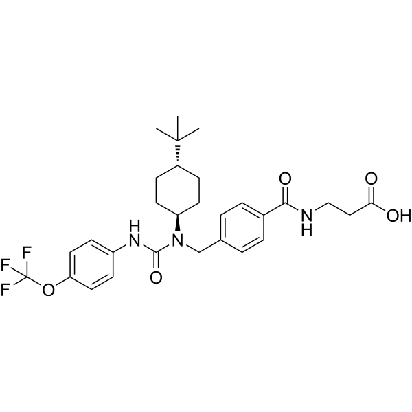
HY-50675
GRA Ex-25GCGR Inhibitor
Publications Citing Use of MedChemExpress (MCE) Cotadutide
More

Biological Activity
Description
IC50 & Target
EC50: 6.9 pM (GLP-1); 10.2 pM (GCGR)[1]
In Vitro
Cotadutide stimulates a concentration-dependent increase in cAMP accumulation in rat (INS-1 832/3) and human (EndoC-βH1) β-cell lines (EC50: 226 pM and 1051 pM, respectively), as well as rat, mouse and human hepatocytes (EC50: 462 pM, 840 pM, 1447 pM, respectively)[1]. Cotadutide (100 pM-1 μM) potentiates glucose-stimulated insulin secretion in the rat (INS-1 832/3) pancreatic β-cell line and increases glucose output in rat hepatocytes[1]. Cotadutide (100 nM, 2 h) induces mitochondrial turnover and enhances mitochondrial function in mouse primary hepatocytes[2].
MedChemExpress (MCE) has not independently confirmed the accuracy of these methods. They are for reference only.
In Vivo
MedChemExpress (MCE) has not independently confirmed the accuracy of these methods. They are for reference only.
-
Animal Model:Diet-induced obesity (DIO) mice[1]
-
Dosage:10 nmol/kg
-
Administration:Subcutaneousinjection (s.c.)
-
Result:Showed a redction of food intake in mice after an acute administration.
-
Animal Model:Diet-induced obesity (DIO) mice[1]
-
Dosage:10 or 30 nmol/kg
-
Administration:Subcutaneousinjection (s.c.)
-
Result:Reduced body weight and food intake, and improved glucose tolerance in DIO mice.


Chemical Information
-
CAS No. 1686108-82-6
-
Appearance Solid
-
Molecular Weight 3728.09
-
Formula C167H252N42O55
-
Color White to off-white
-
Synonyms
MEDI0382
-
Sequence
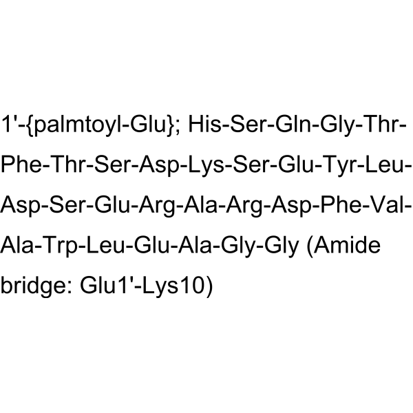
His-Ser-Gln-Gly-Thr-Phe-Thr-Ser-Asp-{Lys(γGlu-C16 acid)}-Ser-Glu-Tyr-Leu-Asp-Ser-Glu-Arg-Ala-Arg-Asp-Phe-Val-Ala-Trp-Leu-Glu-Ala-Gly-Gly
-
Sequence Shortening
HSQGTFTSD-{Lys(γGlu-C16 acid)}-SEYLDSERARDFVAWLEAGG
-
Shipping
Room temperature in continental US; may vary elsewhere.
-
Storage
Sealed storage, away from moisture
Powder -80°C 2 years -20°C 1 year * In solvent : -80°C, 6 months; -20°C, 1 month (sealed storage, away from moisture)


Publications (1)
-
Journal Impact Factor
-
Most Recent
-
Adv Sci (Weinh)
2024 Aug;11(29):e2400819. PMID: 38837628


Solvent & Solubility
In Vitro:
DMSO : 100 mg/mL (26.82 mM; Need ultrasonic; Hygroscopic DMSO has a significant impact on the solubility of product, please use newly opened DMSO)
Please refer to the solubility information to select the appropriate solvent. Once prepared, please aliquot and store the solution to prevent product inactivation from repeated freeze-thaw cycles.
Storage method and period of stock solution: -80°C, 6 months; -20°C, 1 month (sealed storage, away from moisture). When stored at -80°C, please use it within 6 months. When stored at -20°C, please use it within 1 month.
Please refer to the solubility information to select the appropriate solvent. Once prepared, please aliquot and store the solution to prevent product inactivation from repeated freeze-thaw cycles.
Storage method and period of stock solution: -80°C, 6 months; -20°C, 1 month (sealed storage, away from moisture). When stored at -80°C, please use it within 6 months. When stored at -20°C, please use it within 1 month.
Concentration (start) × Volume (start) = Concentration (final) × Volume (final)


Purity & Documentation
-
Data Sheet (287 KB)
-
SDS (252 KB)
- English - EN (252 KB)
- Français - FR (252 KB)
- Deutsch - DE (252 KB)
- Norwegian - NO (252 KB)
- Español - ES (252 KB)
- Swedish - SV (252 KB)
- Italian - IT (252 KB)
- Korean - KR (252 KB)
- Portuguese - PT (252 KB)
-
Handling Instructions (2659 KB)
References
[1]. Henderson SJ,et al. Robust anti-obesity and metabolic effects of a dual GLP-1/glucagon receptor peptide agonist in rodents and non-human primates.Diabetes Obes Metab. 2016 Dec;18(12):1176-1190. [Content Brief]
[2]. Resolution of NASH and hepatic fibrosis by the GLP-1R/GcgR dual-agonist Cotadutide via modulating mitochondrial function and lipogenesis. Nat Metab. 2020 May;2(5):413-431. [Content Brief]


Complete Stock Solution Preparation Table
Please refer to the solubility information to select the appropriate solvent. Once prepared, please aliquot and store the solution to prevent product inactivation from repeated freeze-thaw cycles.
Storage method and period of stock solution: -80°C, 6 months; -20°C, 1 month (sealed storage, away from moisture). When stored at -80°C, please use it within 6 months. When stored at -20°C, please use it within 1 month.
| Optional Solvent | Concentration Solvent Mass | 1 mg | 5 mg | 10 mg | 25 mg |
|---|---|---|---|---|---|
| DMSO | 1 mM | 0.2682 mL | 1.3412 mL | 2.6823 mL | 6.7058 mL |
| 5 mM | 0.0536 mL | 0.2682 mL | 0.5365 mL | 1.3412 mL | |
| 10 mM | 0.0268 mL | 0.1341 mL | 0.2682 mL | 0.6706 mL | |
| 15 mM | 0.0179 mL | 0.0894 mL | 0.1788 mL | 0.4471 mL | |
| 20 mM | 0.0134 mL | 0.0671 mL | 0.1341 mL | 0.3353 mL | |
| 25 mM | 0.0107 mL | 0.0536 mL | 0.1073 mL | 0.2682 mL |


Cotadutide Related Classifications
HY-P2231 Related Classifications
 Powered by Bioz