



BMS-1001
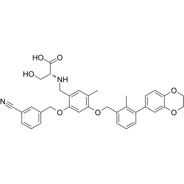
BMS-1001 is an orally active human PD-L1/PD-1 immune checkpoint inhibitor. BMS-1001 exhibits low-toxicity in cells. The IC50 value of BMS-1001 in a homogeneous time-resolved fluorescence (HTRF) binding assay is 2.25 nM.
For research use only. We do not sell to patients.
- CAS No.: 2113650-03-4
- Formula: C35H34N2O7
- Molecular Weight:594.65
-
Storage:
Please store the product under the recommended conditions in the Certificate of Analysis.

To place orders, for customer services and technical support, please contact: MedChemExpress USA
Tel: 609-228-6898 E-mail: [email protected] [email protected]


-
 Biological Activity
-
Chemical Information
- Protocols
- Purity & Documentation
- References
-
 Help & FAQs


-
HY-19745
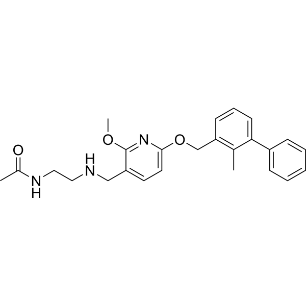
BMS-202 -
HY-102011
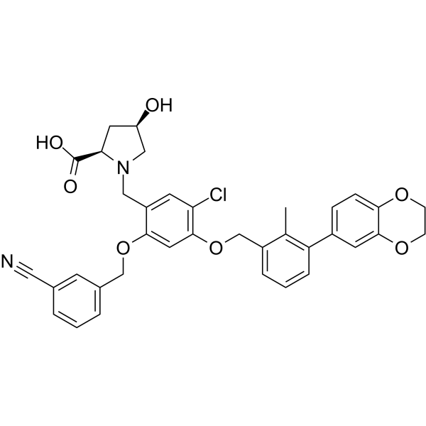
BMS-1166PD-1/PD-L1 Interaction Inhibitor -
HY-101093
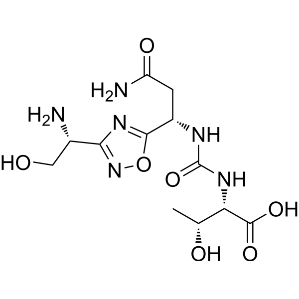
CA-170VISTA/PD-L1 Inhibitor -
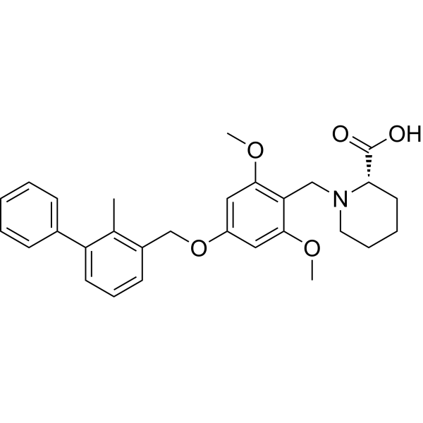
HY-19991
BMS-1PD-1/PD-L1 PPI Inhibitor -
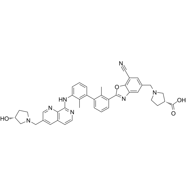
HY-134884
INCB086550PD-L1 Inhibitor -
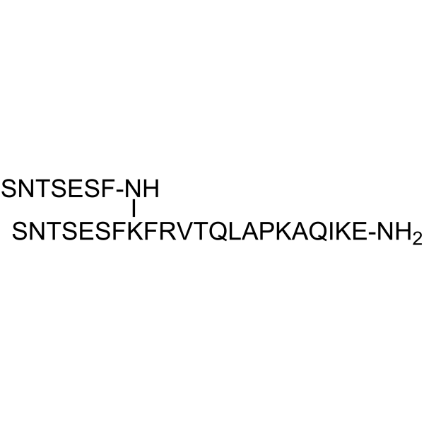
HY-P1812
AUNP-12PD-1 Antagonist -

HY-P9902
PembrolizumabPD-1 Inhibitor -

HY-P9904
AtezolizumabAnti-PD-L1 Antibody -
HY-P9903
NivolumabPD-1 Inhibitor -
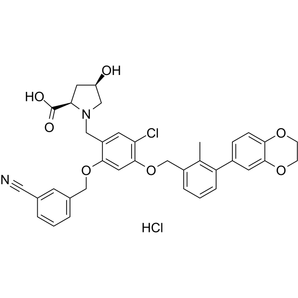
HY-102011A
BMS-1166 hydrochloridePD-1/PD-L1 Interaction Inhibitor -
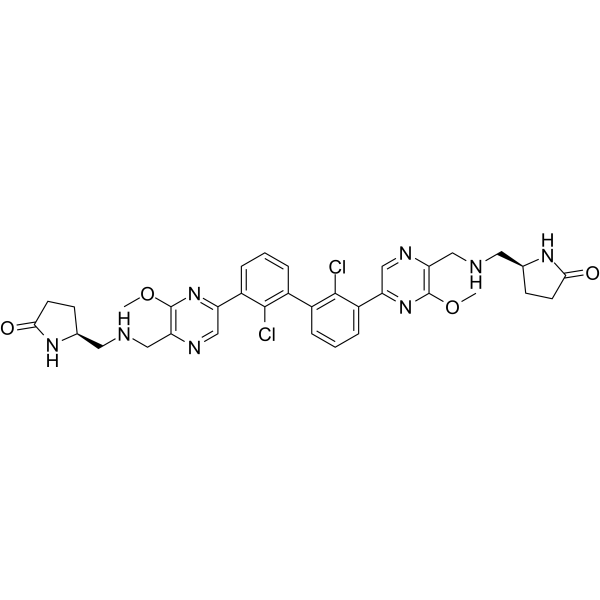
HY-138407
EvixapodlinPD-1/PD-L1 PPI Inhibitor -
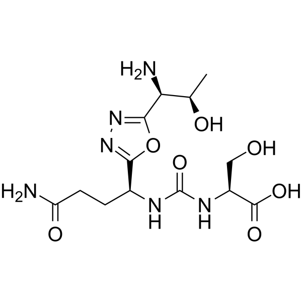
HY-101097
PD-1-IN-17PD-1 Inhibitor


Biological Activity
Description
Cellular Effect
|
Cell Line

|
Type | Value | Description | References |
|---|---|---|---|---|
| Jurkat | IC50 |
<100 nM
Compound: 16; BMS-1001
|
Cytotoxicity against human Jurkat T cells assessed as reduction in cell viability
Cytotoxicity against human Jurkat T cells assessed as reduction in cell viability
|
[PMID: 33517226] |
In Vitro
BMS-1001 binds to human PD-L1 and blocks its interaction with PD-1. BMS-1001 presents low toxicity towards tested cell lines and block the interaction of soluble PD-L1 with the cell surface-expressed PD-1. BMS-1001 alleviates the inhibitory effect of the soluble PD-L1 on the T-cell receptor-mediated activation of T-lymphocytes[1].
MedChemExpress (MCE) has not independently confirmed the accuracy of these methods. They are for reference only. Further protocols information, click here.


Chemical Information
-
CAS No. 2113650-03-4
-
Molecular Weight 594.65
-
Formula C35H34N2O7
-
SMILES
OC[C@H](C(O)=O)NCC1=CC(C)=C(OCC2=CC=CC(C3=CC=C4OCCOC4=C3)=C2C)C=C1OCC5=CC=CC(C#N)=C5
-
Shipping
Room temperature in continental US; may vary elsewhere.
-
Storage
Please store the product under the recommended conditions in the Certificate of Analysis.

Protocols
-
Cell Cytotoxicity Assay
Cytotoxicity assays are usually based on the assessment of cell membrane damage, which can also be indirectly detected by measuring cell viability. Detection methods include MTT assay, CKK-8 assay, LDH assay and ATP assay, etc.
-
Research Protocol for Cancer Immunology
Cancer immunology studies how the immune system recognizes, suppresses, edits, or fails to eliminate malignant cells through tumor antigen release, antigen presentation, T-cell priming, immune trafficking, tumor-cell killing, and feedback inhibition in the tumor microenvironment. The cancer-immunity cycle links tumor antigenicity, dendritic-cell priming, CD8+ T-cell infiltration, cytotoxic function, and immune-checkpoint regulation to tumor rejection or immune escape. Immune-checkpoint pathways such as PD-1/PD-L1 and CTLA-4 suppress antitumor T-cell activity and can be therapeutically blocked, but many tumors remain resistant because of poor antigen presentation, weak T-cell infiltration, suppressive myeloid cells, regulatory T cells, and tumor-intrinsic immune-exclusion programs. Unresolved questions include which immune-cell states predict response, how tumor-intrinsic pathways exclude immune cells, how myeloid suppression limits checkpoint blockade, and which combination strategies


Purity & Documentation
References
[1]. Skalniak L, et al. Small-molecule inhibitors of PD-1/PD-L1 immune checkpoint alleviate the PD-L1-induced exhaustion of T-cells. Oncotarget. 2017 Aug 7;8(42):72167-72181. [Content Brief]
[2]. Guzik K, et al. Small-Molecule Inhibitors of the Programmed Cell Death-1/Programmed Death-Ligand 1 (PD-1/PD-L1) Interaction via Transiently Induced Protein States and Dimerization of PD-L1. J Med Chem. 2017 Jul 13;60(13):5857-5867. [Content Brief]

Calculators
Concentration (start) × Volume (start) = Concentration (final) × Volume (final)


BMS-1001 Related Classifications
HY-120647 Related Classifications
 Powered by Bioz