



2-NBDG (solution)

Based on 1 Customer Validation
2-NBDG (solution) is a fluorescently-labeled deoxyglucose analog that is used primarily to directly monitor glucose uptake by living cells and tissues. It is also used as a topical contrast reagent for the detection of neoplasia. 2-NBDG can be used in real-time confocal, high-resolution, or wide-field fluorescence microscopy as well as in flow cytometry. The probe can be excited by the Argon laser at 488 nm to give the environment-sensitive fluorescence. It has lower photostability than the rhodamine-based fluorescent probes.
Solvent and Concentration: Sterile PBS: 5 mM
Para uso exclusivo en investigación. No vendemos a pacientes.
- No. CAS: 186689-07-6
- Fòrmula: C12H14N4O8
- Peso molecular:342.26
-
Almacenamiento:
Please store the product under the recommended conditions in the Certificate of Analysis.


To place orders, for customer services and technical support, please contact: MedChemExpress USA
Tel: 609-228-6898 E-mail: [email protected] [email protected]


-
 Actividad biológica
-
Chemical Information
- Protocolo
- Pureza y Documentación
- Referencias
-
 Help & FAQs


-
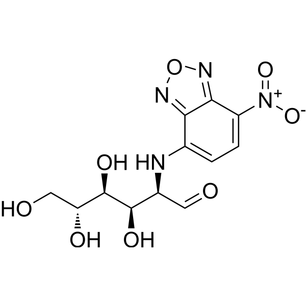
HY-116215
2-NBDGFluorescent Indicator -
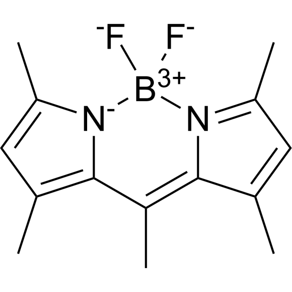
HY-W090090
BODIPY 493/503Neutral Lipid Dye -
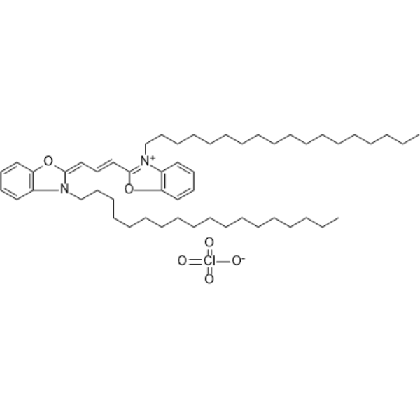
HY-D0969
DiOFluorescent Probe -
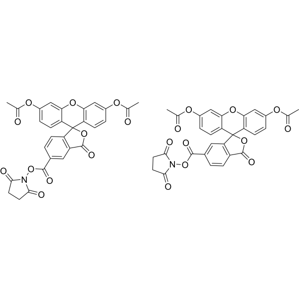
HY-D0938
CFDA-SECell Proliferation Fluorescent Probe -
HY-D0711
Indocyanine greenFluorescent Agent -
HY-D0819
CY5-SEReactive Dye -
HY-D0042
NBD-ClNonfluorescent Reagent -

HY-DY1004
BODIPY 493/503 (solution)Neutral Lipid Dye -
HY-D0073
2,3-DiaminonaphthaleneFluorescent Dye -
HY-D0933
Auramine OFluorescent Dye -

HY-DY1063
CY5-SE (solution)Reactive Dye -
HY-120657
9-PAHSAGPR120 Agonist


Actividad biológica
Descripciòn
Solvent and Concentration: Sterile PBS: 5 mM
In Vitro
Guide (The following is the experimental plan we recommend. This plan serves only as a reference guide. The specific operations should be adjusted according to your actual needs.)
1. Preparation of 2-NBDG Solution
Dilute the stored solution with pre-warmed serum-free cell culture medium or PBS to prepare a 2-NBDG working solution of 10-200 μM.
Note: Please adjust the concentration of the 2-NBDG working solution according to the actual situation, and prepare it as needed.
2. Cell Staining
2.1 Preparation of Cells
Suspended cells: Centrifuge at 1000 g for 3-5 minutes at 4°C. Collect the cell precipitate and wash it twice with PBS, each time for 5 minutes.
Monolayer cells: Remove the culture medium and add trypsin to digest the cells. Centrifuge at 1000 g for 3-5 minutes at 4°C. Collect the cell pellet, add PBS for two washes, each for 5 minutes.
2.2 Add 1 mL of the 2-NBDG working solution and incubate at room temperature for 5 to 60 minutes.
2.3 400 g, centrifuge at 4℃ for 3-4 minutes, then discard the supernatant.
2.4 Add PBS to wash the cells twice, for 5 minutes each time.
2.5 After resuspending the cells in 1 mL of serum-free medium or PBS, observe them using a flow cytometer;
If an activity test is conducted, use an ELISA microplate reader to record the optical density (O.D.) at 540/570 nm. The cell viability is calculated based on the control ratio, and a curve is plotted against the logarithmic concentration of the drug. The IC50 value is then calculated.
MedChemExpress (MCE) has not independently confirmed the accuracy of these methods. They are for reference only.


Chemical Information
-
No. CAS 186689-07-6
-
Appearance Liquid (Density: 1.751±0.06 g/cm3)
-
Peso molecular 342.26
-
Fòrmula C12H14N4O8
-
Color Yellow to orange
-
SMILES
O=C[C@H](NC1=CC=C([N+]([O-])=O)C2=NON=C21)[C@H]([C@@H]([C@@H](CO)O)O)O
-
Envío
Room temperature in continental US; may vary elsewhere.
-
Almacenamiento
Please store the product under the recommended conditions in the Certificate of Analysis.


Pureza y Documentación
-
Ficha de datos (274 KB)
-
SDS (251 KB)
- English - EN (251 KB)
- Français - FR (251 KB)
- Deutsch - DE (251 KB)
- Norwegian - NO (251 KB)
- Español - ES (251 KB)
- Swedish - SV (251 KB)
- Italian - IT (251 KB)
- Korean - KR (251 KB)
- Portuguese - PT (251 KB)
-
Instrucciones de manejo (2659 KB)
Referencias
[1]. Yamada K, et al. A real-time method of imaging glucose uptake in single, living mammalian cells. Nat Protoc. 2007;2(3):753-62. [Content Brief]
[2]. Zou C, et al. 2-NBDG as a fluorescent indicator for direct glucose uptake measurement. J Biochem Biophys Methods. 2005 Sep 30;64(3):207-15. [Content Brief]
[3]. Katsuya Yamada, et al. A real-time method of imaging glucose uptake in single, living mammalian cells. Nat Protoc. 2007;2(3):753-62. [Content Brief]
[4]. Zou C, et al. 2-NBDG as a fluorescent indicator for direct glucose uptake measurement. J Biochem Biophys Methods. 2005 Sep 30;64(3):207-15. [Content Brief]

Calculators
Concentration (start) × Volume (start) = Concentration (final) × Volume (final)


2-NBDG (solution) Related Classifications
HY-DY1019 Related Classifications
 Powered by Bioz