



Dehydroergosterol
Based on 1 publication(s) in Google Scholar
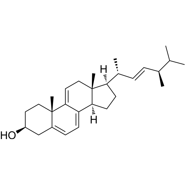
Dehydroergosterol is a fluorescent cholesterol analog (Ex/Em = 325/375 nm) used in cholesterol transport, membrane structure research, and live-cell imaging. Unlike cholesterol, Dehydroergosterol contains three additional double bonds, an extra methyl group, and an ergosterol side chain, forming a conjugated system that produces intrinsic fluorescence. In its monomeric form, Dehydroergosterol exhibits excitation peaks at 310-311, 324, and 338-340 nm, as well as emission peaks at 354, 371-375, 390-394, and 414 nm; in its microcrystalline form, Dehydroergosterol shows enhanced excimer emission around 404 and 426 nm. Dehydroergosterol mimics the properties of cholesterol, integrates into membranes and lipoproteins, and localizes to organelles.
For research use only. We do not sell to patients.
- Purity : ≥99.0%
- CAS No.: 516-85-8
- Formula: C28H42O
- Molecular Weight:394.63
-
Storage:Powder -20°C, 3 years ; In solvent -80°C, 6 months , -20°C, 1 month

To place orders, for customer services and technical support, please contact: MedChemExpress USA
Tel: 609-228-6898 E-mail: [email protected] [email protected]


-
 Biological Activity
-
Chemical Information
- Purity & Documentation
- References
-
 Help & FAQs




Publications Citing Use of MedChemExpress (MCE) Dehydroergosterol
More

Biological Activity
Description
In Vitro
1. Preparation of Working Solution
1.1 Diluent: Cell culture medium; Alternative diluents: PBS, bicarbonate-buffered Ham's F-12 medium supplemented with 5% lipoprotein-deficient serum (LPDS).
1.2 Working concentration: 12.5 μM; Alternative working concentrations/formulations: 20 μg Dehydroergosterol per 1 mL cell culture medium, 20 μg Dehydroergosterol per sample (delivered as DHE-MbCD complex), POPC:DHE at a molar ratio of 65:35 (present in large unilamellar vesicles).
1.3 Notes: Adjust the concentration of the working solution as needed; Prepare it immediately before use.
2. Staining Procedure
2.1 Description of Sample Types
2.1.1 Adherent cultured cells[1][2]:
2.1.1.1 L cell fibroblasts[1]: Cells are cultured on chamber coverslips; No trypsin digestion step is mentioned for staining.
2.1.1.2 CHO cell line TRVb-1/Tac-TGN38[2]: Trypsin digestion is required after incubation.
2.2 Incubation Conditions
2.2.1 Adherent cultured cells[1][2]:
2.2.1.1 Stock solution diluted with medium (ethanol-based system)[1]: Incubate for 2 days, with temperature unspecified.
2.2.1.2 Large unilamellar vesicle system[1]: Incubate for 1 day, with temperature unspecified.
2.2.1.3 DHE-MbCD complex system[1]: Incubate at room temperature for 45 min; Protect the complex from light during preparation.
2.2.1.4 Bicarbonate-buffered Ham's F-12 medium system supplemented with 5% LPDS[2]: Incubate cells in a humidified incubator at 37°C with 5% CO2 for 16-20 h.
2.3 Washing Steps
2.3.1 Adherent cultured cells[1][2]:
2.3.1.1 Stock solution diluted with medium (ethanol-based system)[1]: Wash cells multiple times with an unspecified buffer.
2.3.1.2 DHE-MbCD complex system[1]: Wash cells 3 times with PBS.
2.3.1.3 CHO cell line TRVb-1/Tac-TGN38[2]: Wash the cell monolayer twice with PBS after incubation; After trypsin digestion, resuspend the cells by centrifugation and wash twice with PBS again.
2.4 Post-streatment
2.4.1 CHO cell line TRVb-1/Tac-TGN38[2]: Trypsinize the labeled cells at 37°C for 5 min, centrifuge at approximately 135 × g, resuspend in Ham's F-12 medium containing 5% LPDS, and seed onto coverslips coated with poly-D-lysine or fibronectin; Continue culturing the cells for 16-20 h before imaging.
3. Control Setup
3.1 Set up unlabeled cell controls to evaluate autofluorescence under the same imaging conditions.
4. Detection and Analysis
4.1 Instrument types: Multiphoton laser scanning microscope; Fluorescence microscope equipped with a cooled CCD camera.
4.2 Excitation/Emission Wavelengths
4.2.1 Multiphoton laser scanning microscope[1]: Excitation wavelength: 900 nm; Detect emitted light using D400/100 and UV 440LP dichroic filters.
4.2.2 Monomeric DHE[1][2]: Maximum excitation wavelengths: 311 nm, 324 nm, 340 nm; Maximum emission wavelengths: 354 nm, 371 nm, 375 nm, 390 nm, 394 nm, 414 nm.
4.2.3 Crystalline DHE[1]: Maximum emission wavelengths: 404 nm, 426 nm.
4.2.4 Fluorescence microscope with UV filter set[2]: Excitation filter: 335 nm (20 nm bandpass); Dichroic filter: 365 nm longpass; Emission filter: 405 nm (40 nm bandpass).
4.3 Result Analysis
4.3.1 Fluorescence Intensity and Morphology[1][2]:
4.3.1.1 Crystalline DHE shows strong, bright patchy fluorescence; Monomeric DHE shows moderate-intensity uniform fluorescence.
4.3.1.2 After photobleaching, the DHE fluorescence in the bleached area recovers within 20 min at 37°C, indicating dynamic sterol trafficking.
4.3.2 Fluorescence Localization[1][2]:
4.3.2.1 DHE from ethanol stock solution[1]: Initially accumulates in lysosomes as crystalline DHE; After 2-3 days, monomeric DHE distributes to the plasma membrane, lipid droplets, endoplasmic reticulum and other organelle membranes.
4.3.2.2 DHE from large unilamellar vesicles[1]: Monomeric DHE distributes to the plasma membrane and intracellular compartments, with partial accumulation in lipid storage vesicles.
4.3.2.3 DHE from DHE-MbCD complex[1]: Monomeric DHE strongly labels the plasma membrane (including microvilli and filopodia), with weak labeling of other intracellular membranes.
4.3.2.4 DHE in CHO cell line TRVb-1/Tac-TGN38[2]: Localizes to the plasma membrane and perinuclear regions, including the endosomal recycling compartment (ERC) and trans-Golgi network (TGN).
4.3.3 Fluorescence Distribution[1]: In the plasma membrane of living cells, DHE distributes non-randomly in sterol-rich and sterol-poor regions.
4.3.4 Ratio Analysis[1]: Calculate the ratio of fluorescence intensity at 426 nm to that at 373 nm to distinguish crystalline DHE (ratio approximately 3.4) from monomeric DHE (ratio approximately 0.3).
4.3.5 Colocalization Analysis[2]: DHE shows extensive colocalization with endocytosed transferrin (ERC marker) and Tac-TGN38 (TGN marker).
MedChemExpress (MCE) has not independently confirmed the accuracy of these methods. They are for reference only.


Chemical Information
-
CAS No. 516-85-8
-
Appearance Solid
-
Molecular Weight 394.63
-
Formula C28H42O
-
Color White to off-white
-
SMILES
CC([C@H](/C=C/[C@H]([C@]1(CC[C@]2(C3=CC=C4C[C@H](CC[C@@]4(C3=CC[C@@]21C)C)O)[H])[H])C)C)C
-
Shipping
Room temperature in continental US; may vary elsewhere.
-
Storage
Powder -20°C 3 years In solvent -80°C 6 months -20°C 1 month


Publications (1)
-
Journal Impact Factor
-
Most Recent


Purity & Documentation
-
Data Sheet (289 KB)
-
SDS (418 KB)
- English - EN (418 KB)
- Français - FR (418 KB)
- Deutsch - DE (418 KB)
- Norwegian - NO (418 KB)
- Español - ES (418 KB)
- Swedish - SV (418 KB)
- Italian - IT (418 KB)
- Korean - KR (418 KB)
- Portuguese - PT (418 KB)
-
Handling Instructions (2659 KB)
References

Calculators
Concentration (start) × Volume (start) = Concentration (final) × Volume (final)


Dehydroergosterol Related Classifications
HY-118667 Related Classifications
 Powered by Bioz