



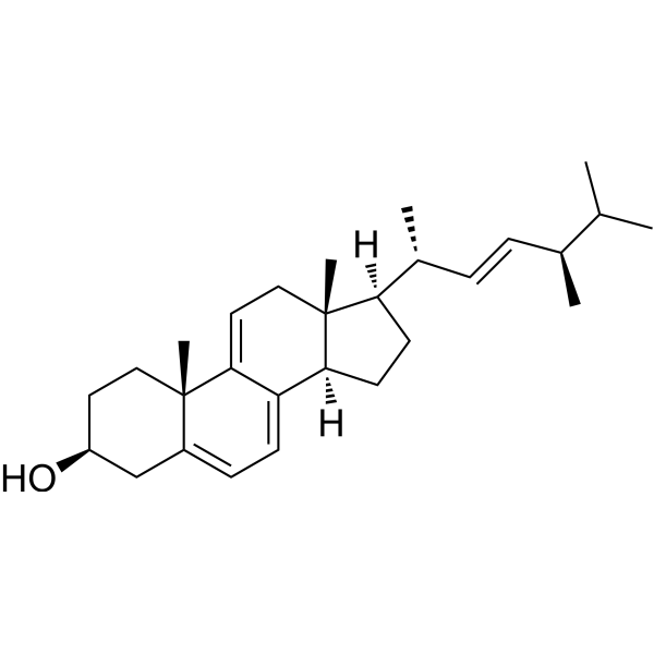
Dehydroergosterol
Based on 1 publication(s) in Google Scholar
Dehydroergosterol is a fluorescent cholesterol analog (Ex/Em = 325/375 nm) used in cholesterol transport, membrane structure research, and live-cell imaging. Unlike cholesterol, Dehydroergosterol contains three additional double bonds, an extra methyl group, and an ergosterol side chain, forming a conjugated system that produces intrinsic fluorescence. In its monomeric form, Dehydroergosterol exhibits excitation peaks at 310-311, 324, and 338-340 nm, as well as emission peaks at 354, 371-375, 390-394, and 414 nm; in its microcrystalline form, Dehydroergosterol shows enhanced excimer emission around 404 and 426 nm. Dehydroergosterol mimics the properties of cholesterol, integrates into membranes and lipoproteins, and localizes to organelles.
商品は「研究用試薬」です。人や動物の医療用・臨床診断用・食品用の製品ではありません。
研究用途以外に使用した場合、当社は一切の責任を負いかねます。
- 純度 : ≥99.0%
- CAS 番号: 516-85-8
- 分子式: C28H42O
- 分子量:394.63
-
保管条件:Powder -20°C, 3 years ; In solvent -80°C, 6 months , -20°C, 1 month

To place orders, for customer services and technical support, please contact: MedChemExpress USA
Tel: 609-228-6898 E-mail: [email protected] [email protected]


-
 生物活性
生物活性
-
化学情報
- 純度とドキュメンテーション
- 参考文献
-
 Help & FAQs




MedChemExpress(MCE)の使用を引用している文献 Dehydroergosterol
More

生物活性
製品説明
体外実験
1. Preparation of Working Solution
1.1 Diluent: Cell culture medium; Alternative diluents: PBS, bicarbonate-buffered Ham's F-12 medium supplemented with 5% lipoprotein-deficient serum (LPDS).
1.2 Working concentration: 12.5 μM; Alternative working concentrations/formulations: 20 μg Dehydroergosterol per 1 mL cell culture medium, 20 μg Dehydroergosterol per sample (delivered as DHE-MbCD complex), POPC:DHE at a molar ratio of 65:35 (present in large unilamellar vesicles).
1.3 Notes: Adjust the concentration of the working solution as needed; Prepare it immediately before use.
2. Staining Procedure
2.1 Description of Sample Types
2.1.1 Adherent cultured cells[1][2]:
2.1.1.1 L cell fibroblasts[1]: Cells are cultured on chamber coverslips; No trypsin digestion step is mentioned for staining.
2.1.1.2 CHO cell line TRVb-1/Tac-TGN38[2]: Trypsin digestion is required after incubation.
2.2 Incubation Conditions
2.2.1 Adherent cultured cells[1][2]:
2.2.1.1 Stock solution diluted with medium (ethanol-based system)[1]: Incubate for 2 days, with temperature unspecified.
2.2.1.2 Large unilamellar vesicle system[1]: Incubate for 1 day, with temperature unspecified.
2.2.1.3 DHE-MbCD complex system[1]: Incubate at room temperature for 45 min; Protect the complex from light during preparation.
2.2.1.4 Bicarbonate-buffered Ham's F-12 medium system supplemented with 5% LPDS[2]: Incubate cells in a humidified incubator at 37°C with 5% CO2 for 16-20 h.
2.3 Washing Steps
2.3.1 Adherent cultured cells[1][2]:
2.3.1.1 Stock solution diluted with medium (ethanol-based system)[1]: Wash cells multiple times with an unspecified buffer.
2.3.1.2 DHE-MbCD complex system[1]: Wash cells 3 times with PBS.
2.3.1.3 CHO cell line TRVb-1/Tac-TGN38[2]: Wash the cell monolayer twice with PBS after incubation; After trypsin digestion, resuspend the cells by centrifugation and wash twice with PBS again.
2.4 Post-streatment
2.4.1 CHO cell line TRVb-1/Tac-TGN38[2]: Trypsinize the labeled cells at 37°C for 5 min, centrifuge at approximately 135 × g, resuspend in Ham's F-12 medium containing 5% LPDS, and seed onto coverslips coated with poly-D-lysine or fibronectin; Continue culturing the cells for 16-20 h before imaging.
3. Control Setup
3.1 Set up unlabeled cell controls to evaluate autofluorescence under the same imaging conditions.
4. Detection and Analysis
4.1 Instrument types: Multiphoton laser scanning microscope; Fluorescence microscope equipped with a cooled CCD camera.
4.2 Excitation/Emission Wavelengths
4.2.1 Multiphoton laser scanning microscope[1]: Excitation wavelength: 900 nm; Detect emitted light using D400/100 and UV 440LP dichroic filters.
4.2.2 Monomeric DHE[1][2]: Maximum excitation wavelengths: 311 nm, 324 nm, 340 nm; Maximum emission wavelengths: 354 nm, 371 nm, 375 nm, 390 nm, 394 nm, 414 nm.
4.2.3 Crystalline DHE[1]: Maximum emission wavelengths: 404 nm, 426 nm.
4.2.4 Fluorescence microscope with UV filter set[2]: Excitation filter: 335 nm (20 nm bandpass); Dichroic filter: 365 nm longpass; Emission filter: 405 nm (40 nm bandpass).
4.3 Result Analysis
4.3.1 Fluorescence Intensity and Morphology[1][2]:
4.3.1.1 Crystalline DHE shows strong, bright patchy fluorescence; Monomeric DHE shows moderate-intensity uniform fluorescence.
4.3.1.2 After photobleaching, the DHE fluorescence in the bleached area recovers within 20 min at 37°C, indicating dynamic sterol trafficking.
4.3.2 Fluorescence Localization[1][2]:
4.3.2.1 DHE from ethanol stock solution[1]: Initially accumulates in lysosomes as crystalline DHE; After 2-3 days, monomeric DHE distributes to the plasma membrane, lipid droplets, endoplasmic reticulum and other organelle membranes.
4.3.2.2 DHE from large unilamellar vesicles[1]: Monomeric DHE distributes to the plasma membrane and intracellular compartments, with partial accumulation in lipid storage vesicles.
4.3.2.3 DHE from DHE-MbCD complex[1]: Monomeric DHE strongly labels the plasma membrane (including microvilli and filopodia), with weak labeling of other intracellular membranes.
4.3.2.4 DHE in CHO cell line TRVb-1/Tac-TGN38[2]: Localizes to the plasma membrane and perinuclear regions, including the endosomal recycling compartment (ERC) and trans-Golgi network (TGN).
4.3.3 Fluorescence Distribution[1]: In the plasma membrane of living cells, DHE distributes non-randomly in sterol-rich and sterol-poor regions.
4.3.4 Ratio Analysis[1]: Calculate the ratio of fluorescence intensity at 426 nm to that at 373 nm to distinguish crystalline DHE (ratio approximately 3.4) from monomeric DHE (ratio approximately 0.3).
4.3.5 Colocalization Analysis[2]: DHE shows extensive colocalization with endocytosed transferrin (ERC marker) and Tac-TGN38 (TGN marker).
MedChemExpress (MCE) has not independently confirmed the accuracy of these methods. They are for reference only.


化学情報
-
CAS 番号 516-85-8
-
性状 Solid
-
分子量 394.63
-
分子式 C28H42O
-
Color White to off-white
-
SMILES
CC([C@H](/C=C/[C@H]([C@]1(CC[C@]2(C3=CC=C4C[C@H](CC[C@@]4(C3=CC[C@@]21C)C)O)[H])[H])C)C)C
-
輸送条件
Room temperature in continental US; may vary elsewhere.
-
保管条件
Powder -20°C 3 years In solvent -80°C 6 months -20°C 1 month


Publications (1)
-
Journal Impact Factor
-
Most Recent


純度とドキュメンテーション
-
データシート (289 KB)
-
SDS (418 KB)
- English - EN (418 KB)
- Français - FR (418 KB)
- Deutsch - DE (418 KB)
- Norwegian - NO (418 KB)
- Español - ES (418 KB)
- Swedish - SV (418 KB)
- Italian - IT (418 KB)
- Korean - KR (418 KB)
- Portuguese - PT (418 KB)
-
取扱説明書 (2659 KB)
参考文献

Calculators
濃度 (開始) × 体積 (開始) = 濃度 (終了) × 体積 (終了)


Dehydroergosterol 関連分類
HY-118667 関連分類
 Powered by Bioz