



SLMP53-1

Based on 1 Customer Validation
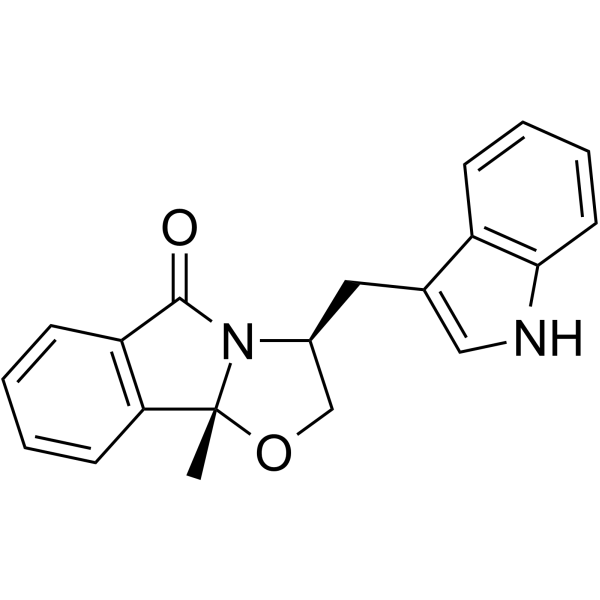
SLMP53-1 is a wild-type and mutant p53 reactivator with promising antitumor activity. SLMP53-1 mediates the reprograming of glucose metabolism in cancer cells. SLMP53-1 depletes angiogenesis, decreasing endothelial cell tube formation and vascular endothelial growth factor (VEGF) expression levels.
商品は「研究用試薬」です。人や動物の医療用・臨床診断用・食品用の製品ではありません。
研究用途以外に使用した場合、当社は一切の責任を負いかねます。
- 純度 : 99.25%
- CAS 番号: 1643469-17-3
- 分子式: C20H18N2O2
- 分子量:318.37
-
保管条件:Powder -20°C, 3 years , 4°C, 2 years ; In solvent -80°C, 6 months , -20°C, 1 month

To place orders, for customer services and technical support, please contact: MedChemExpress USA
Tel: 609-228-6898 E-mail: [email protected] [email protected]


-
 生物活性
生物活性
-
化学情報
-
溶剤 & 溶解度
- プロトコル
- 純度とドキュメンテーション
- 参考文献
-
 Help & FAQs


-
HY-18343A
CP-31398 dihydrochloridep53 Stabilizer -
HY-123076
Pifithrin-α, p-Nitro, Cyclicp53 Inhibitor -
HY-18935
CBL0137FACT Inhibitor -
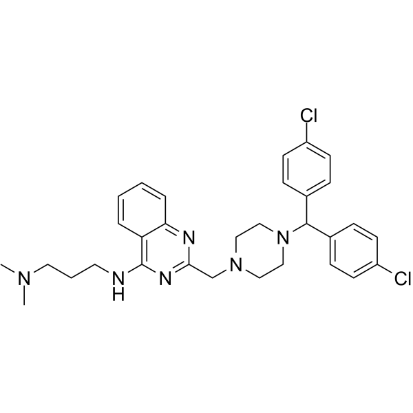
HY-110088
SCH529074P53 Activator -
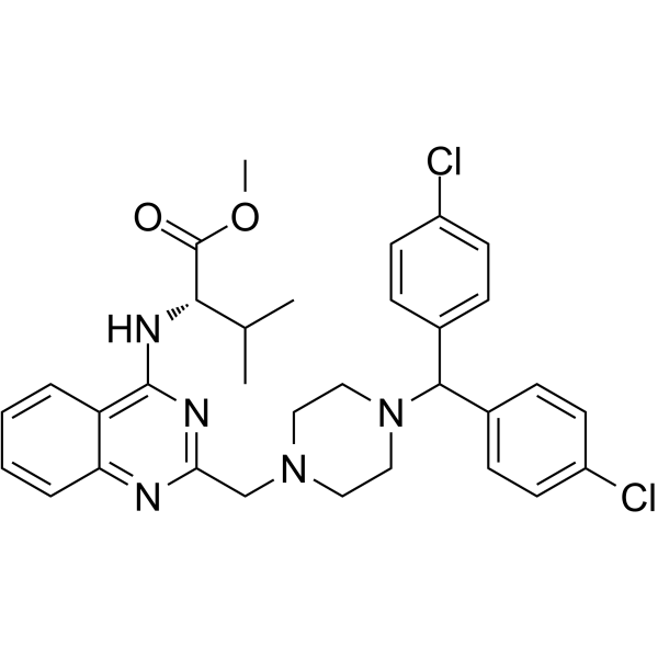
HY-122578
P53R3p53 Reactivator -
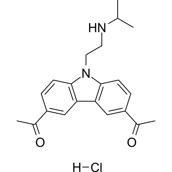
HY-18935A
CBL0137 hydrochlorideNF-κB Inhibitor -
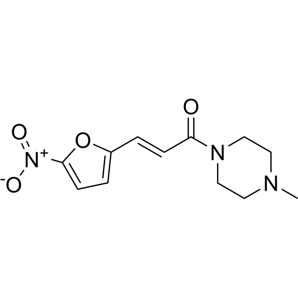
HY-19726
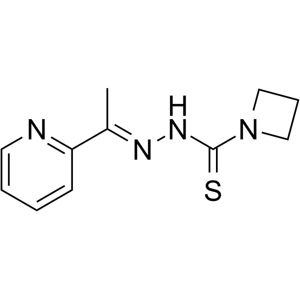
NSC59984MDM-2/p53 p53 Activator -
HY-18634
NSC319726MDM-2/p53 p53 Activator -
HY-137864
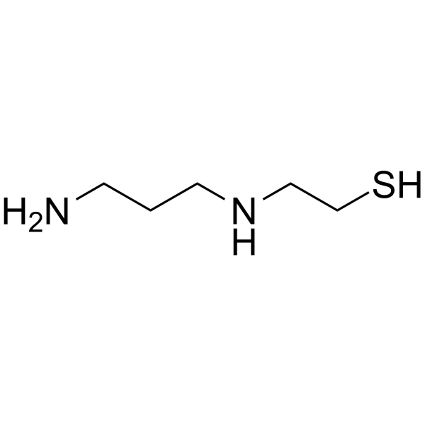
Amifostine thiolRadioprotector/p53 Activator -
HY-156967
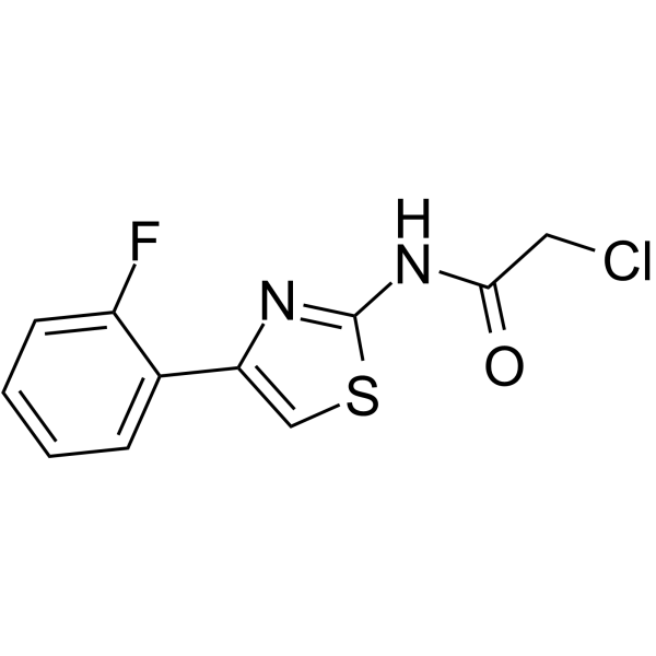
BAY 1892005Modulator -
HY-P4210
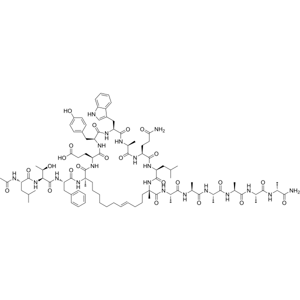
Sulanemadlinp53-MDM2/MDMX Inhibitor -
HY-108637
PhiKan 083MDM-2/p53 p53 Activator


生物活性
製品説明


化学情報
-
CAS 番号 1643469-17-3
-
性状 Solid
-
分子量 318.37
-
分子式 C20H18N2O2
-
Color White to off-white
-
SMILES
O=C1N2[C@@](OC[C@@H]2CC3=CNC4=C3C=CC=C4)(C)C5=C1C=CC=C5
-
輸送条件
Room temperature in continental US; may vary elsewhere.
-
保管条件
Powder -20°C 3 years 4°C 2 years In solvent -80°C 6 months -20°C 1 month


溶剤 & 溶解度
体外:
DMSO : 250 mg/mL (785.25 mM; ultrasonic and warming and heat to 80°C; Hygroscopic DMSO has a significant impact on the solubility of product, please use newly opened DMSO)
Please refer to the solubility information to select the appropriate solvent. Once prepared, please aliquot and store the solution to prevent product inactivation from repeated freeze-thaw cycles.
Storage method and period of stock solution: -80°C, 6 months; -20°C, 1 month. When stored at -80°C, please use it within 6 months. When stored at -20°C, please use it within 1 month.
Please refer to the solubility information to select the appropriate solvent. Once prepared, please aliquot and store the solution to prevent product inactivation from repeated freeze-thaw cycles.
Storage method and period of stock solution: -80°C, 6 months; -20°C, 1 month. When stored at -80°C, please use it within 6 months. When stored at -20°C, please use it within 1 month.
濃度 (開始) × 体積 (開始) = 濃度 (終了) × 体積 (終了)

プロトコル
-
RNA extraction experimental
By lysing cells, releasing RNA, and removing impurities such as proteins and DNA, high-purity RNA products are finally obtained. The commonly used traditional method is the guanidine isothiocyanate/phenol/chloroform method (Trizol), which is suitable for a variety of animal materials including animal tissues, microorganisms, cultured cells, etc., and most plant materials.
-
Human pluripotent stem cell endothelial-cell differentiation
Human pluripotent stem cell endothelial differentiation is based on stepwise developmental patterning: early activation of WNT/GSK3β inhibition promotes mesodermal or vascular progenitor entry, followed by endothelial specification using VEGF-related signaling, BMP4, FGF2, Notch modulation, or cAMP depending on the published protocol. Endothelial differentiation is read out by acquisition of CD31, CD34, VE-cadherin/CD144, KDR/VEGFR2, vWF, Tie2, NOS3, acetylated LDL uptake, tube/network formation, barrier function, and in vivo vessel-forming capacity where tested.
-
Vascular/Branching Fractal Analysis
Vascular/branching fractal analysis quantifies the geometric complexity of vessel trees or vascular networks from segmented 2D images, commonly by converting vessels into binary and/or skeletonized maps and estimating fractal dimension using box-counting or related approaches. Fractal dimension is interpreted as an image-derived readout of vascular branching complexity, space filling, or density, and has been applied to retinal photographs, fluorescein angiography, OCT angiography, capillary perfusion maps, and in vitro Matrigel angiogenesis networks. The assay readout is generated from vessel-positive pixels after image preprocessing, vessel segmentation, binarization, and optional skeletonization; reported outputs include fractal dimension, vessel density, branchpoint density, endpoint density, vessel length density, tortuosity, and generation-based branching metrics when VESGEN-style analysis is used. The biological interpretation is limited to quantitative vascular patterning and s
-
Endothelial Tube Formation Assay
Endothelial tube formation assay evaluates the ability of endothelial cells to attach, migrate, align, and organize into capillary-like networks when cultured on gelled basement membrane extract or Matrigel; the readout is the morphology and quantity of tube-like networks, which reflects an in vitro endothelial morphogenesis step related to angiogenesis. Basement membrane extract/Matrigel provides laminin-rich extracellular matrix cues that support endothelial differentiation into capillary-like structures, but it can contain biologically active growth factors, so growth-factor-reduced matrix is preferred when testing defined angiogenic stimulators or inhibitors.
-
Subcutaneous Cell-Line-Derived Xenograft
Subcutaneous cell-line-derived xenograft (CDX) models are established by implanting cultured human cancer cell lines into immunodeficient mice, where the injected cells form localized tumors that can be monitored in vivo as a measure of tumorigenic potential, growth kinetics, and treatment response. These models are widely used in oncology research because they allow reproducible tumor formation and enable comparative assessment of tumor growth between different cell lines or genetic manipulations in a controlled in vivo microenvironment. Subcutaneous implantation of cancer cells in immunodeficient mice is a standard approach for evaluating tumor growth behavior and therapeutic response across multiple cancer types, including prostate, esophageal, pancreatic, and colon cancer models.
-
Endothelial Cell Migration/Angiogenic Sprouting Assay
Endothelial cell migration and angiogenic sprouting assays are in vitro (and partially ex vivo-adapted) functional models that quantify the ability of endothelial cells to undergo coordinated migration, extracellular matrix invasion, and multicellular organization into capillary-like sprouts in response to pro-angiogenic stimuli such as VEGF, bFGF, or conditioned microenvironments. These assays are used to model early angiogenic events including tip-cell formation, directional migration, and lumen-like sprout extension, which collectively reflect angiogenic activation and vascular morphogenesis processes observed in vivo.


純度とドキュメンテーション
-
データシート (275 KB)
-
SDS (251 KB)
- English - EN (251 KB)
- Français - FR (251 KB)
- Deutsch - DE (251 KB)
- Norwegian - NO (251 KB)
- Español - ES (251 KB)
- Swedish - SV (251 KB)
- Italian - IT (251 KB)
- Korean - KR (251 KB)
- Portuguese - PT (251 KB)
-
取扱説明書 (2659 KB)
参考文献
[1]. Gomes S, et al. SLMP53-2 Restores Wild-Type-Like Function to Mutant p53 through Hsp70: Promising Activity in Hepatocellular Carcinoma. Cancers (Basel). 2019 Aug 10;11(8):1151. [Content Brief]
[2]. Ramos H, et al. SLMP53-1 Inhibits Tumor Cell Growth through Regulation of Glucose Metabolism and Angiogenesis in a P53-Dependent Manner. Int J Mol Sci. 2020 Jan 17;21(2):596. [Content Brief]


Complete Stock Solution Preparation Table
Please refer to the solubility information to select the appropriate solvent. Once prepared, please aliquot and store the solution to prevent product inactivation from repeated freeze-thaw cycles.
Storage method and period of stock solution: -80°C, 6 months; -20°C, 1 month. When stored at -80°C, please use it within 6 months. When stored at -20°C, please use it within 1 month.
| Optional Solvent | Concentration Solvent Mass | 1 mg | 5 mg | 10 mg | 25 mg |
|---|---|---|---|---|---|
| DMSO | 1 mM | 3.1410 mL | 15.7050 mL | 31.4100 mL | 78.5250 mL |
| 5 mM | 0.6282 mL | 3.1410 mL | 6.2820 mL | 15.7050 mL | |
| 10 mM | 0.3141 mL | 1.5705 mL | 3.1410 mL | 7.8525 mL | |
| 15 mM | 0.2094 mL | 1.0470 mL | 2.0940 mL | 5.2350 mL | |
| 20 mM | 0.1570 mL | 0.7852 mL | 1.5705 mL | 3.9262 mL | |
| 25 mM | 0.1256 mL | 0.6282 mL | 1.2564 mL | 3.1410 mL | |
| 30 mM | 0.1047 mL | 0.5235 mL | 1.0470 mL | 2.6175 mL | |
| 40 mM | 0.0785 mL | 0.3926 mL | 0.7852 mL | 1.9631 mL | |
| 50 mM | 0.0628 mL | 0.3141 mL | 0.6282 mL | 1.5705 mL | |
| 60 mM | 0.0523 mL | 0.2617 mL | 0.5235 mL | 1.3087 mL | |
| 80 mM | 0.0393 mL | 0.1963 mL | 0.3926 mL | 0.9816 mL | |
| 100 mM | 0.0314 mL | 0.1570 mL | 0.3141 mL | 0.7852 mL |


SLMP53-1 関連分類
HY-122753 関連分類
 Powered by Bioz