



Macrocarpal H

Based on 1 Customer Validation
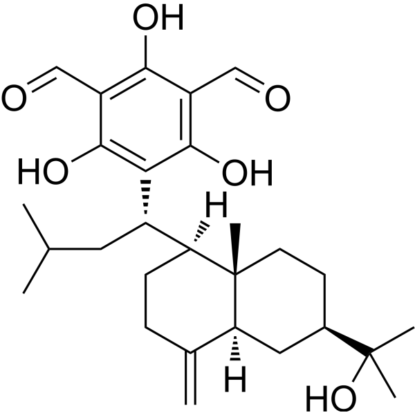
Macrocarpal H is a natural compound that can be isolated from the Leaves of Eucalyptus globulus. Macrocarpal H exhibits antibacterial activity against Gram-positive cariogenic bacteria and Gram-negative periodontopathic bacteria. Macrocarpal H inhibits adherent water-insoluble glucan synthesis via glucosyltransferase. Macrocarpal H can be used for the research of caries, periodontal disease, dental caries, periodontal disorders.
연구목적의 판매만을 진행합니다. 환자를 대상으로 한 판매는 하지 않습니다.
- Purity : 73.0%
- CAS No.: 179388-53-5
- 화학식: C28H40O6
- 분자량:472.61
-
보관:
-20°C, sealed storage, away from moisture
* In solvent : -80°C, 6 months; -20°C, 1 month (sealed storage, away from moisture)

To place orders, for customer services and technical support, please contact: MedChemExpress USA
Tel: 609-228-6898 E-mail: [email protected] [email protected]


-
 Biological Activity
-
Chemical Information
- Protocol
- 순도&문서
- References
-
 Help & FAQs




Biological Activity
제품 설명
In Vitro
Macrocarpal H (compound 7) (24-48 h) potently inhibits the growth of cariogenic bacteria including S. mutans Ingbritt, S. mutans LA7, S. sobrinus 6715, S. sobrinus B13, and A. viscosus 15987, and periodontopathic bacteria including P. gingivalis 33277, P. melaninogenica 25845, P. intermedia 25611, and C. ochracea 33596, with MIC values ranging from 0.39 to 3.13 µg/mL, and shows weaker activity against F. nucleatum 25586 with an MIC of 6.25 µg/mL[2].
Macrocarpal H (1-100 µg/mL; 16 h) strongly inhibits glucosyltransferase activity from S. sobrinus 6715, reducing adherent water-insoluble glucan synthesis by 97.6% at 100 µg/mL and 64.0% at 10 µg/mL[2].
MedChemExpress (MCE) has not independently confirmed the accuracy of these methods. They are for reference only.


Chemical Information
-
CAS No. 179388-53-5
-
Appearance Solid
-
분자량 472.61
-
화학식 C28H40O6
-
Color Light yellow to yellow
-
SMILES
CC(C)C[C@H](C1=C(C(C=O)=C(O)C(C=O)=C1O)O)[C@@]2([H])[C@]3([C@@](C(CC2)=C)([H])C[C@H](C(C)(O)C)CC3)C
-
선적
Room temperature in continental US; may vary elsewhere.
-
보관
-20°C, sealed storage, away from moisture
* In solvent : -80°C, 6 months; -20°C, 1 month (sealed storage, away from moisture)


순도&문서
-
Data Sheet (277 KB)
-
SDS (252 KB)
- English - EN (252 KB)
- Français - FR (252 KB)
- Deutsch - DE (252 KB)
- Norwegian - NO (252 KB)
- Español - ES (252 KB)
- Swedish - SV (252 KB)
- Italian - IT (252 KB)
- Korean - KR (252 KB)
- Portuguese - PT (252 KB)
-
Handling Instructions (2659 KB)
References

Calculators
Concentration (start) × Volume (start) = Concentration (final) × Volume (final)


Macrocarpal H Related Classifications
HY-N3341 Related Classifications
 Powered by Bioz
Keywords
- Macrocarpal H
- 179388-53-5
- Glucosylceramide Synthase (GCS)
- Bacterial
- periodontal disease
- Gram-negative periodontopathic bacteria
- Gram-positive cariogenic bacteria
- glucosyltransferase
- periodontopathic bacteria
- caries
- S. mutans Ingbritt
- periodontal disorders
- cariogenic bacteria
- dental caries
- Inhibitor
- inhibitor
- inhibit