



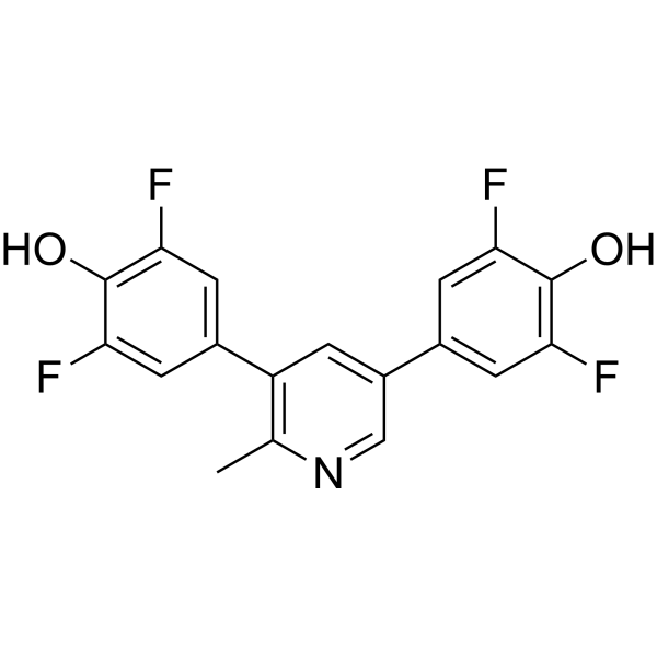
VRK-IN-1
Based on 6 publication(s) in Google Scholar
VRK-IN-1 is a potent and selective inhibitor of vaccinia-related kinases 1 (VRK1), with an IC50 of 150 nM. VRK1 is human Ser/Thr protein kinases associated with increased cell division and neurological disorders.
연구목적의 판매만을 진행합니다. 환자를 대상으로 한 판매는 하지 않습니다.
- Purity : 99.47%
- CAS No.: 2378855-09-3
- 화학식: C18H11F4NO2
- 분자량:349.28
-
보관:Powder -20°C, 3 years , 4°C, 2 years ; In solvent -80°C, 2 years , -20°C, 1 year

To place orders, for customer services and technical support, please contact: MedChemExpress USA
Tel: 609-228-6898 E-mail: [email protected] [email protected]


-
 Biological Activity
-
Chemical Information
-
용액&용해도
- Protocol
- 순도&문서
- References
-
 Help & FAQs


-
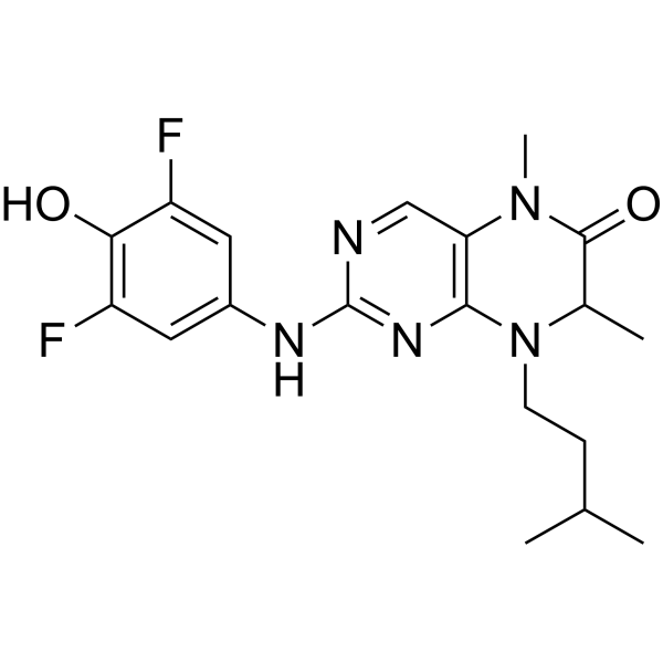
HY-10510
BI-D1870RSK Inhibitor -
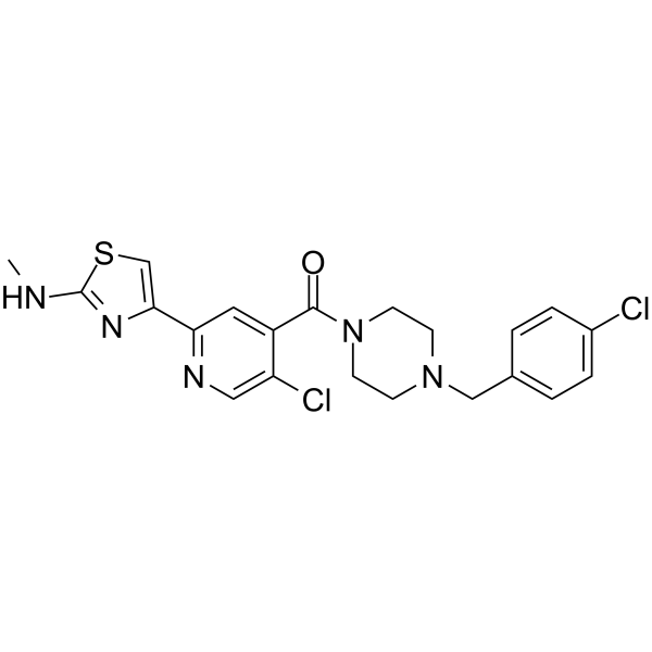
HY-112094
WNK-IN-11WNK Inhibitor -

HY-15141
StaurosporinePKC/PKA Inhibitor -
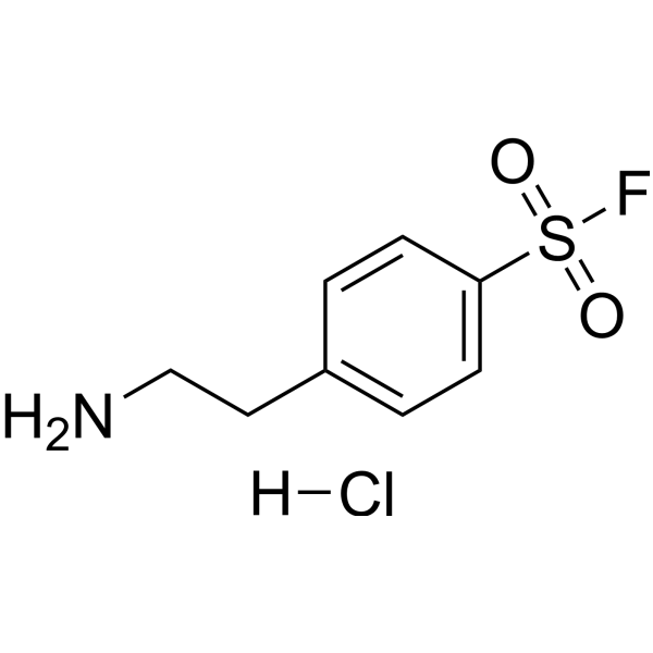
HY-12821
AEBSF hydrochlorideThrombin Inhibitor -

HY-158975
VRK1/CK1-IN-1VRK1/CK1 Family Inhibitor -
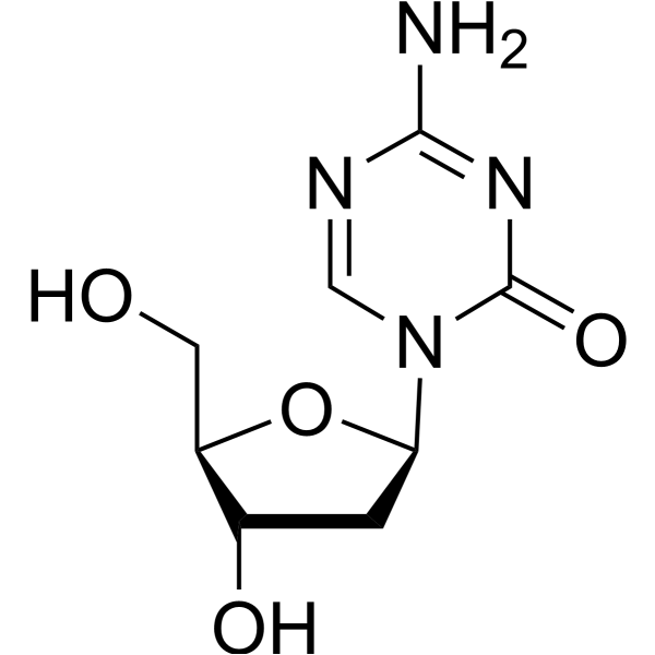
HY-A0004
DecitabineDNMT Inhibitor -
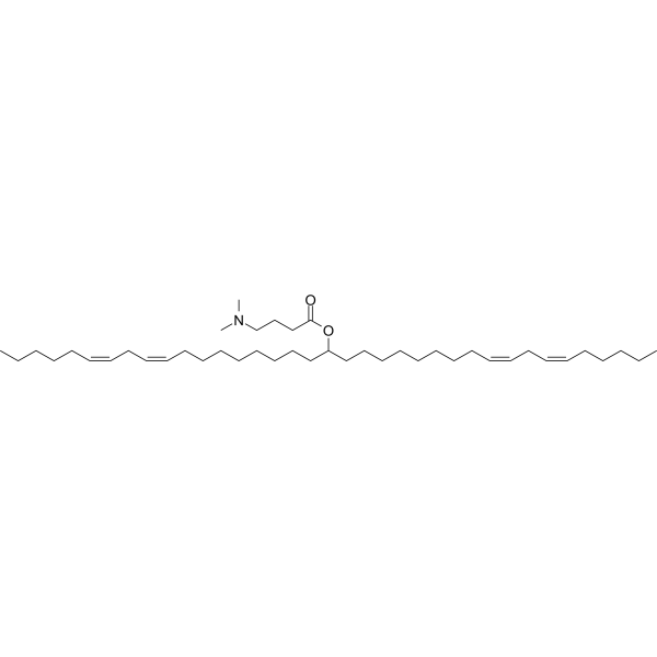
HY-112251
D-Lin-MC3-DMAsiRNA Delivery Vehicle -
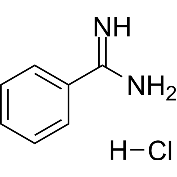
HY-W018781
Benzamidine hydrochlorideSerine Proteases Inhibitor -
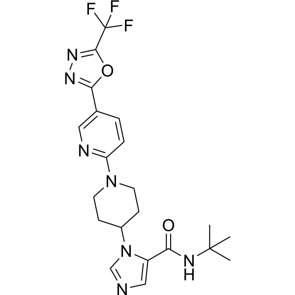
HY-100626
WNK463WNK Inhibitor -

HY-10227
BortezomibProteasome Inhibitor -

HY-13259
MG-132Proteasome Inhibitor -

HY-10431
SB-431542TGF-β Receptor Kinase Inhibitor
Publications Citing Use of MedChemExpress (MCE) VRK-IN-1
More Customer Validation & Images

-
WB

-
Cell Imaging/Staining

-
IF

-
WB

-
IF


Biological Activity
제품 설명
IC50 & Target
[1]|
VRK1 150 nM (IC50) |
In Vitro
VRK-IN-1 binds to VRK1-FL by enthalpy-driven, with a Kd of 190 nM[1].
VRK-IN-1 (0.4-25 μM; 24 h) slightly decreases the viability of human HeLa cells at the concentration of 3.2 μM[1].
MedChemExpress (MCE) has not independently confirmed the accuracy of these methods. They are for reference only. Further protocols information, click here.


Chemical Information
-
CAS No. 2378855-09-3
-
Appearance Solid
-
분자량 349.28
-
화학식 C18H11F4NO2
-
Color White to off-white
-
SMILES
FC1=C(O)C(F)=CC(C2=C(C)N=CC(C3=CC(F)=C(O)C(F)=C3)=C2)=C1
-
선적
Room temperature in continental US; may vary elsewhere.
-
보관
Powder -20°C 3 years 4°C 2 years In solvent -80°C 2 years -20°C 1 year


Publications (6)
-
Journal Impact Factor
-
Most Recent
-
EMBO J
2024 Oct;43(20):4752-4785. PMID: 39256562 -
Int J Mol Sci
VRK1 Kinase Activity Modulating Histone H4K16 Acetylation Inhibited by SIRT2 and VRK-IN-1. [Abstract]2023 Mar 3;24(5):4912. PMID: 36902348
VRK-IN-1 purchased from MedChemExpress. Usage Cited in: Int J Mol Sci. 2023 Mar 3;24(5):4912. [Abstract]
GST-VRK1 and human histone H3 were incubated with increasing concentrations of the VRK-IN-1 inhibitor (250 nM–1 µM) for 2 h at 37 °C. Then, an in vitro kinase assay was carried out and phospho-specific signal were identified by WB with H3T3Ph antibody. As a negative control, a kinase-dead VRK1 (K179E) was used.
VRK-IN-1 purchased from MedChemExpress. Usage Cited in: Int J Mol Sci. 2023 Mar 3;24(5):4912. [Abstract]
VRK-IN-1 treatment induces an accumulation of 3′-OH DNA ends resulting from DNA strand-breaks. A549 cells were treated with 600 nM VRK-IN-1 for 24 h and 3 µM doxorubicin for 2 h. The available free 3′-DNA ends were detected by labeling with terminal deoxynucleotidyl transferase (TdT) in TUNEL assays.
VRK-IN-1 purchased from MedChemExpress. Usage Cited in: Int J Mol Sci. 2023 Mar 3;24(5):4912. [Abstract]
VRK1 was knocked down using a specific siRNA (siVRK1-02). A549 cells were deprived of serum for 48 h and incubated with different concentrations of VRK-IN-1 as indicated for 24 h. Image panels showing the levels of H4K16 acetylation after VRK1 depletion or VRK-IN-1 treatment. The effect was detected by immunofluorescence with a specific antibody anti-H4K16ac. The VRK-IN-1 inhibitor was used at the indicated doses. The box indicates the individual cell shown to the right for detail.
-
Epigenetics Chromatin
The pattern of histone H3 epigenetic posttranslational modifications is regulated by the VRK1 chromatin kinase. [Abstract]2023 May 13;16(1):18. PMID: 37179361
VRK-IN-1 purchased from MedChemExpress. Usage Cited in: Epigenetics Chromatin. 2023 May 13;16(1):18. [Abstract]
Effect of VRK1 depletion or inhibition with VRK-IN-1 on epigenetic modifications of H3K4 in A549 cells the presence or absence of serum. Effect of VRK-IN-1 (600 nM; 24 h) on histone H3K4m3 levels.
VRK-IN-1 purchased from MedChemExpress. Usage Cited in: Epigenetics Chromatin. 2023 May 13;16(1):18. [Abstract]
Effect of VRK1 depletion or inhibition with VRK-IN-1 on epigenetic modifications of H3K9 in A549 cells the presence or absence of serum. Effect of VRK-IN-1 (600 nM; 24 h) on histone H3K9ac and histone H3K9m3 levels.
-
Exp Cell Res
Inactivation of VRK1 sensitizes ovarian cancer to PARP inhibition through regulating DNA-PK stability. [Abstract]2024 Apr 16;438(1):114036. PMID: 38614421 -
Mol Biol Rep
2024 Mar 27;51(1):453. PMID: 38536553 -


용액&용해도
In Vitro:
DMSO : 100 mg/mL (286.30 mM; Need ultrasonic; Hygroscopic DMSO has a significant impact on the solubility of product, please use newly opened DMSO)
Please refer to the solubility information to select the appropriate solvent. Once prepared, please aliquot and store the solution to prevent product inactivation from repeated freeze-thaw cycles.
Storage method and period of stock solution: -80°C, 2 years; -20°C, 1 year. When stored at -80°C, please use it within 2 years. When stored at -20°C, please use it within 1 year.
Please refer to the solubility information to select the appropriate solvent. Once prepared, please aliquot and store the solution to prevent product inactivation from repeated freeze-thaw cycles.
Storage method and period of stock solution: -80°C, 2 years; -20°C, 1 year. When stored at -80°C, please use it within 2 years. When stored at -20°C, please use it within 1 year.
Concentration (start) × Volume (start) = Concentration (final) × Volume (final)
In Vivo:
Select the appropriate dissolution method based on your experimental animal and administration route.
- For the following dissolution methods, please ensure to first prepare a clear stock solution using an In Vitro approach and then sequentially add co-solvents:
- To ensure reliable experimental results, the clarified stock solution can be appropriately stored based on storage conditions. As for the working solution for In Vivo experiments, it is recommended to prepare freshly and use it on the same day.
- The percentages shown for the solvents indicate their volumetric ratio in the final prepared solution. If precipitation or phase separation occurs during preparation, heat and/or sonication can be used to aid dissolution.
In Vivo Dissolution Calculator
Please enter the basic information of animal experiments:
-
-
-
-
Recommended: Prepare an additional quantity of animals to account for potential losses during experiments.
Please enter your animal formula composition:
-
%DMSO +
Recommended: Keep the proportion of DMSO in working solution below 2% if your animal is weak.
-
%+
-
+%Tween-80 + +
-
%Saline +
The co-solvents required include: DMSO, . All of co-solvents are available by MedChemExpress (MCE). , Tween 80. All of co-solvents are available by MedChemExpress (MCE).
Working solution concentration: 0.22 mg/mL
Method for preparing stock solution: mg drug dissolved in μL DMSO. Stock solution concentration: mg/mL.
1. Take μL DMSO stock solution;
2. Add μL .
μL , mix evenly;
3. Then add μL Tween 80, mix evenly;
4. Then add μL
Please ensure that the stock solution in the first step is dissolved to a clear state, and add co-solvents in sequence. You can use ultrasonic heating (ultrasonic cleaner, recommended frequency 20-40 kHz), vortexing, etc. to assist dissolution.

Protocol
-
Cell Cytotoxicity Assay
Cytotoxicity assays are usually based on the assessment of cell membrane damage, which can also be indirectly detected by measuring cell viability. Detection methods include MTT assay, CKK-8 assay, LDH assay and ATP assay, etc.
-
CFSE Dye Dilution Proliferation Assay
The CFSE (carboxyfluorescein diacetate succinimidyl ester) dye dilution proliferation assay is based on the covalent labeling of intracellular proteins by a cell-permeant fluorescent dye that becomes fluorescent upon intracellular ester cleavage and then is stably retained within cells. As labeled cells divide, the dye is partitioned equally between daughter cells, resulting in a stepwise halving of fluorescence intensity that can be quantified by flow cytometry to determine the number of cell divisions undergone by each cell population. This fluorescence dilution approach enables quantitative tracking of lymphocyte proliferation at the single-cell level over multiple rounds of division. CFSE-based proliferation analysis has been widely applied to measure antigen-driven lymphocyte expansion in vitro, where discrete fluorescence peaks correspond to successive cell divisions and allow reconstruction of proliferative history within heterogeneous populations.
-
Dye-dilution cell tracking and proliferation staining
Dye-dilution cell tracking assays quantify cell proliferation by covalently labeling intracellular proteins with a stable fluorescent dye that is equally partitioned between daughter cells during mitosis, resulting in stepwise halving of fluorescence intensity with each cell division as measured by flow cytometry histograms. Carboxyfluorescein diacetate succinimidyl ester (CFSE) is a prototypical dye that diffuses into cells, is enzymatically converted into a fluorescent compound, and then covalently binds intracellular amine groups, producing long-lived fluorescence suitable for tracking multiple rounds of division in vitro and in vivo. Successive generations of dividing cells form discrete peaks of decreasing fluorescence intensity, enabling estimation of proliferation history, precursor frequency, and division index within heterogeneous populations. Alternative dyes such as CellTrace Violet (CTV) and far-red membrane dyes (e. g. , PKH26) follow the same dilution principle but differ


순도&문서
-
Data Sheet (272 KB)
-
SDS (252 KB)
- English - EN (252 KB)
- Français - FR (252 KB)
- Deutsch - DE (252 KB)
- Norwegian - NO (252 KB)
- Español - ES (252 KB)
- Swedish - SV (252 KB)
- Italian - IT (252 KB)
- Korean - KR (252 KB)
- Portuguese - PT (252 KB)
-
Handling Instructions (2659 KB)
References


Complete Stock Solution Preparation Table
Please refer to the solubility information to select the appropriate solvent. Once prepared, please aliquot and store the solution to prevent product inactivation from repeated freeze-thaw cycles.
Storage method and period of stock solution: -80°C, 2 years; -20°C, 1 year. When stored at -80°C, please use it within 2 years. When stored at -20°C, please use it within 1 year.
| Optional Solvent | Concentration Solvent Mass | 1 mg | 5 mg | 10 mg | 25 mg |
|---|---|---|---|---|---|
| DMSO | 1 mM | 2.8630 mL | 14.3152 mL | 28.6303 mL | 71.5758 mL |
| 5 mM | 0.5726 mL | 2.8630 mL | 5.7261 mL | 14.3152 mL | |
| 10 mM | 0.2863 mL | 1.4315 mL | 2.8630 mL | 7.1576 mL | |
| 15 mM | 0.1909 mL | 0.9543 mL | 1.9087 mL | 4.7717 mL | |
| 20 mM | 0.1432 mL | 0.7158 mL | 1.4315 mL | 3.5788 mL | |
| 25 mM | 0.1145 mL | 0.5726 mL | 1.1452 mL | 2.8630 mL | |
| 30 mM | 0.0954 mL | 0.4772 mL | 0.9543 mL | 2.3859 mL | |
| 40 mM | 0.0716 mL | 0.3579 mL | 0.7158 mL | 1.7894 mL | |
| 50 mM | 0.0573 mL | 0.2863 mL | 0.5726 mL | 1.4315 mL | |
| 60 mM | 0.0477 mL | 0.2386 mL | 0.4772 mL | 1.1929 mL | |
| 80 mM | 0.0358 mL | 0.1789 mL | 0.3579 mL | 0.8947 mL | |
| 100 mM | 0.0286 mL | 0.1432 mL | 0.2863 mL | 0.7158 mL |


VRK-IN-1 Related Classifications
HY-126542 Related Classifications
 Powered by Bioz