



Licoflavone C
Based on 1 publication(s) in Google Scholar
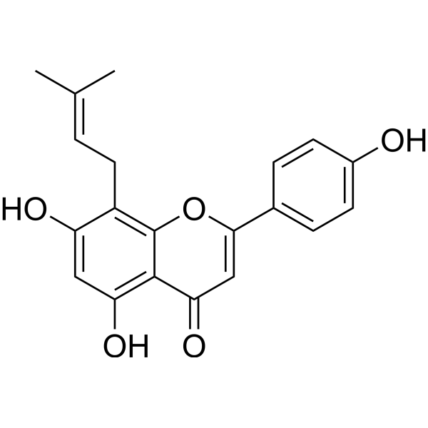
Licoflavone C is a broad-spectrum antiviral inhibitor with estrogen-like properties. Licoflavone C binds to viral endonuclease (CEN) and inhibits the replication of various bunyaviruses including severe fever with thrombocytopenia syndrome virus (SFTSV) and lymphocytic choriomeningitis virus in a non-substrate competitive manner. The IC50 values of Licoflavone C against SFTSV CEN and SFTSV CEN are 35.5 μM and 135.8 μM, respectively, and its Kd value against SFTSV CEN is 9.53 μM. After viral entry into cells, Licoflavone C reduces viral loads in mouse tissues in a dose-dependent manner, and exhibits extremely low cytotoxicity and genotoxicity. Licoflavone C induces apoptosis by increasing caspase 3/7 activity, blocks the cell cycle, and alleviates chemotherapy-induced chromosomal damage. Licoflavone C is applicable to the research on severe fever with thrombocytopenia syndrome and related viral infection mechanisms.
Nos produits utilisent uniquement pour la recherche. Nous ne vendons pas aux patients.
- Pureté : 99.83%
- CAS No.: 72357-31-4
- Formule: C20H18O5
- Masse moléculaire:338.35
-
Stockage:
4°C, protect from light
* In solvent : -80°C, 6 months; -20°C, 1 month (protect from light)

To place orders, for customer services and technical support, please contact: MedChemExpress USA
Tel: 609-228-6898 E-mail: [email protected] [email protected]


-
 Activité biologique
-
Chemical Information
-
Solvant et solubilité
- Protocole
- Pureté et documentation
- Références
-
 Help & FAQs




Publications Citing Use of MedChemExpress (MCE) Licoflavone C
MoreVoir tous les produits spécifiques à Isoform Caspase
More

Activité biologique
Description
IC50 & Target
|
Caspase 3 |
Caspase-7 |
Cellular Effect
|
Cell Line

|
Type | Value | Description | References |
|---|---|---|---|---|
| COS-7 | IC50 |
1.29 μM
Compound: 8-PA
|
Inhibition of human recombinant PDE5A1 expressed in COS7 cells
Inhibition of human recombinant PDE5A1 expressed in COS7 cells
|
[PMID: 18778098] |
| HeLa | IC50 |
>40 μM
Compound: 2
|
Cytotoxicity against human HeLa cells assessed as inhibition of cell proliferation by MTT assay
Cytotoxicity against human HeLa cells assessed as inhibition of cell proliferation by MTT assay
|
[PMID: 33609662] |
| HepG2 | IC50 |
>40 μM
Compound: 2
|
Cytotoxicity against human HepG2 cells assessed as inhibition of cell proliferation by MTT assay
Cytotoxicity against human HepG2 cells assessed as inhibition of cell proliferation by MTT assay
|
[PMID: 33609662] |
| MCF7 | IC50 |
>40 μM
Compound: 2
|
Cytotoxicity against human MCF7 cells assessed as inhibition of cell proliferation by MTT assay
Cytotoxicity against human MCF7 cells assessed as inhibition of cell proliferation by MTT assay
|
[PMID: 33609662] |
| NCI-H460 | IC50 |
>40 μM
Compound: 2
|
Cytotoxicity against human NCI-H460 cells assessed as inhibition of cell proliferation by MTT assay
Cytotoxicity against human NCI-H460 cells assessed as inhibition of cell proliferation by MTT assay
|
[PMID: 33609662] |
| RAW264.7 | IC50 |
3.83 μM
Compound: 43
|
Antiinflammatory activity in mouse RAW264.7 cells assessed as inhibition of LPS-induced NO production after 24 hrs by Griess reagent based assay
Antiinflammatory activity in mouse RAW264.7 cells assessed as inhibition of LPS-induced NO production after 24 hrs by Griess reagent based assay
|
[PMID: 28522265] |
In Vitro
Licoflavone C (50 μM; 0.5 h) potently inhibits the enzymatic activity of purified SFTSV CEN protein in vitro[1].
Licoflavone C (15 μM) effectively inhibits the proliferation of SFTSV in Vero cells at the post-entry stage, with the strongest activity when administered within 12 h post-infection. Its EC50 value is 1.90 μM for post-infection administration and 1.84 μM for full-course administration[1].
Licoflavone C (incubated with SFTSV CEN for 30 min; 50-200 μM) inhibits SFTSV CEN-mediated RNA cleavage in a concentration- and time-dependent manner in vitro, with an IC50 of 35.5 μM[1].
Licoflavone C potently inhibits infections by HRTV, GTV, and LCMV in vitro, with EC50 values of 9.21 μM, 4.21 μM, and 2.61 μM, respectively[1].
Licoflavone C (0.1-300 μM; 48 h) does not induce chromosome damage in cultured human peripheral blood lymphocytes even at concentrations up to 300 μM, but it triggers dose-dependent cell cycle disturbances starting from 10 μM and exhibits strong toxicity at 600 μM[2].
Licoflavone C (0.1-1.0 μM; 48 h) significantly reduces the chromosomal damage induced by Daunorubicin (HY-13062A) and Mitomycin C (HY-13316) in cultured human peripheral blood lymphocytes in vitro, but fails to ameliorate the cell cycle progression impairment caused by the mutagens[2].
MedChemExpress (MCE) has not independently confirmed the accuracy of these methods. They are for reference only.
-
Cell Line:Vero cells infected with SFTSV
-
Concentration:Gradient dilutions
-
Incubation Time:1 h pre-incubation, then continuous treatment for 36 h total
-
Result:Exhibited an EC50 of 1.85 μM against SFTSV infection in Vero cells, with a CC50 >300.0 μM and a selectivity index (SI) >162.16.
-
Cell Line:Vero cells infected with HRTV or GTV; BHK-21 cells infected with LCMV
-
Concentration:Gradient dilutions
-
Incubation Time:1 h pre-incubation, then continuous treatment for 36 h total
-
Result:Exhibited EC50 values of 9.21 μM against HRTV in Vero cells, 4.21 μM against GTV in Vero cells, and 2.61 μM against LCMV in BHK-21 cells.
Parmacokinetics
In Vivo
Licoflavone C (5-20 mg/kg; intravenous injection; once every 12 hours; for 2 consecutive days) dose-dependently reduces the SFTSV viral load in the liver, spleen, kidney and blood of C57BL/6 J mice infected with SFTSV[1].
MedChemExpress (MCE) has not independently confirmed the accuracy of these methods. They are for reference only.
-
Animal Model:C57BL/6 J (female, 6-8 weeks of age, challenged with SFTSV via intraperitoneal injection)[1]
-
Dosage:20 mg/kg
-
Administration:i.v.; once daily; 3 consecutive days
-
Result:Reduced SFTSV viral loads in the liver, spleen, and blood significantly compared to vehicle control.
Showed no significant reduction in kidney viral loads.
-
Animal Model:C57BL/6 J (female, 6-8 weeks of age, challenged with SFTSV via intraperitoneal injection)[1]
-
Dosage:5 mg/kg; 10 mg/kg; 20 mg/kg
-
Administration:i.v.; every 12 hours; 2 days
-
Result:Reduced SFTSV viral loads in the liver, spleen, kidney, and blood in a dose-dependent manner.
Reduced SFTSV viral loads across all four analyzed tissues significantly at 20 mg/kg.
Reduced SFTSV viral loads in the spleen, kidney, and blood significantly at 10 mg/kg.
Reduced SFTSV viral load in the spleen only significantly at 5 mg/kg.
Reduced SFTSV nucleoprotein fluorescence intensity in spleen tissue, consistent with viral load reductions.
- Research Protocol for Infectious Diseases
- Mammalian live/dead viability and cytotoxicity staining
- Annexin V plus membrane-impermeant dye apoptosis staining
- BrdU Incorporation Assay
- Protocol for Cell Cycle
- Cell Viability Determination by MTT Colorimetric Assay
- Detection of Apoptosis
- TUNEL staining for apoptotic DNA fragmentation
- Cell Cytotoxicity Assay
- Flow cytometric DNA-content cell-cycle staining
- Research Protocol for Endocrine Diseases
- Apoptosis Solutions
- Genotoxicity/Mutagenicity Study


Chemical Information
-
CAS No. 72357-31-4
-
Appearance Solid
-
Masse moléculaire 338.35
-
Formule C20H18O5
-
Color Light yellow to brown
-
SMILES
O=C1C=C(C2=CC=C(O)C=C2)OC3=C(C/C=C(C)\C)C(O)=CC(O)=C13
-
Structure Classification
-
Initial Source
-
Livraison
Room temperature in continental US; may vary elsewhere.
-
Stockage
4°C, protect from light
* In solvent : -80°C, 6 months; -20°C, 1 month (protect from light)


Publications (1)
-
Journal Impact Factor
-
Most Recent
-
Food Res Int
Glycyrrhiza uralensis Fisch: A novel source of analgesic activity through NaV1.8 sodium channel modulation. [Abstract]2025 Dec;222(Pt 1):117620. PMID: 41267240


Solvant et solubilité
In Vitro:
DMSO : 62.5 mg/mL (184.72 mM; Need ultrasonic; Hygroscopic DMSO has a significant impact on the solubility of product, please use newly opened DMSO)
Please refer to the solubility information to select the appropriate solvent. Once prepared, please aliquot and store the solution to prevent product inactivation from repeated freeze-thaw cycles.
Storage method and period of stock solution: -80°C, 6 months; -20°C, 1 month (protect from light). When stored at -80°C, please use it within 6 months. When stored at -20°C, please use it within 1 month.
Please refer to the solubility information to select the appropriate solvent. Once prepared, please aliquot and store the solution to prevent product inactivation from repeated freeze-thaw cycles.
Storage method and period of stock solution: -80°C, 6 months; -20°C, 1 month (protect from light). When stored at -80°C, please use it within 6 months. When stored at -20°C, please use it within 1 month.
Concentration (start) × Volume (start) = Concentration (final) × Volume (final)


Pureté et documentation
-
Fiche technique (280 KB)
-
SDS (394 KB)
- English - EN (394 KB)
- Français - FR (394 KB)
- Deutsch - DE (394 KB)
- Norwegian - NO (394 KB)
- Español - ES (394 KB)
- Swedish - SV (394 KB)
- Italian - IT (394 KB)
- Korean - KR (394 KB)
- Portuguese - PT (394 KB)
-
Instruction de manipulation (2659 KB)
Références
[1]. Gao X, et al. Identification of Licoflavone C as a cap-dependent endonuclease inhibitor against severe fever with thrombocytopenia syndrome virus. Acta Pharmacol Sin. 2025;46(9):2482-2495. [Content Brief]
[2]. Scarpato R, et al. Licoflavone C attenuates the genotoxicity of cancer drugs in human peripheral lymphocytes. Phytother Res. 2008;22(12):1650-1654. [Content Brief]


Complete Stock Solution Preparation Table
Please refer to the solubility information to select the appropriate solvent. Once prepared, please aliquot and store the solution to prevent product inactivation from repeated freeze-thaw cycles.
Storage method and period of stock solution: -80°C, 6 months; -20°C, 1 month (protect from light). When stored at -80°C, please use it within 6 months. When stored at -20°C, please use it within 1 month.
| Optional Solvent | Concentration Solvent Mass | 1 mg | 5 mg | 10 mg | 25 mg |
|---|---|---|---|---|---|
| DMSO | 1 mM | 2.9555 mL | 14.7776 mL | 29.5552 mL | 73.8880 mL |
| 5 mM | 0.5911 mL | 2.9555 mL | 5.9110 mL | 14.7776 mL | |
| 10 mM | 0.2956 mL | 1.4778 mL | 2.9555 mL | 7.3888 mL | |
| 15 mM | 0.1970 mL | 0.9852 mL | 1.9703 mL | 4.9259 mL | |
| 20 mM | 0.1478 mL | 0.7389 mL | 1.4778 mL | 3.6944 mL | |
| 25 mM | 0.1182 mL | 0.5911 mL | 1.1822 mL | 2.9555 mL | |
| 30 mM | 0.0985 mL | 0.4926 mL | 0.9852 mL | 2.4629 mL | |
| 40 mM | 0.0739 mL | 0.3694 mL | 0.7389 mL | 1.8472 mL | |
| 50 mM | 0.0591 mL | 0.2956 mL | 0.5911 mL | 1.4778 mL | |
| 60 mM | 0.0493 mL | 0.2463 mL | 0.4926 mL | 1.2315 mL | |
| 80 mM | 0.0369 mL | 0.1847 mL | 0.3694 mL | 0.9236 mL | |
| 100 mM | 0.0296 mL | 0.1478 mL | 0.2956 mL | 0.7389 mL |


Licoflavone C Related Classifications
HY-N4183 Related Classifications
 Powered by Bioz