



Common Questions and Solutions for WB
2024-05-09
Protein blotting or immunoblotting (Western blot, WB) is a common laboratory method used to detect proteins and assess their expression levels. The protein of interest is identified based on its molecular weight and immunoreactivity with specific antibodies. Protein blotting consists of a series of interconnected steps (Figure 1).
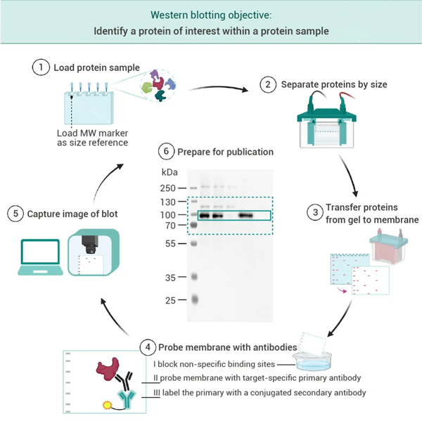
(2) Gel electrophoresis is used to separate proteins based on their molecular weights.
(3) Transfer or "blot" the proteins onto a membrane.
(4) Block the membrane to reduce non-specific binding, then probe with primary and secondary antibodies that specifically bind to the target protein in sequence. The latter binds to the primary antibody and carries an enzyme or fluorescent group that allows subsequent detection.
(5) Detect signals through chemiluminescence reaction or fluorescence respectively.
(6) Prepare protein blot images for publication.
Due to the long duration and intricate details of this experiment, from sample preparation to development, problems may arise at each step, resulting in bands that are often peculiar and unsatisfactory... In this article, we will introduce some common issues in WB experiments and their corresponding solutions[2].



01. Smiling Bands

• Reason: Too fast migration, high temperature of the electrophoresis buffer, protein overload, or insufficient running buffer inside the well.
• Suggestion: Reduce migration speed, pre-cool the buffer, decrease the amount of protein loaded, ensure the buffer fully covers the well and that buffer in the inner chamber does not leak[4][6].
02. Frowning Bands

• Reason: Improper equipment setup, which may be due to bubbles at the bottom of the gel and glass plates, or incomplete polymerization on both sides.
• Suggestion: This issue can be avoided by adjusting the setup.
03. Trailing Bands

• Reason: Poor sample dissolution; certain degree of protein degradation; repeated use of electrophoresis buffer.
• Suggestion: Load samples after they are fully dissolved and mixed; try to use fresh samples; use freshly prepared electrophoresis buffer.
04. Dumbbell-shaped Uneven Bands

• Reason: Issues with gel formulation, unevenness after the gel solidifies; sample may contain too many impurities.
• Suggestion: Reconfigure the gel and make sure there's no problem with its quality to avoid this phenomenon; centrifuge the sample before using.
05. Sticking Bands

• Reason: It could be due to excessive sample loading, or issues with gel preparation. There might be a gap between the separating gel and the stacking gel causing sample leakage.
• Suggestion: This can be avoided by reducing the sample loading amount and improving the quality of gel preparation.
06. Background Bubbles

• Reason: There are bubbles on the membrane during transfer.
• Suggestion: Ensure all bubbles between the gel and blotting membrane are removed when assembling the "sandwich".
07. Uneven Black Spots on the Background

• Reason: The blocking solution is not fully dissolved, causing insoluble particles to adhere to the membrane and form black spots when luminescence occurs; or uneven distribution of the antibody on the membrane.
• Suggestion: Ensure the blocking solution is fully dissolved. After blocking, wash three times with TBST before adding the primary antibody. Keep shaking during antibody incubation.
08. Blank Spots in Bands

• Reason: There's a blank spot in the middle of the target protein band, but the surrounding background is normal. This phenomenon may be due to excessively high concentrations of primary and secondary antibodies, which causes rapid substrate consumption, leading to the formation of blank spots when chemiluminescence is performed as the substrate is exhausted.
• Suggestion: You can reduce the amount of protein loaded; lower the concentration of primary and secondary antibodies; or dilute the developing solution.
Through this article, we believe that common problems are no longer a challenge for you! If the content above does not solve your problem, please feel free to leave a comment below. Little M welcomes everyone to actively discuss various challenging issues encountered in experiments~
| More Product Recommendations: |
| HY-P80993 One of the commonly used internal reference antibodies, it can detect endogenous levels of Beta Actin total protein. |
| HY-P80538 It can detect the endogenous levels of alpha-tubulin total protein and will not cross-react with recombinant beta-tubulin protein. |
| HY-P80626 This antibody can detect the endogenous levels of c-Myc total protein. |
| HY-P8004 Goat-derived anti-mouse IgG antibody, used for WB, ELISA, and IHC experiments under mouse backgrounds. |
| HY-P8001 Goat-derived anti-rabbit IgG antibody, used for WB, IHC-P, and ELISA experiments under rabbit backgrounds. |
| HY-P80447 The antibody mainly labels endothelial cells and is used to identify benign/malignant vascular tumors as well as the status of angiogenesis in tumor stroma. |
| HY-P80982 The antibody combined with CK7 is used for differential diagnosis of the primary site of metastatic cancer. |
[2] Cui Y. Anal Biochem. 2020 Mar 15;593:113598.
[3] Begum H, et al. Biotechniques. 2022 Jun;73(1):58-69.
[4] Mary Johnson,MATER METHODS 2013;3:195.
[5] Gilda JE, et al. PLoS One. 2015 Aug 19;10(8):e0135392.
[6] Sule R, et al. Biotechniques. 2023 Sep;75(3):99-114.
 LinkedIn
 Researchgate
 Instagram
 X
X Facebook
 WhatsApp
 Reddit
 Mail
Mail