



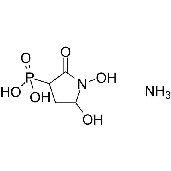
SF2312 ammonium

Based on 1 Customer Validation
SF2312 ammonium is a phosphonate Antibiotic and Enolase inhibitor, with an IC50 of 18.4 nM against E. coli enolase. SF2312 ammonium reduces intracellular ATP levels and inhibits glycolytic flux. SF2312 ammonium exhibits activity against both Gram-positive and Gram-negative bacteria, and exerts synergistic effects against E. coli when combined with Fosfomycin (HY-B1075A). SF2312 ammonium shows selective toxicity toward gliomas with ENO1 gene knockout. SF2312 ammonium can be used in research related to bacterial infections and gliomas.
For research use only. We do not sell to patients.
- Purity : 98.0%
- Formula: C4H11N2O6P
- Molecular Weight:214.11
-
Storage:
-20°C, protect from light, stored under nitrogen
* In solvent : -80°C, 6 months; -20°C, 1 month (protect from light, stored under nitrogen)

To place orders, for customer services and technical support, please contact: MedChemExpress USA
Tel: 609-228-6898 E-mail: [email protected] [email protected]


-
 Biological Activity
-
Chemical Information
-
Solvent & Solubility
- Protocols
- Purity & Documentation
- References
-
 Help & FAQs


All Antibiotic Isoforms
More

Biological Activity
Description
In Vitro
SF2312 (2 nM-20 μM; 5 min preincubation) ammonium potently inhibits purified recombinant E. coli enolase with an IC50 of 18.4 nM and a Ki of 3.4 nM[1].
SF2312 (0.2-300 μg/mL) ammonium exhibits antibacterial activity against E. coli and S. aureus, with improved potency (MICs of 100 μg/mL and 20 μg/mL, respectively) when cultures are supplemented with glucose-6-phosphate, while activity against P. aeruginosa and A. baumannii is not enhanced by glucose-6-phosphate[1].
SF2312 (0.05-100 μg/mL) ammonium acts synergistically with fosfomycin against E. coli ATCC 25922, reducing the MIC of SF2312 by up to 16-fold and yielding an FIC index ≤0.5[1].
SF2312 (low μM to >200 μM; 2 weeks) ammonium selectively inhibits the proliferation of ENO1-deleted D423 and Gli56 glioma cells, with potent activity in the low μM range under normoxia, and SF2312 (>6.25 μM; 72 hours) causes eradication of ENO1-deleted D423 cells under hypoxia, while ENO1-rescued cells are resistant until concentrations exceed 200 μM[3].
SF2312 (12.5-400 μM; ≥24 hours) ammonium selectively induces cell death in ENO1-deleted D423 glioma cells starting at 12.5 μM, whereas ENO1-rescued D423 cells only undergo cell death at 400 μM[3].
SF2312 (10 μM; 4 days) ammonium selectively inhibits glycolytic flux in ENO1-deleted D423 and Gli56 glioma cells, with 10 μM causing near-complete inhibition of glucose-to-lactate conversion after 4 days, while ENO1-rescued cells show no such inhibition[3].
MedChemExpress (MCE) has not independently confirmed the accuracy of these methods. They are for reference only. Further protocols information, click here.
-
Cell Line:ENO1-deleted D423 glioma cells, isogenic ENO1-rescued D423 glioma cells, isogenic ENO2-overexpressing D423 glioma cells, ENO1-deleted Gli56 glioma cells
-
Concentration:6.25-400 μM (hypoxia); low μM to >200 μM (normoxia)
-
Incubation Time:72 h (hypoxia); 2 weeks (normoxia)
-
Result:Inhibited proliferation of ENO1-deleted D423 glioma cells in the low μM range over 2 weeks, while isogenic ENO1-rescued D423 cells only showed proliferation inhibition at concentrations above 200 μM.
Isogenic ENO2-overexpressing D423 cells were more sensitive than ENO1-rescued cells but far more resistant than parental ENO1-deleted D423 cells.
Under hypoxia (72-hour treatment), ENO1-deleted D423 cells were essentially eradicated at SF2312 concentrations >6.25 μM, while normoxic treatment at 100 μM did not cause eradication over the same timeframe.
Showed selective proliferation inhibition against ENO1-deleted Gli56 glioma cells relative to ENO1-intact controls.


Chemical Information
-
Appearance Solid
-
Molecular Weight 214.11
-
Formula C4H11N2O6P
-
Color White to off-white
-
SMILES
O=C1N(C(CC1P(O)(O)=O)O)O.N
-
Shipping
Room temperature in continental US; may vary elsewhere.
-
Storage
-20°C, protect from light, stored under nitrogen
* In solvent : -80°C, 6 months; -20°C, 1 month (protect from light, stored under nitrogen)


Solvent & Solubility
In Vitro:
H2O : ≥ 30 mg/mL (140.11 mM)
* "≥" means soluble, but saturation unknown.
Please refer to the solubility information to select the appropriate solvent. Once prepared, please aliquot and store the solution to prevent product inactivation from repeated freeze-thaw cycles.
Storage method and period of stock solution: -80°C, 6 months; -20°C, 1 month (protect from light, stored under nitrogen). When stored at -80°C, please use it within 6 months. When stored at -20°C, please use it within 1 month.
* Note: If you choose water as the stock solution, please dilute it to the working solution, then filter and sterilize it with a 0.22 μm filter before use.
Please refer to the solubility information to select the appropriate solvent. Once prepared, please aliquot and store the solution to prevent product inactivation from repeated freeze-thaw cycles.
Storage method and period of stock solution: -80°C, 6 months; -20°C, 1 month (protect from light, stored under nitrogen). When stored at -80°C, please use it within 6 months. When stored at -20°C, please use it within 1 month.
* Note: If you choose water as the stock solution, please dilute it to the working solution, then filter and sterilize it with a 0.22 μm filter before use.
Concentration (start) × Volume (start) = Concentration (final) × Volume (final)

Protocols
-
Gene Editing
Gene editing modify specific sites within the genome through gene deletions, insertions or conversions to study functionally unknown genes or conduct gene therapy. It is also used to change the biological traits of organisms to establish new varieties. Gene editing techniques include zinc finger nuclease (ZFN), transcription activator-like effector nuclease (TALEN), and clustered regularly interspaced short palindromic repeats (CRISPR)/CRISPR-associated protein 9 (Cas 9) (CRISPR/Cas9).
-
CRISPR-Cas9 knockout in cultured mammalian cells
CRISPR-Cas9 knockout in cultured mammalian cells uses an sgRNA to direct Cas9 to a complementary genomic sequence adjacent to a compatible PAM; Cas9 creates a targeted DNA double-strand break, and repair by non-homologous end joining can introduce insertions or deletions that disrupt the coding sequence or functional genomic element. The readout of knockout is detection of edited alleles and loss of gene product or phenotype, commonly by PCR/Sanger-sequence trace decomposition, targeted sequencing, immunoblotting, immunostaining, or flow cytometry when the target protein is detectable at the cell surface.
-
CRISPR-Cas9 zebrafish embryo editing
CRISPR-Cas9 zebrafish embryo editing introduces targeted double-strand breaks in genomic DNA by delivering Cas9 nuclease with a guide RNA into one-cell-stage embryos; repair by endogenous DNA-repair pathways produces indels or donor-mediated insertions that can be detected by phenotype, PCR-based genotyping, heteroduplex assays, Sanger sequencing, or amplicon sequencing. The readout reflects the frequency and spectrum of edited alleles in mosaic F0 embryos or transmitted F1 animals; because injected embryos can carry multiple alleles, founder screening and sequence confirmation are required before establishing stable mutant lines.
-
Constitutive Germline Knockout Model
Constitutive germline knockout models are generated by producing a heritable loss-of-function allele in the mouse genome, typically through complete gene disruption in embryonic stem (ES) cells followed by germline transmission or through CRISPR/Cas-mediated editing of zygotes, resulting in offspring that carry a stable null allele in all tissues. Classical approaches rely on homologous recombination in ES cells to introduce targeted gene disruptions, which are then transmitted through chimeric mice to the germline. More recent genome editing strategies use CRISPR/Cas systems to induce double-strand breaks and non-homologous end joining (NHEJ), frequently generating frameshift mutations that abolish gene function, enabling faster generation of knockout alleles directly in embryos. Germline transmission or direct germline editing ensures that the mutation is present in all cells of the resulting animal, allowing systemic functional analysis of gene loss.
-
Gram Staining of Tissue Sections
Gram staining of tissue sections is a histochemical technique used to differentiate Gram-positive and Gram-negative bacteria within histological specimens based on differences in bacterial cell wall structure and dye retention, adapted from classical bacteriological Gram staining into tissue-compatible “histological Gram stain” variants. In tissue applications, modifications of the Brown-Hopps and Brown-Brenn methods are commonly used to improve differentiation of microorganisms embedded within host connective tissue and to reduce overstaining or loss of Gram-negative signal, which are known limitations of earlier approaches. The principle relies on crystal violet-iodine complex retention in Gram-positive organisms and subsequent decolorization and counterstaining steps that allow contrast visualization of Gram-negative organisms against tissue background.
-
CRISPR/Cas9 Knockout Animal Model
CRISPR/Cas9 knockout animal modeling uses guide RNA to direct Cas9 to a genomic target, where Cas9 creates a DNA double-strand break; repair by error-prone non-homologous end joining generates insertions or deletions that can disrupt coding sequence and produce knockout alleles. Classic animal-model workflows deliver Cas9 mRNA or Cas9 protein with sgRNA into fertilized zygotes by microinjection or electroporation, then transfer edited embryos into pseudopregnant recipients and genotype founders for target-site mutations.


Purity & Documentation
-
Data Sheet (274 KB)
-
SDS (252 KB)
- English - EN (252 KB)
- Français - FR (252 KB)
- Deutsch - DE (252 KB)
- Norwegian - NO (252 KB)
- Español - ES (252 KB)
- Swedish - SV (252 KB)
- Italian - IT (252 KB)
- Korean - KR (252 KB)
- Portuguese - PT (252 KB)
-
Handling Instructions (2659 KB)
References
[1]. Krucinska J, et al. Functional and structural basis of E. coli enolase inhibition by SF2312: a mimic of the carbanion intermediate. Sci Rep. 2019;9(1):17106. Published 2019 Nov 19. [Content Brief]
[2]. Milanes JE, et al. Enolase inhibitors as therapeutic leads for Naegleria fowleri infection. PLoS Pathog. 2024;20(8):e1012412. Published 2024 Aug 1. [Content Brief]
[3]. Leonard PG, et al. SF2312 is a natural phosphonate inhibitor of enolase. Nat Chem Biol. 2016;12(12):1053-1058. [Content Brief]


Complete Stock Solution Preparation Table
Please refer to the solubility information to select the appropriate solvent. Once prepared, please aliquot and store the solution to prevent product inactivation from repeated freeze-thaw cycles.
Storage method and period of stock solution: -80°C, 6 months; -20°C, 1 month (protect from light, stored under nitrogen). When stored at -80°C, please use it within 6 months. When stored at -20°C, please use it within 1 month.
| Optional Solvent | Concentration Solvent Mass | 1 mg | 5 mg | 10 mg | 25 mg |
|---|---|---|---|---|---|
| H2O | 1 mM | 4.6705 mL | 23.3525 mL | 46.7050 mL | 116.7624 mL |
| 5 mM | 0.9341 mL | 4.6705 mL | 9.3410 mL | 23.3525 mL | |
| 10 mM | 0.4670 mL | 2.3352 mL | 4.6705 mL | 11.6762 mL | |
| 15 mM | 0.3114 mL | 1.5568 mL | 3.1137 mL | 7.7842 mL | |
| 20 mM | 0.2335 mL | 1.1676 mL | 2.3352 mL | 5.8381 mL | |
| 25 mM | 0.1868 mL | 0.9341 mL | 1.8682 mL | 4.6705 mL | |
| 30 mM | 0.1557 mL | 0.7784 mL | 1.5568 mL | 3.8921 mL | |
| 40 mM | 0.1168 mL | 0.5838 mL | 1.1676 mL | 2.9191 mL | |
| 50 mM | 0.0934 mL | 0.4670 mL | 0.9341 mL | 2.3352 mL | |
| 60 mM | 0.0778 mL | 0.3892 mL | 0.7784 mL | 1.9460 mL | |
| 80 mM | 0.0584 mL | 0.2919 mL | 0.5838 mL | 1.4595 mL | |
| 100 mM | 0.0467 mL | 0.2335 mL | 0.4670 mL | 1.1676 mL |
* Note: If you choose water as the stock solution, please dilute it to the working solution, then filter and sterilize it with a 0.22 μm filter before use.


SF2312 ammonium Related Classifications
HY-117778B Related Classifications
 Powered by Bioz