



SLMP53-1

Based on 1 Customer Validation
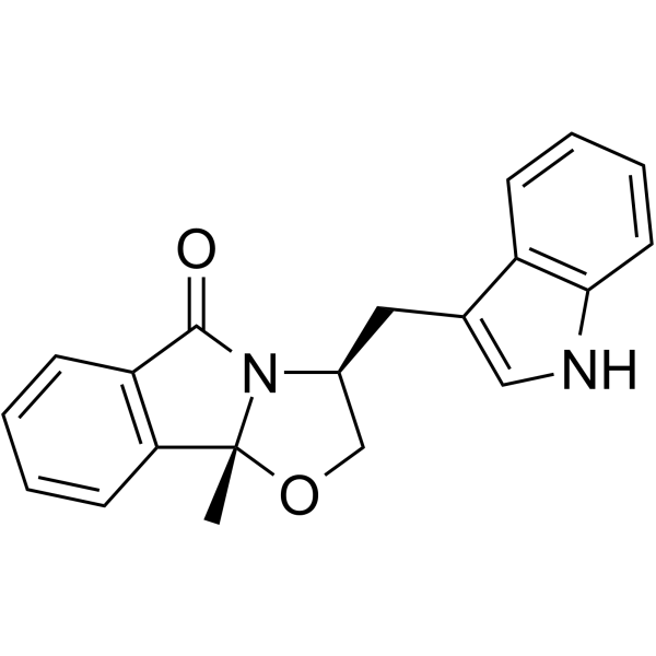
SLMP53-1 is a wild-type and mutant p53 reactivator with promising antitumor activity. SLMP53-1 mediates the reprograming of glucose metabolism in cancer cells. SLMP53-1 depletes angiogenesis, decreasing endothelial cell tube formation and vascular endothelial growth factor (VEGF) expression levels.
For research use only. We do not sell to patients.
- Purity : 99.25%
- CAS No.: 1643469-17-3
- Formula: C20H18N2O2
- Molecular Weight:318.37
-
Storage:Powder -20°C, 3 years , 4°C, 2 years ; In solvent -80°C, 6 months , -20°C, 1 month

To place orders, for customer services and technical support, please contact: MedChemExpress USA
Tel: 609-228-6898 E-mail: [email protected] [email protected]


-
 Biological Activity
-
Chemical Information
-
Solvent & Solubility
- Protocols
- Purity & Documentation
- References
-
 Help & FAQs


-
HY-18343A
CP-31398 dihydrochloridep53 Stabilizer -
HY-123076
Pifithrin-α, p-Nitro, Cyclicp53 Inhibitor -
HY-18935
CBL0137FACT Inhibitor -
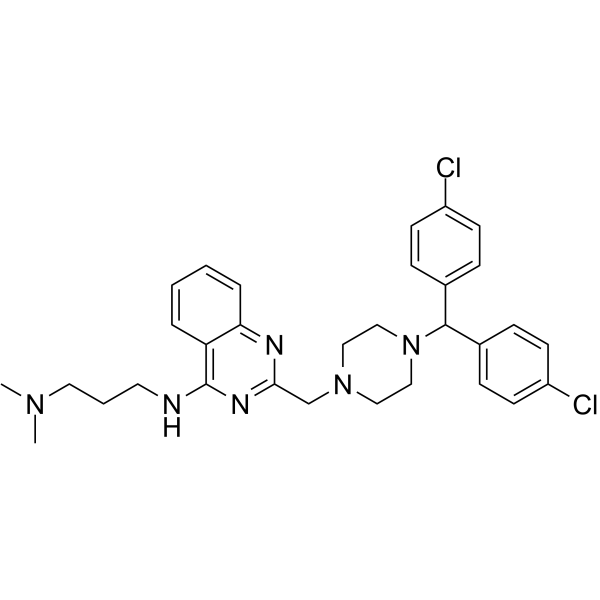
HY-110088
SCH529074P53 Activator -
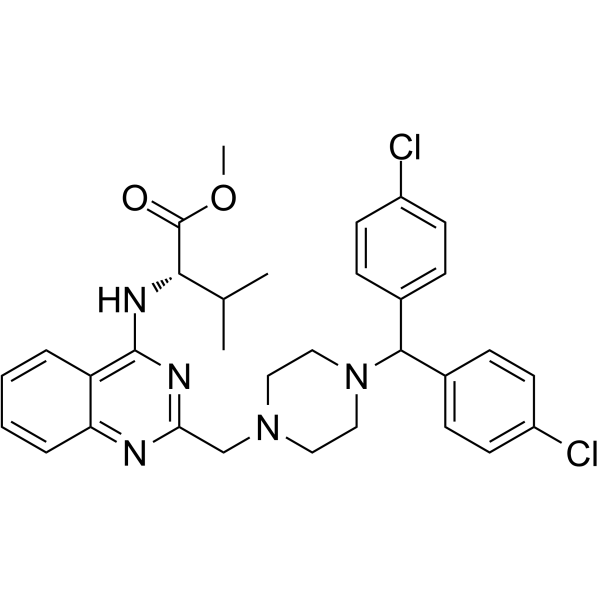
HY-122578
P53R3p53 Reactivator -
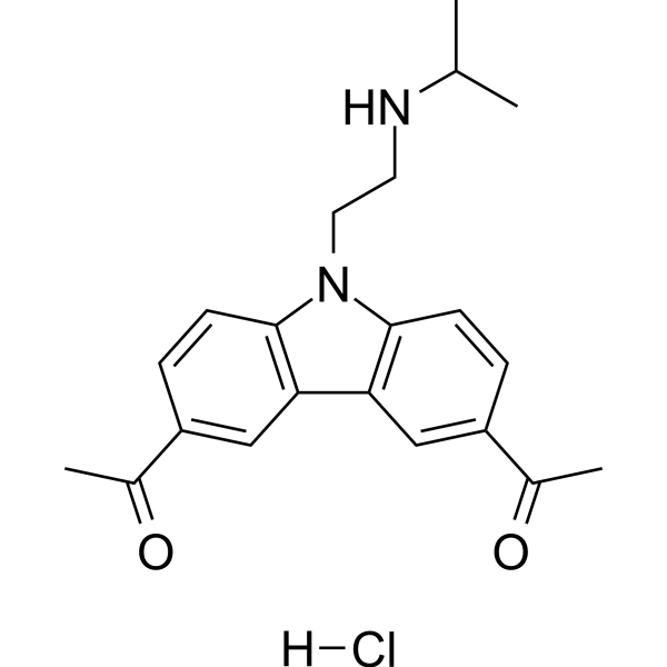
HY-18935A
CBL0137 hydrochlorideNF-κB Inhibitor -
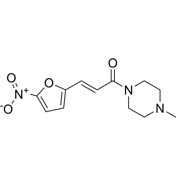
HY-19726
NSC59984MDM-2/p53 p53 Activator -
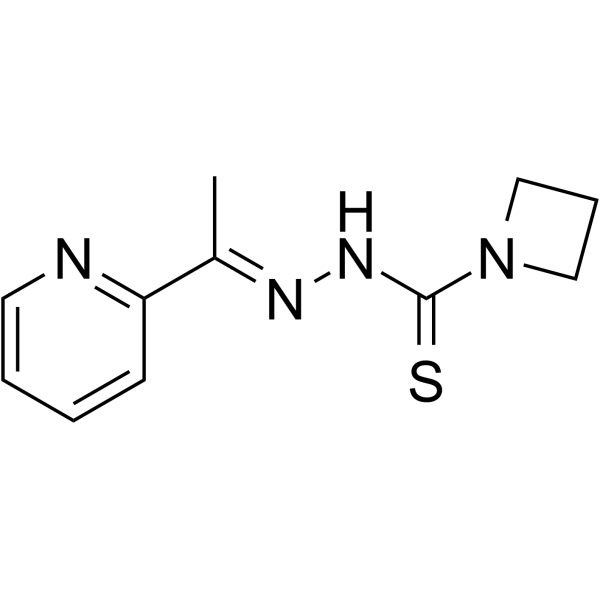
HY-18634
NSC319726MDM-2/p53 p53 Activator -
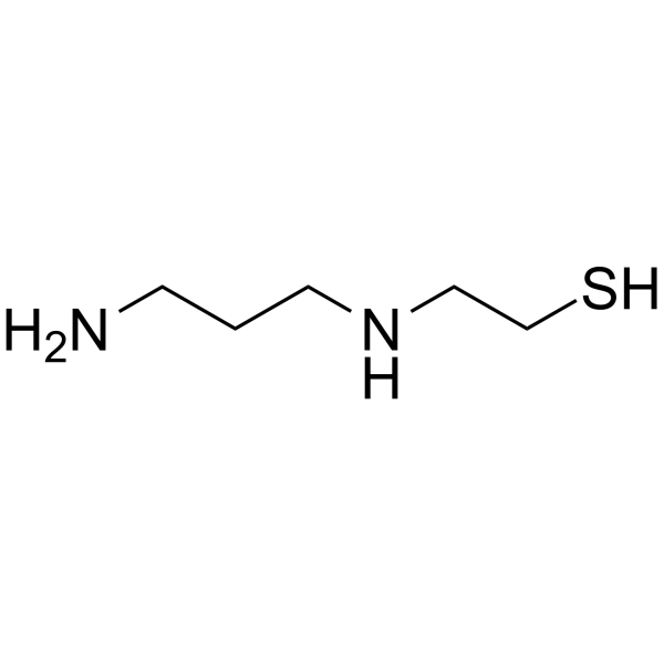
HY-137864
Amifostine thiolRadioprotector/p53 Activator -
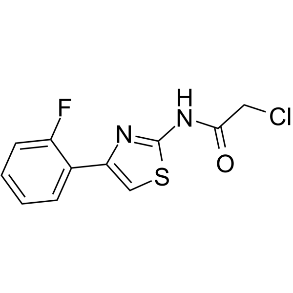
HY-156967
BAY 1892005Modulator -
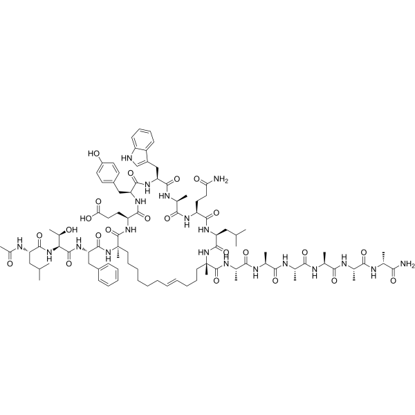
HY-P4210
Sulanemadlinp53-MDM2/MDMX Inhibitor -
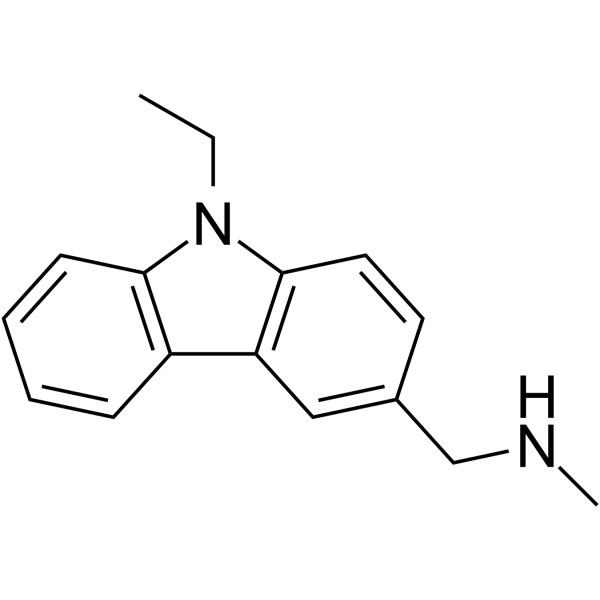
HY-108637
PhiKan 083MDM-2/p53 p53 Activator


Biological Activity
Description


Chemical Information
-
CAS No. 1643469-17-3
-
Appearance Solid
-
Molecular Weight 318.37
-
Formula C20H18N2O2
-
Color White to off-white
-
SMILES
O=C1N2[C@@](OC[C@@H]2CC3=CNC4=C3C=CC=C4)(C)C5=C1C=CC=C5
-
Shipping
Room temperature in continental US; may vary elsewhere.
-
Storage
Powder -20°C 3 years 4°C 2 years In solvent -80°C 6 months -20°C 1 month


Solvent & Solubility
In Vitro:
DMSO : 250 mg/mL (785.25 mM; ultrasonic and warming and heat to 80°C; Hygroscopic DMSO has a significant impact on the solubility of product, please use newly opened DMSO)
Please refer to the solubility information to select the appropriate solvent. Once prepared, please aliquot and store the solution to prevent product inactivation from repeated freeze-thaw cycles.
Storage method and period of stock solution: -80°C, 6 months; -20°C, 1 month. When stored at -80°C, please use it within 6 months. When stored at -20°C, please use it within 1 month.
Please refer to the solubility information to select the appropriate solvent. Once prepared, please aliquot and store the solution to prevent product inactivation from repeated freeze-thaw cycles.
Storage method and period of stock solution: -80°C, 6 months; -20°C, 1 month. When stored at -80°C, please use it within 6 months. When stored at -20°C, please use it within 1 month.
Concentration (start) × Volume (start) = Concentration (final) × Volume (final)


Purity & Documentation
-
Data Sheet (275 KB)
-
SDS (251 KB)
- English - EN (251 KB)
- Français - FR (251 KB)
- Deutsch - DE (251 KB)
- Norwegian - NO (251 KB)
- Español - ES (251 KB)
- Swedish - SV (251 KB)
- Italian - IT (251 KB)
- Korean - KR (251 KB)
- Portuguese - PT (251 KB)
-
Handling Instructions (2659 KB)
References
[1]. Gomes S, et al. SLMP53-2 Restores Wild-Type-Like Function to Mutant p53 through Hsp70: Promising Activity in Hepatocellular Carcinoma. Cancers (Basel). 2019 Aug 10;11(8):1151. [Content Brief]
[2]. Ramos H, et al. SLMP53-1 Inhibits Tumor Cell Growth through Regulation of Glucose Metabolism and Angiogenesis in a P53-Dependent Manner. Int J Mol Sci. 2020 Jan 17;21(2):596. [Content Brief]


Complete Stock Solution Preparation Table
Please refer to the solubility information to select the appropriate solvent. Once prepared, please aliquot and store the solution to prevent product inactivation from repeated freeze-thaw cycles.
Storage method and period of stock solution: -80°C, 6 months; -20°C, 1 month. When stored at -80°C, please use it within 6 months. When stored at -20°C, please use it within 1 month.
| Optional Solvent | Concentration Solvent Mass | 1 mg | 5 mg | 10 mg | 25 mg |
|---|---|---|---|---|---|
| DMSO | 1 mM | 3.1410 mL | 15.7050 mL | 31.4100 mL | 78.5250 mL |
| 5 mM | 0.6282 mL | 3.1410 mL | 6.2820 mL | 15.7050 mL | |
| 10 mM | 0.3141 mL | 1.5705 mL | 3.1410 mL | 7.8525 mL | |
| 15 mM | 0.2094 mL | 1.0470 mL | 2.0940 mL | 5.2350 mL | |
| 20 mM | 0.1570 mL | 0.7852 mL | 1.5705 mL | 3.9262 mL | |
| 25 mM | 0.1256 mL | 0.6282 mL | 1.2564 mL | 3.1410 mL | |
| 30 mM | 0.1047 mL | 0.5235 mL | 1.0470 mL | 2.6175 mL | |
| 40 mM | 0.0785 mL | 0.3926 mL | 0.7852 mL | 1.9631 mL | |
| 50 mM | 0.0628 mL | 0.3141 mL | 0.6282 mL | 1.5705 mL | |
| 60 mM | 0.0523 mL | 0.2617 mL | 0.5235 mL | 1.3087 mL | |
| 80 mM | 0.0393 mL | 0.1963 mL | 0.3926 mL | 0.9816 mL | |
| 100 mM | 0.0314 mL | 0.1570 mL | 0.3141 mL | 0.7852 mL |


SLMP53-1 Related Classifications
HY-122753 Related Classifications
 Powered by Bioz