



TMA-DPH (solution)

Based on 1 Customer Validation
TMA-DPH (solution) is a hydrophobic fluorescent membrane probe (Ex=355 nm; Em=430 nm). TMA-DPH is able to anchor on the cell surface and localize to different regions of the phospholipid bilayer. By analyzing the fluorescence polarization values of TMA-DPH in the plasma membrane and membrane substructures, the fluidity of the cell membrane can be determined.
Solvent and concentration: DMSO: 10 mM
For research use only. We do not sell to patients.
- CAS No.: 115534-33-3
- Formula: C28H31NO3S
- Molecular Weight:461.62
-
Storage:
Please store the product under the recommended conditions in the Certificate of Analysis.


To place orders, for customer services and technical support, please contact: MedChemExpress USA
Tel: 609-228-6898 E-mail: [email protected] [email protected]


-
 Biological Activity
-
Chemical Information
- Purity & Documentation
- References
-
 Help & FAQs


-

HY-DY1025
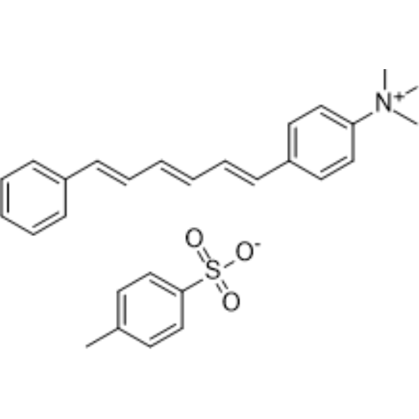
Laurdan (solution)Fluorescent Probe -
HY-D0986
TMA-DPHMembrane Fluidity Fluorescent Probe -
HY-D0085
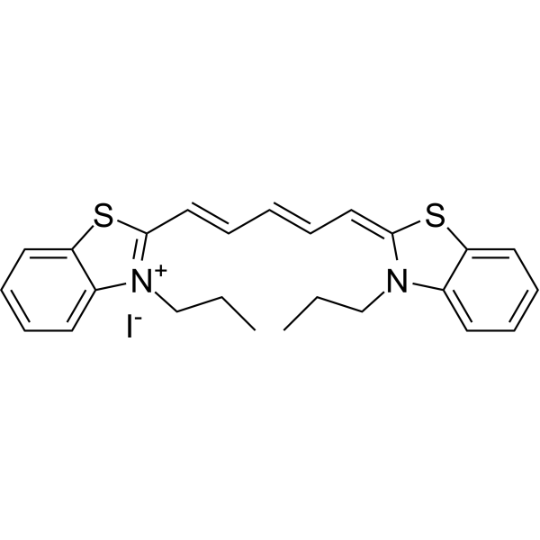
DiSC3(5)Fluorescent Probe -
HY-D0080
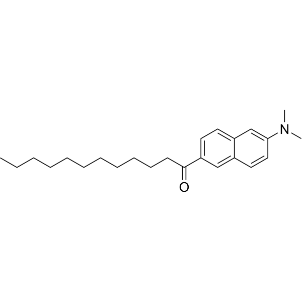
LaurdanFluorescent Probe -
HY-D0940
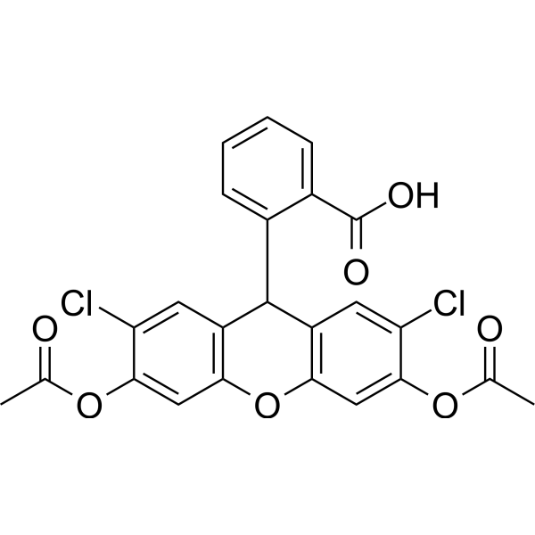
H2DCFDAROS-Sensitive Probe -
HY-D0079
DihydroethidiumFluorescent -

HY-DY1021
DiSC3(5) (solution)Fluorescent Probe -

HY-DY1015
Dihydroethidium (solution)Fluorescent


Biological Activity
Description
Solvent and concentration: DMSO: 10 mM
In Vitro
Guide (The following is our recommended protocol. This protocol is only a guide and should be modified according to your specific needs).
1. Preparation of working solution
Dilute with PBS or serum-free medium (pH 7.4) (optimized according to the experiment). The corresponding stock solution can be diluted according to the actual situation. Note that if the solvent is DMSO, the cytotoxicity of DMSO must be considered, and a solvent control should be prepared; if the solvent is pure water, the working solution needs to be filtered and sterilized before adding cells.
Note: The working solution should be prepared and used immediately. Keep it away from light.
2. Cell staining (suspended cells)
2.1 Collect cells by centrifugation and wash twice with PBS for 5 minutes each time.
2.2 TMA-DPH (0.5-5 μM) in Hanks and 20 mM Hepes buffer pH 7.4, and incubate it at 37°C, for 5-30 minutes (optimized according to the experiment).
2.3 Centrifuge at 400 g for 3-4 minutes and discard the supernatant.
2.4 Add PBS to wash the cells twice, 5 minutes each time.
2.5 Wash the cells and resuspended in the appropriate Hepes buffer pH 7.4.
2.6 Observe using a fluorescence microscope or flow cytometer.
3. Cell staining (adherent cells)
3.1 Culture the adherent cells on a sterile coverslip.
3.2 Remove the coverslip from the culture medium and remove the excess culture medium.
3.3 Add TMA-DPH (0.5-5 μM) in Hanks and 20 mM Hepes buffer pH 7.4, and incubate it at 37°C, for 5-30 minutes (optimized according to the experiment).
3.4 Wash the cells and resuspended in the appropriate Hepes buffer pH 7.4.
3.5 Fluorescence microscopy detection (Ex/Em = 355/430 nm).
Note: If flow cytometry is required, the cells need to be digested with trypsin and resuspended before staining.
MedChemExpress (MCE) has not independently confirmed the accuracy of these methods. They are for reference only.


Chemical Information
-
CAS No. 115534-33-3
-
Appearance Liquid
-
Molecular Weight 461.62
-
Formula C28H31NO3S
-
Color Colorless to light yellow
-
SMILES
C[N+](C)(C)C1=CC=C(/C=C/C=C/C=C/C2=CC=CC=C2)C=C1.[O-]S(=O)(C3=CC=C(C)C=C3)=O
-
Shipping
Room temperature in continental US; may vary elsewhere.
-
Storage
Please store the product under the recommended conditions in the Certificate of Analysis.


Purity & Documentation
-
Data Sheet (268 KB)
-
SDS (251 KB)
- English - EN (251 KB)
- Français - FR (251 KB)
- Deutsch - DE (251 KB)
- Norwegian - NO (251 KB)
- Español - ES (251 KB)
- Swedish - SV (251 KB)
- Italian - IT (251 KB)
- Korean - KR (251 KB)
- Portuguese - PT (251 KB)
-
Handling Instructions (2659 KB)
References
[1]. Illinger D, et al. The kinetic aspects of intracellular fluorescence labeling with TMA-DPH support the maturation model for endocytosis in L929 cells. J Cell Biol. 1994 May;125(4):783-94. [Content Brief]
[2]. Illinger D, et al. A comparison of the fluorescence properties of TMA-DPH as a probe for plasma membrane and for endocytic membrane. Biochim Biophys Acta. 1995 Oct 4;1239(1):58-66. [Content Brief]
[3]. Benedetti A, et al. Plasma membrane fluidity in isolated rat hepatocytes: comparative study using DPH and TMA-DPH as fluorescent probes. J Gastroenterol Hepatol. 1989 May-Jun;4(3):221-7. [Content Brief]

Calculators
Concentration (start) × Volume (start) = Concentration (final) × Volume (final)


TMA-DPH (solution) Related Classifications
HY-DY1057 Related Classifications
 Powered by Bioz