



Arundic Acid
Based on 2 publication(s) in Google Scholar
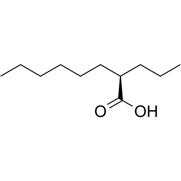
Arundic Acid is an orally effective astrocyte function modulator and neuroprotective agent. Arundic Acid increases the expression and function of the astrocytic glutamate transporter EAAT1 by activating the ERK, Akt and NF-κB pathways. Arundic Acid attenuates retinal ganglion cell death in a normal-tension glaucoma model. Arundic Acid exerts neuroprotective effects in a mouse model of Parkinson's disease. Arundic Acid is a S100β protein synthesis inhibitor that prevents neurological deficits and brain tissue damage after intracerebral hemorrhage in rats. Arundic Acid downregulates neuroinflammation and astrocytic dysfunction after status epilepticus in immature rats. Arundic Acid is applicable to research related to Parkinson's disease, cerebral ischemia, glaucoma, intracerebral hemorrhage and epilepsy.
For research use only. We do not sell to patients.
- Purity : 98.0%
- CAS No.: 185517-21-9
- Formula: C11H22O2
- Molecular Weight:186.29
-
Storage:Pure form -20°C, 3 years , 4°C, 2 years ; In solvent -80°C, 6 months , -20°C, 1 month

To place orders, for customer services and technical support, please contact: MedChemExpress USA
Tel: 609-228-6898 E-mail: [email protected] [email protected]


-
 Biological Activity
-
Chemical Information
-
Solvent & Solubility
- Protocols
- Purity & Documentation
- References
-
 Help & FAQs


-

HY-107661R
Arundic Acid (Standard)Astrocyte-modulating Agent -
HY-P1240
MOG (35-55) mouse, ratCNS Myelin Component -
HY-P1242
NEP(1-40)NgR Antagonist -
HY-107660
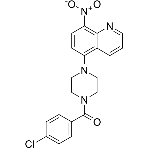
SIRT2-IN-8SIRT2 Inhibitor -

HY-W096171
D-DopaGCPII Inhibitor -
HY-17587
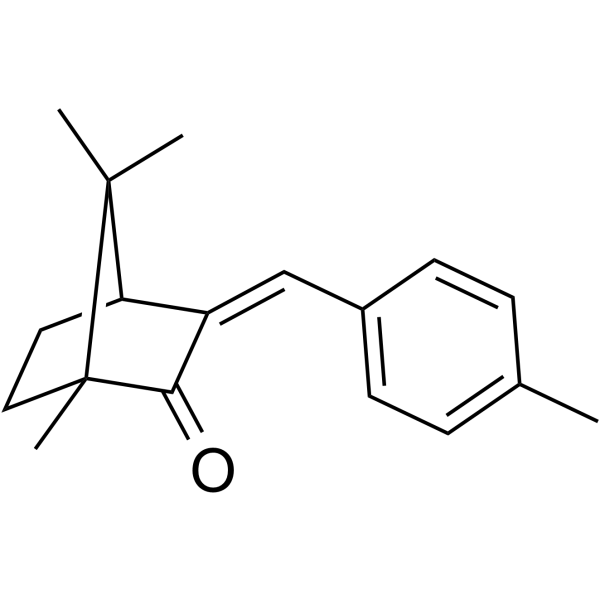
4-Methylbenzylidene camphorEstrogen-Like Drug -
HY-107662
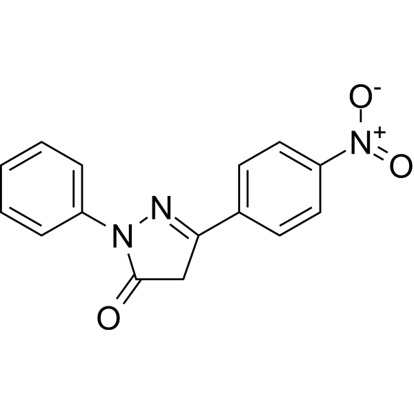
TCS PrP Inhibitor 13PrP C Inhibitor -

HY-17587R
4-Methylbenzylidene camphor (Standard)Estrogen-Like Drug -
![[SER140]-PLP(139-151)](https://file.medchemexpress.com/product_pic/hy-p1038.gif)
HY-P1038
[SER140]-PLP(139-151) -

HY-107662R
TCS PrP Inhibitor 13 (Standard)Reference Standard
Publications Citing Use of MedChemExpress (MCE) Arundic Acid
More

Biological Activity
Description
In Vitro
Arundic acid (0-100 μM; 3-24 h) enhances the promoter activity of EAAT1/EAAT2 in the human astrocytic H4 cell line[1].
Arundic acid (6.25-100 μM; 24 h) mediates the upregulation of EAAT1 promoter activity, mRNA and protein levels, and glutamate uptake function in human astrocytic H4 cells via the Akt, ERK and NF-κB pathways[1].
Arundic acid (50 μM; 3-24 h) activates the NF-κB pathway in human astrocyte H4 cells by reducing the cytoplasmic level of IκBα and inducing nuclear translocation of NF-κB p65[1].
Arundic acid (50 μM; 5 min-24 h) activates the Akt and ERK signaling pathways within 5 min of treatment[1].
Arundic acid (50 μM; 24 h) attenuates Mn-induced inhibition of EAAT1 expression by suppressing Mn-activated YY1 expression in human astrocyte H4 cells[1].
Arundic acid (100 μM; 14 days) enhances the glutamate uptake rate of primary cultured mouse Müller cells by increasing Vmax without altering glutamate affinity[2].
Arundic acid (0-100 μM; 14 days) upregulates the expression of GLAST mRNA in primary cultured mouse Müller cells[2].
Arundic acid modulates multiple astrocyte activation responses in cultured rat cerebral astrocytes, including inhibition of S-100β upregulation and pro-inflammatory gene expression, without altering GFAP levels[3].
MedChemExpress (MCE) has not independently confirmed the accuracy of these methods. They are for reference only. Further protocols information, click here.
-
Cell Line:Primary cultured mouse Müller cells
-
Concentration:0, 12.5, 25, 50, 100 μM
-
Incubation Time:14 days
-
Result:Increased endogenous GLAST mRNA expression.
Detected GLAST mRNA induction as early as 24 h post-treatment.
-
Cell Line:human astrocyte H4
cell -
Concentration:0, 6.25, 12.5, 25, 50, 100 μM
-
Incubation Time:24 h
-
Result:Increased EAAT1 protein expression.
-
Cell Line:human astrocyte H4
cell -
Concentration:50 μM
-
Incubation Time:0, 6, 12, 24 h
-
Result:Enhanced NF-κB reporter gene activity and reduced IκBα levels in cytoplasmic components
In Vivo
Arundic acid (30 mg/kg; i.p.; 5 post-MPTP doses) protects dopaminergic neurons in mice from the neurotoxicity of MPTP (HY-W114750) in the MPTP-induced Parkinson's disease mouse model, maintains 52% of dopamine levels in the striatum and 44% of dopaminergic neurons in the substantia nigra, and ameliorates motor function deficits[3].
Arundic acid (0.02-20 μg/μL; i.c.v.; single dose) reduces the levels of S100B and GFAP in the striatum of healthy male Wistar rats[4].
A single intracerebroventricular (i.c.v.) dose of Arundic acid (2 μg/μL) reverses intracerebral hemorrhage (ICH)-induced neurological deficits, reduces striatal lesion volume, inhibits reactive astrogliosis, prevents neuronal apoptosis, normalizes central and peripheral S100B levels, and enhances early antioxidant defense capacity in male Wistar rats[4].
Arundic acid (10 mg/kg; i.p.; single injection; 6 h or 24 h post-SE induction) exerts neuroprotective effects on rats with lithium-pilocarpine-induced status epilepticus (SE) by alleviating hippocampal neuroinflammation, reversing astrocytic dysfunction, restoring glutamate homeostasis and improving glucose uptake, with administration at 24 h post-SE induction showing more stable efficacy across all endpoint indicators[5].
MedChemExpress (MCE) has not independently confirmed the accuracy of these methods. They are for reference only.
-
Animal Model:C57BL/6J (backcrossed for ≥10 generations; GLAST heterozygous; postnatal day 22 to 35)[2]
-
Dosage:10 mg/kg
-
Administration:p.o.; daily; 14 days
-
Result:Increased retinal GLAST mRNA levels relative to vehicle controls.
Increased retinal GLAST protein expression relative to vehicle controls.
Increased retinal glutamate uptake velocity by 1.23-fold relative to vehicle-treated GLAST+/- mice.
Increased GCL neuron count to 438 cells per retina, compared with 366 cells in vehicle-treated GLAST+/- mice.
Showed no effect on glutamate uptake in GLAST-/- mice.
-
Animal Model:C57BL/6 (male, 10 weeks old, 22-28 g, MPTP-induced Parkinson’s disease)[3]
-
Dosage:30 mg/kg
-
Administration:i.p.; 5 doses at 1 minute, 6 h, 24 h, 48 h, 72 h after last MPTP injection
-
Result:Preserved striatal dopamine levels at 52% of normal control values.
Preserved striatal DOPAC and HVA levels at 47% and 60% of normal control values, respectively.
Significantly reduced motor function deficits in the pole test (T-turn and T-LA) and catalepsy test.
Preserved the number of tyrosine hydroxylase-positive dopaminergic neurons in the substantia nigra pars compacta at 42% of normal control values 3 days after MPTP injection and 44% of normal control values 7 days after MPTP injection.
Induced earlier appearance of GFAP-positive reactive astrocytes (by 3 days post-MPTP) compared to MPTP-only mice, with GFAP immunoreactivity comparable to MPTP-only mice at 7 days.
Reduced S-100 immunoreactivity in the substantia nigra at 3 days and in the striatum at 7 days post-MPTP compared to MPTP-only mice.
Showed no significant effect on microglial activation relative to MPTP-only mice.
-
Animal Model:Wistar rats (adult male, 90 days-old, 300-350 g)[4]
-
Dosage:0.02 μg/μL; 0.2 μg/μL; 2 μg/μL; 20 μg/μL
-
Administration:i.c.v.; single dose
-
Result:Significantly reduced striatal S100B immunocontent at 0.2 μg/μL and 2 μg/μL doses compared to control groups.
Significantly reduced striatal GFAP immunocontent at 2 μg/μL dose compared to control groups.
Showed no inhibitory effect on either S100B or GFAP levels at 20 μg/μL dose.
-
Animal Model:Wistar rats (adult male, 90 days-old, 300-350 g, intracerebral hemorrhage induced by intrastriatal injection of type-IV collagenase)[4]
-
Dosage:2 μg/μL
-
Administration:i.c.v.; single dose; administered immediately before ICH induction
-
Result:Reversed motor dysfunction completely at 7 days post-injury, with neurological scores and significantly.
Reduced striatal lesion volume significantly at 7 days post-injury.
Lowered striatal GFAP and S100B fluorescence intensity and immunostained surface area significantly at 7 days post-injury.
Increased NeuN fluorescence intensity and number of NeuN-positive cells significantly at 7 days post-injury.
Decreased cleaved caspase-3 fluorescence intensity and number of cleaved caspase-3-positive cells significantly at 7 days post-injury.
Lowered striatal GFAP levels significantly at 7 days post-injury.
Lowered striatal S100B levels significantly at 7 days post-injury; lowered cerebrospinal fluid S100B levels significantly at 72 hours post-injury; lowered serum S100B levels significantly at 24 hours and 7 days post-injury.
Increased striatal GSH levels significantly at 24 hours post-injury.
Decreased striatal GS activity significantly at 24 hours post-injury.
-
Animal Model:Wistar rats (male, postnatal day 27)[5]
-
Dosage:10 mg/kg (6 h post-SE induction); 10 mg/kg (24 h post-SE induction)
-
Administration:i.p.; single injection
-
Result:Partially reversed elevated hippocampal IL-1β levels, abolished elevated hippocampal TLR4 protein expression, partially reversed elevated hippocampal COX2 protein expression, increased hippocampal TGF-β levels, and did not alter TNF-α, RAGE, or Iba1 levels at 6 h post-SE.
Prevented elevated hippocampal IL-1β levels, decreased hippocampal TNF-α levels, prevented elevated hippocampal RAGE and TLR4 protein expression, completely abolished elevated hippocampal COX2 protein expression, partially reversed elevated hippocampal Iba1 levels, and increased hippocampal TGF-β levels at 24 h post-SE.
Prevented elevated hippocampal GFAP levels at 6 h and 24 h post-SE.
Partially reversed elevated hippocampal S100B levels, reversed reduced hippocampal connexin-43 (Cx-43) protein expression, partially reversed elevated CSF S100B levels, and partially reversed reduced serum S100B levels at 6 h post-SE.
Abolished elevated hippocampal S100B levels, and did not alter reduced hippocampal Cx-43 levels or CSF/serum S100B levels at 24 h post-SE.
Reversed reduced hippocampal GLAST protein expression, partially reversed reduced hippocampal GLT1 protein expression, reversed increased hippocampal glutamate uptake, did not alter reduced hippocampal GSH levels, partially reversed reduced hippocampal glutamine synthetase (GS) activity, and reversed reduced hippocampal GS protein expression at 6 h post-SE.
Partially reversed reduced hippocampal GLAST protein expression, partially reversed reduced hippocampal GLT1 protein expression, reversed increased hippocampal glutamate uptake, prevented reduced hippocampal GSH levels, did not alter reduced hippocampal GS activity, and reversed reduced hippocampal GS protein expression at 24 h post-SE.
Abolished reduced hippocampal glucose uptake, and did not alter hippocampal GLUT1 protein expression at 6 h and 24 h post-SE.
Clinical Trial
| NCT Number | Sponsor | Condition | Start Date |
Phase

|
|---|---|---|---|---|
| NCT01329991 | Plexxikon| | 2011-05 | PHASE1 |


Chemical Information
-
CAS No. 185517-21-9
-
Appearance Liquid (Density: 0.908 g/cm3)
-
Molecular Weight 186.29
-
Formula C11H22O2
-
Color Colorless to light yellow
-
SMILES
CCCCCC[C@H](C(O)=O)CCC
-
Synonyms
ONO-2506; (R)-2-Propyloctanoic acid
-
Shipping
Room temperature in continental US; may vary elsewhere.
-
Storage
Pure form -20°C 3 years 4°C 2 years In solvent -80°C 6 months -20°C 1 month


Publications (2)
-
Journal Impact Factor
-
Most Recent
-
Int Immunopharmacol
2021 Aug:97:107792. PMID: 34051593 -
Behav Brain Res
Overexpression of S100B promotes depressive-like behaviors in stroke-induced rats by modulating the PI3K/AKT/NF-κB pathway. [Abstract]2025 Jun 25:488:115597. PMID: 40250529


Solvent & Solubility
In Vitro:
DMSO : 100 mg/mL (536.80 mM; Need ultrasonic; Hygroscopic DMSO has a significant impact on the solubility of product, please use newly opened DMSO)
Please refer to the solubility information to select the appropriate solvent. Once prepared, please aliquot and store the solution to prevent product inactivation from repeated freeze-thaw cycles.
Storage method and period of stock solution: -80°C, 6 months; -20°C, 1 month. When stored at -80°C, please use it within 6 months. When stored at -20°C, please use it within 1 month.
Please refer to the solubility information to select the appropriate solvent. Once prepared, please aliquot and store the solution to prevent product inactivation from repeated freeze-thaw cycles.
Storage method and period of stock solution: -80°C, 6 months; -20°C, 1 month. When stored at -80°C, please use it within 6 months. When stored at -20°C, please use it within 1 month.
Concentration (start) × Volume (start) = Concentration (final) × Volume (final)
In Vivo:
Select the appropriate dissolution method based on your experimental animal and administration route.
- For the following dissolution methods, please ensure to first prepare a clear stock solution using an In Vitro approach and then sequentially add co-solvents:
- To ensure reliable experimental results, the clarified stock solution can be appropriately stored based on storage conditions. As for the working solution for In Vivo experiments, it is recommended to prepare freshly and use it on the same day.
- The percentages shown for the solvents indicate their volumetric ratio in the final prepared solution. If precipitation or phase separation occurs during preparation, heat and/or sonication can be used to aid dissolution.
Add each solvent one by one: 10% DMSO 40% PEG300 5% Tween-80 45% Saline
Solubility: 3.75 mg/mL (20.13 mM); Suspended solution; Need ultrasonic
This protocol yields a suspended solution of 3.75 mg/mL. Suspended solution can be used for oral and intraperitoneal injection.
Taking 1 mL working solution as an example, add 100 μL DMSO stock solution (37.5 mg/mL) to 400 μL PEG300, and mix evenly; then add 50 μL Tween-80 and mix evenly; then add 450 μL Saline to adjust the volume to 1 mL.
Preparation of Saline: Dissolve 0.9 g sodium chloride in ddH₂O and dilute to 100 mL to obtain a clear Saline solution.
Add each solvent one by one: 10% DMSO 90% (20% SBE-β-CD in Saline)
Solubility: ≥ 3.75 mg/mL (20.13 mM); Clear solution
This protocol yields a clear solution of ≥ 3.75 mg/mL (saturation unknown).
Taking 1 mL working solution as an example, add 100 μL DMSO stock solution (37.5 mg/mL) to 900 μL 20% SBE-β-CD in Saline, and mix evenly.
Preparation of 20% SBE-β-CD in Saline (4°C, storage for one week): 2 g SBE-β-CD powder is dissolved in 10 mL Saline, completely dissolve until clear.
In Vivo Dissolution Calculator
Please enter the basic information of animal experiments:
-
-
-
-
Recommended: Prepare an additional quantity of animals to account for potential losses during experiments.
Please enter your animal formula composition:
-
%DMSO +
Recommended: Keep the proportion of DMSO in working solution below 2% if your animal is weak.
-
%+
-
+%Tween-80 + +
-
%Saline +
The co-solvents required include: DMSO, . All of co-solvents are available by MedChemExpress (MCE). , Tween 80. All of co-solvents are available by MedChemExpress (MCE).
Working solution concentration: 0.22 mg/mL
Method for preparing stock solution: mg drug dissolved in μL DMSO. Stock solution concentration: mg/mL.
1. Take μL DMSO stock solution;
2. Add μL .
μL , mix evenly;
3. Then add μL Tween 80, mix evenly;
4. Then add μL
Please ensure that the stock solution in the first step is dissolved to a clear state, and add co-solvents in sequence. You can use ultrasonic heating (ultrasonic cleaner, recommended frequency 20-40 kHz), vortexing, etc. to assist dissolution.

Protocols
-
RNA extraction experimental
By lysing cells, releasing RNA, and removing impurities such as proteins and DNA, high-purity RNA products are finally obtained. The commonly used traditional method is the guanidine isothiocyanate/phenol/chloroform method (Trizol), which is suitable for a variety of animal materials including animal tissues, microorganisms, cultured cells, etc., and most plant materials.
-
Cell Cytotoxicity Assay
Cytotoxicity assays are usually based on the assessment of cell membrane damage, which can also be indirectly detected by measuring cell viability. Detection methods include MTT assay, CKK-8 assay, LDH assay and ATP assay, etc.
-
Mammalian live/dead viability and cytotoxicity staining
Live/dead viability and cytotoxicity staining assays are based on the simultaneous detection of intracellular esterase activity in metabolically active (viable) cells and membrane integrity loss in non-viable cells. In commonly used dual-staining approaches, membrane-permeant fluorogenic substrates are converted by intracellular esterases into fluorescent products in live cells, while impermeant DNA-binding dyes selectively enter cells with compromised plasma membranes and label nucleic acids in dead or dying cells, enabling discrimination between viable and non-viable populations by fluorescence microscopy or flow cytometry.
-
Human pluripotent stem cell midbrain dopaminergic neuron differentiation
Human pluripotent stem cells are directed toward midbrain dopaminergic neurons by first inducing a neural floor-plate-like progenitor state, then patterning cells with ventralizing SHH signaling and midbrain/WNT-FGF cues, and finally maturing progenitors into neurons expressing dopaminergic markers such as TH, NURR1/NR4A2, PITX3, DAT/SLC6A3, VMAT2/SLC18A2, GIRK2/KCNJ6, FOXA2, LMX1A, and EN1. The main readouts are loss of pluripotency, acquisition of FOXA2+/LMX1A+ midbrain floor-plate progenitors, emergence of βIII-tubulin+/MAP2+ neurons, and production of TH+ dopaminergic neurons with molecular, dopamine-release, and electrophysiological features of midbrain dopaminergic identity.
-
Apoptosis Solutions
Apoptosis is a regulated, generally non-lytic cell-death pathway that removes unwanted, damaged, infected, or abnormal cells through coordinated morphological changes, caspase activation, DNA fragmentation, and membrane remodeling. The intrinsic apoptosis pathway is controlled mainly by mitochondrial outer membrane permeabilization, BCL-2 family proteins, cytochrome c release, apoptosome formation, caspase-9 activation, and downstream executioner caspase-3/7 activation. The extrinsic apoptosis pathway is initiated by death receptors such as Fas, TNFR, and TRAIL receptors, which recruit adaptor proteins and activate caspase-8 before engaging executioner caspases or mitochondrial amplification through BID cleavage. Apoptosis is linked to many phenotypes, including cancer cell killing, tissue homeostasis, immune regulation, neurodegeneration, infection response, and treatment-induced cytotoxicity; unresolved questions include how apoptosis interacts with necroptosis, pyroptosis, ferroptos
-
Research Protocol for Inflammation-related Diseases
The NLRP3 inflammasome is a cytosolic innate immune signaling platform that integrates priming signals and danger-signal activation to promote caspase-1 activation, maturation of IL-1β and IL-18, and gasdermin D-mediated pyroptotic cell death. The core experimental logic is to determine whether inflammatory disease phenotypes are driven by increased NLRP3 expression, ASC-containing inflammasome assembly, caspase-1 cleavage, GSDMD cleavage, and extracellular release of IL-1β/IL-18 rather than by nonspecific cell injury alone. The pathway is strongly linked to inflammation-related disease phenotypes because monosodium urate crystals activate NALP3/NLRP3 inflammasome signaling in gout-like crystal inflammation, cholesterol crystals activate NLRP3 inflammasomes in atherogenesis models, and DSS-induced intestinal inflammation has been reported to involve NLRP3 inflammasome activity. However, experimental colitis studies also show context-dependent protective effects of NLRP3 inflammasome co
-
Research Protocol for Neurological Diseases
PINK1/Parkin-mediated mitophagy pathway is a mitochondrial quality-control signaling axis in which mitochondrial depolarization stabilizes PINK1 on damaged mitochondria, activates Parkin recruitment and E3 ubiquitin ligase activity, promotes ubiquitination of outer mitochondrial membrane proteins, recruits selective autophagy adaptors, and drives lysosomal degradation of damaged mitochondria. In neurological disease research, this pathway is experimentally important because neurons, especially dopaminergic neurons, are highly dependent on mitochondrial integrity, and defective mitochondrial turnover can lead to mitochondrial dysfunction, oxidative stress, impaired neuronal survival, α-synuclein accumulation, and neuroinflammatory damage-associated signals. The genetic disease link is strongest in Parkinson’s disease because mutations in PRKN/parkin cause autosomal recessive juvenile parkinsonism, mutations in PINK1 cause hereditary early-onset Parkinson’s disease, and Drosophila studie
-
How to Choose the Right Model Animal
Choosing the right model animal is a validity-driven decision in which the species, strain, sex, age, genetic background, disease-induction method, outcome measures, and welfare burden must match the scientific question rather than laboratory tradition or convenience. A model should be selected by judging face validity, construct validity, and predictive validity: whether it resembles the human phenotype, whether it reproduces relevant mechanisms, and whether results are likely to predict human biology or treatment response. Animal studies often fail to translate because of species differences, weak disease resemblance, poor experimental design, inadequate reporting, publication bias, and underuse of randomization, blinding, and sample-size justification. Unresolved questions include how to rank competing models objectively, how much human-disease complexity must be reproduced for a given objective, and when non-animal systems such as organoids, ex vivo tissue, or computational models


Purity & Documentation
-
Data Sheet (288 KB)
-
SDS (396 KB)
- English - EN (396 KB)
- Français - FR (396 KB)
- Deutsch - DE (396 KB)
- Norwegian - NO (396 KB)
- Español - ES (396 KB)
- Swedish - SV (396 KB)
- Italian - IT (396 KB)
- Korean - KR (396 KB)
- Portuguese - PT (396 KB)
-
Handling Instructions (2659 KB)
References
[1]. Karki P, et al. Arundic Acid Increases Expression and Function of Astrocytic Glutamate Transporter EAAT1 Via the ERK, Akt, and NF-κB Pathways. Mol Neurobiol. 2018;55(6):5031-5046. [Content Brief]
[2]. Yanagisawa M, et al. Arundic acid attenuates retinal ganglion cell death by increasing glutamate/aspartate transporter expression in a model of normal tension glaucoma. Cell Death Dis. 2015;6(3):e1693. Published 2015 Mar 19. [Content Brief]
[3]. Kato H, et al. Arundic acid, an astrocyte-modulating agent, protects dopaminergic neurons against MPTP neurotoxicity in mice. Brain Res. 2004;1030(1):66-73. [Content Brief]
[4]. Cordeiro JL, et al. Arundic Acid (ONO-2506), an Inhibitor of S100B Protein Synthesis, Prevents Neurological Deficits and Brain Tissue Damage Following Intracerebral Hemorrhage in Male Wistar Rats. Neuroscience. 2020 Aug 1;440:97-112. [Content Brief]
[5]. Vizuete AFK, et al. Arundic acid (ONO-2506) downregulates neuroinflammation and astrocyte dysfunction after status epilepticus in young rats induced by Li-pilocarpine. Prog Neuropsychopharmacol Biol Psychiatry. 2023;123:110704. [Content Brief]


Complete Stock Solution Preparation Table
Please refer to the solubility information to select the appropriate solvent. Once prepared, please aliquot and store the solution to prevent product inactivation from repeated freeze-thaw cycles.
Storage method and period of stock solution: -80°C, 6 months; -20°C, 1 month. When stored at -80°C, please use it within 6 months. When stored at -20°C, please use it within 1 month.
| Optional Solvent | Concentration Solvent Mass | 1 mg | 5 mg | 10 mg | 25 mg |
|---|---|---|---|---|---|
| DMSO | 1 mM | 5.3680 mL | 26.8399 mL | 53.6797 mL | 134.1994 mL |
| 5 mM | 1.0736 mL | 5.3680 mL | 10.7359 mL | 26.8399 mL | |
| 10 mM | 0.5368 mL | 2.6840 mL | 5.3680 mL | 13.4199 mL | |
| 15 mM | 0.3579 mL | 1.7893 mL | 3.5786 mL | 8.9466 mL | |
| 20 mM | 0.2684 mL | 1.3420 mL | 2.6840 mL | 6.7100 mL | |
| 25 mM | 0.2147 mL | 1.0736 mL | 2.1472 mL | 5.3680 mL | |
| 30 mM | 0.1789 mL | 0.8947 mL | 1.7893 mL | 4.4733 mL | |
| 40 mM | 0.1342 mL | 0.6710 mL | 1.3420 mL | 3.3550 mL | |
| 50 mM | 0.1074 mL | 0.5368 mL | 1.0736 mL | 2.6840 mL | |
| 60 mM | 0.0895 mL | 0.4473 mL | 0.8947 mL | 2.2367 mL | |
| 80 mM | 0.0671 mL | 0.3355 mL | 0.6710 mL | 1.6775 mL | |
| 100 mM | 0.0537 mL | 0.2684 mL | 0.5368 mL | 1.3420 mL |


Arundic Acid Related Classifications
HY-107661 Related Classifications
 Powered by Bioz