



Safranin

Based on 1 Customer Validation
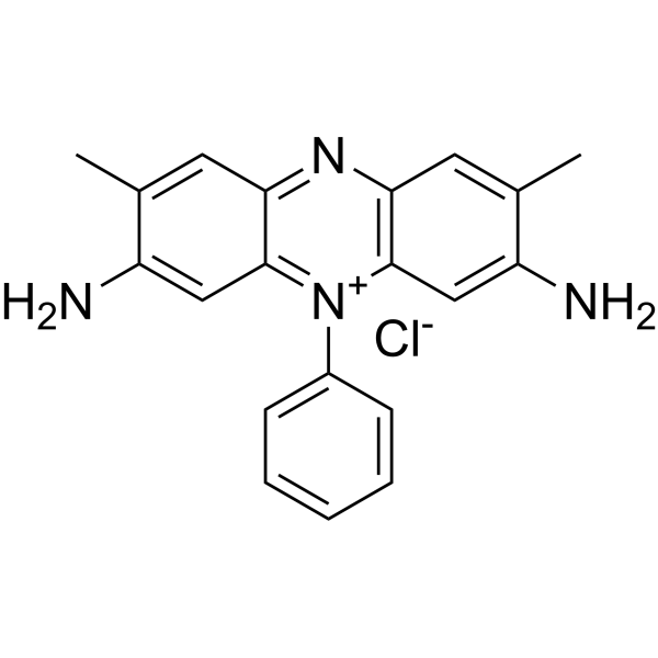
Safranin (Safranin T) is an important and classical phenazinium dye. Safranin has been extensively used in the academic field as a spectroscopic probe and indicator. Safranin possesses a planar structure and cationic charge. Safranin can readily intercalate into biological macromolecules, including DNA and proteins. Safranin has antibacterial effects against gram-positive bacteria (S. aureus). Safranin can be used as a redox indicator in the determination of metal ion concentration.
For research use only. We do not sell to patients.
- CAS No.: 477-73-6
- Formula: C20H19ClN4
- Molecular Weight:350.85
-
Storage:
RT, sealed storage, away from moisture and light.
In solvent -80°C, 1 year , -20°C, 6 months

To place orders, for customer services and technical support, please contact: MedChemExpress USA
Tel: 609-228-6898 E-mail: [email protected] [email protected]


-
 Biological Activity
-
Chemical Information
-
Solvent & Solubility
- Protocols
- Purity & Documentation
- References
-
 Help & FAQs


-
HY-D0505
Eosin YDye Molecule -
HY-D0236
Congo RedAzo Dye -
HY-D0166
Neutral RedBiochemical Assay Reagent -
HY-D0166A
Neutral Red (IND)Biochemical Assay Reagent -
HY-B0324A
Crystal VioletFluorochrome -
HY-D0162
Malachite green hemioxalateIKBKE Inhibitor -

HY-DY2001
Crystal Violet (solution)Fluorochrome -
HY-D0214
Acid Red 94 sodiumPhotosensitizer -
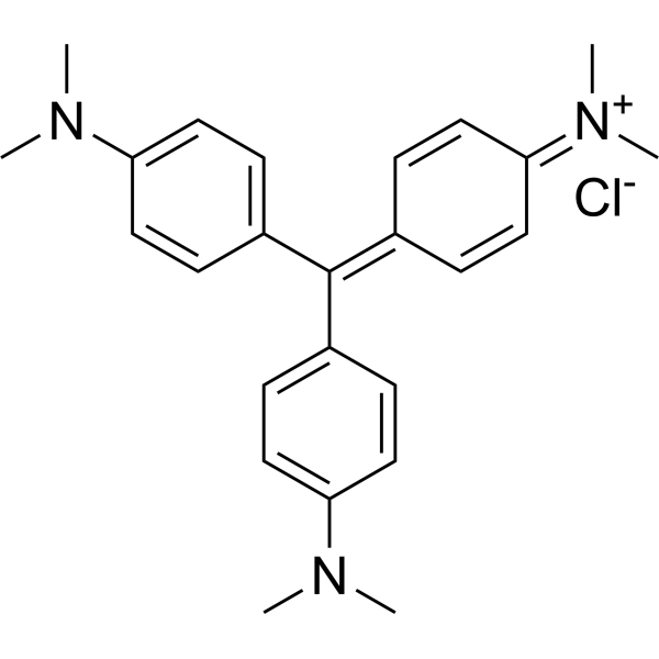
HY-W110794
Crystal violet, IND (0.0-2.0)Biochemical Assay Reagent -

HY-D0214R
Acid Red 94 sodium (Standard)Biological Dye -
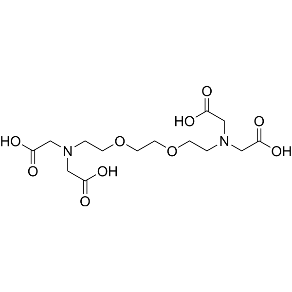
HY-D0861
EGTACalcium Ion Chelator -
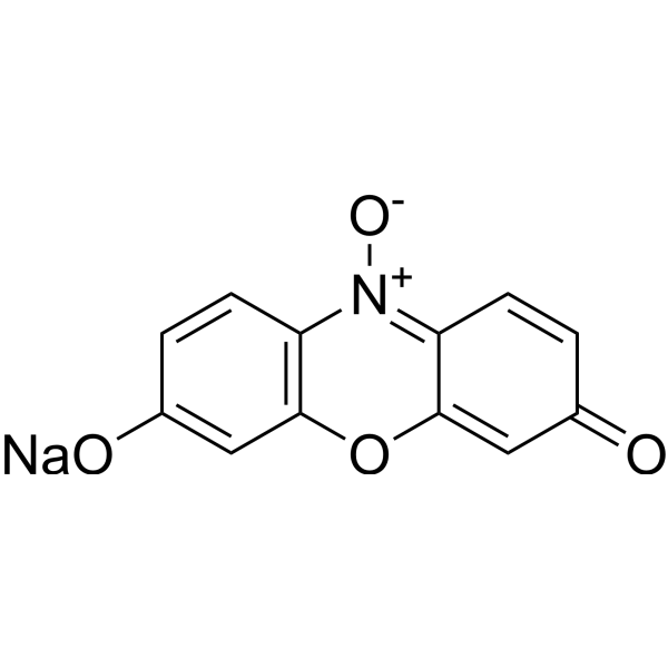
HY-111391
Resazurin sodiumBacterial Inhibitor


Biological Activity
Description
In Vitro
Safranin (0.1% (w/v), 1 min) differentially stains the cell cycle of plant cells[2].
Safranin (up to 2 μM) inhibits Complex I-driven OXPHOS (in mouse brain homogenates) capacity[3].
Safranin (0.025%) shows antibacterial activity against gram-positive bacteria (S. aureus)[4].
MedChemExpress (MCE) has not independently confirmed the accuracy of these methods. They are for reference only. Further protocols information, click here.


Chemical Information
-
CAS No. 477-73-6
-
Appearance Solid
-
Molecular Weight 350.85
-
Formula C20H19ClN4
-
Color Purple to black
-
SMILES
CC1=CC2=NC3=C([N+](C4=CC=CC=C4)=C2C=C1N)C=C(N)C(C)=C3.[Cl-]
-
Synonyms
Safranine T
-
Shipping
Room temperature in continental US; may vary elsewhere.
-
Storage
RT, sealed storage, away from moisture and light
In solvent -80°C 1 year -20°C 6 months


Solvent & Solubility
In Vitro:
DMSO : 25 mg/mL (71.26 mM; ultrasonic and warming and heat to 60°C; Hygroscopic DMSO has a significant impact on the solubility of product, please use newly opened DMSO)
Please refer to the solubility information to select the appropriate solvent. Once prepared, please aliquot and store the solution to prevent product inactivation from repeated freeze-thaw cycles.
Storage method and period of stock solution: -80°C, 1 year; -20°C, 6 months. When stored at -80°C, please use it within 1 year. When stored at -20°C, please use it within 6 months.
Please refer to the solubility information to select the appropriate solvent. Once prepared, please aliquot and store the solution to prevent product inactivation from repeated freeze-thaw cycles.
Storage method and period of stock solution: -80°C, 1 year; -20°C, 6 months. When stored at -80°C, please use it within 1 year. When stored at -20°C, please use it within 6 months.
Concentration (start) × Volume (start) = Concentration (final) × Volume (final)

Protocols
-
Research Protocol for Infectious Diseases
Infectious-disease experiments test how pathogens interact with host barriers, innate immune receptors, inflammatory signaling, pathogen replication, and tissue injury; pattern-recognition receptors such as TLRs, RIG-I-like receptors, NOD-like receptors, and inflammasomes detect microbial molecules and activate NF-κB, interferon, and cytokine responses. The central hypothesis is that infection severity reflects the balance between pathogen burden and host response: protective inflammation restricts pathogen growth, whereas excessive or mislocalized inflammation contributes to tissue damage and disease phenotype. Unresolved questions include which host pathways are protective versus pathogenic, why some infection models fail to translate to human disease, and which combined readouts best predict clinically relevant infection outcomes.
-
ROS/oxidative-stress fluorescent staining
ROS/oxidative-stress fluorescent staining uses cell-permeant fluorogenic probes that become fluorescent after oxidation inside cells or tissues; commonly used examples include DCFH-DA/DCFDA for broad cellular oxidant detection, DHE for superoxide-related signal detection, MitoSOX for mitochondrial superoxide-related signal detection, and CellROX probes for oxidative-stress-associated fluorescence readouts. The assay detects probe oxidation rather than a single ROS species unless the probe and analysis method have been chemically validated for that species. DCFH-DA enters cells, is deacetylated by intracellular esterases to DCFH, and produces fluorescent DCF after oxidation, so the readout is used as an operational measure of total cellular oxidative stress rather than a species-specific ROS measurement. DHE and MitoSOX can report superoxide-related oxidation, but red fluorescence alone can include non-specific ethidium-like oxidation products; HPLC or optimized spectral approaches are
-
Gram Staining of Tissue Sections
Gram staining of tissue sections is a histochemical technique used to differentiate Gram-positive and Gram-negative bacteria within histological specimens based on differences in bacterial cell wall structure and dye retention, adapted from classical bacteriological Gram staining into tissue-compatible “histological Gram stain” variants. In tissue applications, modifications of the Brown-Hopps and Brown-Brenn methods are commonly used to improve differentiation of microorganisms embedded within host connective tissue and to reduce overstaining or loss of Gram-negative signal, which are known limitations of earlier approaches. The principle relies on crystal violet-iodine complex retention in Gram-positive organisms and subsequent decolorization and counterstaining steps that allow contrast visualization of Gram-negative organisms against tissue background.


Purity & Documentation
-
Data Sheet (274 KB)
-
SDS (477 KB)
- English - EN (477 KB)
- Français - FR (477 KB)
- Deutsch - DE (477 KB)
- Norwegian - NO (477 KB)
- Español - ES (477 KB)
- Swedish - SV (477 KB)
- Italian - IT (477 KB)
- Korean - KR (477 KB)
- Portuguese - PT (477 KB)
-
Handling Instructions (2659 KB)
References
[1]. Wan H, et al. Structure characterization and optical properties investigation of the four main components of the classical phenazinium dye Safranin O. Analyst. 2019;144(24):7149-7156. [Content Brief]
[2]. Swain D, et al. Differential staining of the cell cycle of plant cells using safranin and indigo-picrocarmine. Stain Technol. 1990;65(4):197-204. [Content Brief]
[3]. Krumschnabel G, et al. Use of safranin for the assessment of mitochondrial membrane potential by high-resolution respirometry and fluorometry. Methods Enzymol. 2014;542:163-81. [Content Brief]
[4]. Al-Khikani F, et al. The Antibacterial Action of Safranin and Gentian Violet. Rambam Maimonides Med J. 2022 Jul 31;13(3):e0018. [Content Brief]


Complete Stock Solution Preparation Table
Please refer to the solubility information to select the appropriate solvent. Once prepared, please aliquot and store the solution to prevent product inactivation from repeated freeze-thaw cycles.
Storage method and period of stock solution: -80°C, 1 year; -20°C, 6 months. When stored at -80°C, please use it within 1 year. When stored at -20°C, please use it within 6 months.
| Optional Solvent | Concentration Solvent Mass | 1 mg | 5 mg | 10 mg | 25 mg |
|---|---|---|---|---|---|
| DMSO | 1 mM | 2.8502 mL | 14.2511 mL | 28.5022 mL | 71.2555 mL |
| 5 mM | 0.5700 mL | 2.8502 mL | 5.7004 mL | 14.2511 mL | |
| 10 mM | 0.2850 mL | 1.4251 mL | 2.8502 mL | 7.1256 mL | |
| 15 mM | 0.1900 mL | 0.9501 mL | 1.9001 mL | 4.7504 mL | |
| 20 mM | 0.1425 mL | 0.7126 mL | 1.4251 mL | 3.5628 mL | |
| 25 mM | 0.1140 mL | 0.5700 mL | 1.1401 mL | 2.8502 mL | |
| 30 mM | 0.0950 mL | 0.4750 mL | 0.9501 mL | 2.3752 mL | |
| 40 mM | 0.0713 mL | 0.3563 mL | 0.7126 mL | 1.7814 mL | |
| 50 mM | 0.0570 mL | 0.2850 mL | 0.5700 mL | 1.4251 mL | |
| 60 mM | 0.0475 mL | 0.2375 mL | 0.4750 mL | 1.1876 mL |


Safranin Related Classifications
HY-D0215 Related Classifications
 Powered by Bioz