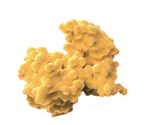


477-73-6

Chemical Structure
Safranin
Synonym(s): Safranine T
- CAS No.: 477-73-6
- Formula:C20H19ClN4
- Molecular Weight:350.85
IUPAC Name: 3,7-diamino-2,8-dimethyl-5-phenylphenazin-5-ium chloride
InChIKey: OARRHUQTFTUEOS-UHFFFAOYSA-N
SMILES: CC1=CC2=NC3=C([N+](C4=CC=CC=C4)=C2C=C1N)C=C(N)C(C)=C3.[Cl-]
Biological Activity: Safranin (Safranin T) is an important and classical phenazinium dye. Safranin has been extensively used in the academic field as a spectroscopic probe and indicator. Safranin possesses a planar structure and cationic charge. Safranin can readily intercalate into biological macromolecules, including DNA and proteins. Safranin has antibacterial effects against gram-positive bacteria (S. aureus). Safranin can be used as a redox indicator in the determination of metal ion concentration[1][2][3][4].
| Cat. No. | Product Name | Purity | Description | Pricing | |||||||||||||||||||
|---|---|---|---|---|---|---|---|---|---|---|---|---|---|---|---|---|---|---|---|---|---|---|---|
|
|
Safranin | Safranin (Safranin T) is an important and classical phenazinium dye. Safranin has been extensively used in the academic field as a spectroscopic probe and indicator. Safranin possesses a planar structure and cationic charge. Safranin can readily intercalate into biological macromolecules, including DNA and proteins. Safranin has antibacterial effects against gram-positive bacteria (S. aureus). Safranin can be used as a redox indicator in the determination of metal ion concentration. | |||||||||||||||||||||
|
loading...
/
|
|||||||||||||||||||||||
|
|
AG 1295 | 99.86% | AG 1295 is a selective platelet-derived growth factor receptor (PDGFR) tyrosine-kinase inhibitor. AG1295 abolishes autophosphorylation of the PDGFR whereas not affects the autophosphorylation of the EGF receptor. | ||||||||||||||||||||
|
loading...
/
|
|||||||||||||||||||||||
|
|
Benzoflavine | Benzoflavine is an acridine dye and photodynamic sensitizer that regulates embryonic development. Benzoflavine can adsorb to the surface of Arbacia sea urchin egg cell membranes, enhance the sensitivity of highly physiologically active embryonic regions to visible light radiation, and induce anterior-posterior developmental differences; at high concentrations, it can independently inhibit embryonic development without illumination, and its photosensitive response range covers the blue-violet region of visible light. Benzoflavine can be used in research fields such as developmental biology, organic synthesis, and photobiochemistry. | |||||||||||||||||||||
|
loading...
/
|
|||||||||||||||||||||||
|
|
Neutral Red (IND) | 96.32% | Neutral Red (IND) is an organic dye commonly used in biology and cytology laboratories. It can be used to stain living cells, secreted proteins and other molecular structures, etc., and has a wide range of applications in cell imaging and staining. In addition, Neutral Red (IND) is widely used in industrial fields such as water treatment, food processing and paper manufacturing, for example as an indicator or colorant. | ||||||||||||||||||||
|
loading...
/
|
|||||||||||||||||||||||
|
|
Toluidine Blue | Toluidine Blue (Toluidine Blue O) is an alkaline quinonimine dye (vivo dye) with high affinity for acidic tissue components, staining nuclei blue and polysaccharides purple. Toluidine Blue shows heterostaining properties for mast cells, mucins and chondrocytes. Toluidine Blue can stain different components of plant tissues and cells in different colours. Toluidine Blue is also used as a diagnostic aid to identify malignant lesions, such as cancer. | |||||||||||||||||||||
|
loading...
/
|
|||||||||||||||||||||||
|
|
Acridine yellow G | Acridine yellow G is a multifunctional dye. Dyes are important tools in biological experiments. They can help researchers observe and analyze cell structures, track biomolecules, evaluate cell functions, distinguish cell types, detect biomolecules, study tissue pathology and monitor microorganisms. Their applications range from basic scientific research to clinical A wide range of diagnostics. Dyes are also widely used in traditional fields such as textile dyeing, as well as in emerging fields such as functional textile processing, food pigments and dye-sensitized solar cells. | |||||||||||||||||||||
|
loading...
/
|
|||||||||||||||||||||||
|
|
Indoine blue | Indoine blue is a basic dye. | |||||||||||||||||||||
|
loading...
/
|
|||||||||||||||||||||||
|
|
Janus green B | 98.75% | Janus green B is a supravital stain. Janus green B staining reaction is oxygen dependent, and is reversibly inhibited by cyanide. Janus Green B has been used to stain structures such as peripheral nerves in living insects, rabbit lymphatic vessels, mitochondria, and mast cells. | ||||||||||||||||||||
|
loading...
/
|
|||||||||||||||||||||||
|
|
Janus green | Janus green is a basic dye. | |||||||||||||||||||||
|
loading...
/
|
|||||||||||||||||||||||
|
|
Phenosafranine | 90.77% | Phenosafranine is a phenazine dye. Phenosafranine exhibits higher binding affinity for triple-stranded RNA than for double-stranded RNA, and binds to both types of RNA via intercalation. Phenosafranine interacts with hemoglobin. Phenosafranine can be used for plant cell staining, as well as the detection of hemoglobin, dopamine, serotonin, etc. | ||||||||||||||||||||
|
loading...
/
|
|||||||||||||||||||||||
|
|
Methylene Violet 3RAX | 97.24% | Methylene Violet 3RAX is a phenazine dye to stain the mitochondria of cells. Methylene Violet 3RAX can change the molecular structure of DNA, undermine the module of DNA, and induce the generation of the reactive singlet oxygen. Methylene Violet 3RAX shows inhibition for human erythrocyte AChE and human plasma BChE with an Kis of 1.58, 0.51 μM, respectively. Methylene Violet 3RAX has the potential for the research of potential photosensitizers for mitochondrial targeting action in PDT (photodynamic therapy). | ||||||||||||||||||||
|
loading...
/
|
|||||||||||||||||||||||
References
- [1]. Wan H, et al. Structure characterization and optical properties investigation of the four main components of the classical phenazinium dye Safranin O. Analyst. 2019;144(24):7149-7156. [Content Brief]
- [2]. Swain D, et al. Differential staining of the cell cycle of plant cells using safranin and indigo-picrocarmine. Stain Technol. 1990;65(4):197-204. [Content Brief]
- [3]. Krumschnabel G, et al. Use of safranin for the assessment of mitochondrial membrane potential by high-resolution respirometry and fluorometry. Methods Enzymol. 2014;542:163-81. [Content Brief]
- [4]. Al-Khikani F, et al. The Antibacterial Action of Safranin and Gentian Violet. Rambam Maimonides Med J. 2022 Jul 31;13(3):e0018. [Content Brief]