



Halofuginone
Based on 23 publication(s) in Google Scholar
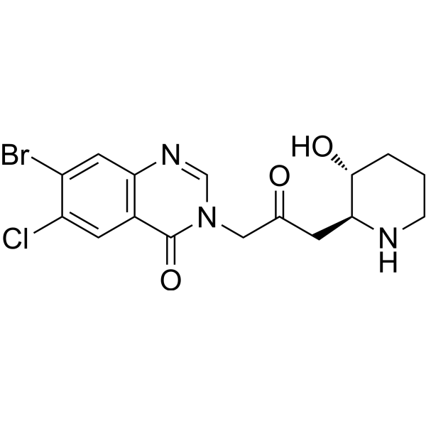
Halofuginone (RU-19110), a Febrifugine derivative, is a competitive prolyl-tRNA synthetase inhibitor with a Ki of 18.3 nM. Halofuginone is a specific inhibitor of type-I collagen synthesis and attenuates osteoarthritis (OA) by inhibition of TGF-β activity. Halofuginone is also a potent pulmonary vasodilator by activating Kv channels and blocking voltage-gated, receptor-operated and store-operated Ca2+ channels. Halofuginone has anti-malaria, anti-inflammatory, anti-cancer, anti-fibrosis effects.
Nos produits utilisent uniquement pour la recherche. Nous ne vendons pas aux patients.
- Pureté : 99.28%
- CAS No.: 55837-20-2
- Formule: C16H17BrClN3O3
- Masse moléculaire:414.68
-
Stockage:Powder -20°C, 3 years , 4°C, 2 years ; In solvent -80°C, 6 months , -20°C, 1 month

To place orders, for customer services and technical support, please contact: MedChemExpress USA
Tel: 609-228-6898 E-mail: [email protected] [email protected]


-
 Activité biologique
-
Chemical Information
-
Solvant et solubilité
- Protocole
- Pureté et documentation
- Références
-
 Help & FAQs


-
HY-13433
ThapsigarginER Stress Inducer -

HY-10219
RapamycinmTOR Inhibitor -
HY-112654
GCN2iBGCN2 Kinase Inhibitor -
HY-N6742
BorrelidinCDK Inhibitor -
HY-K0301
Cell Counting Kit-8 -
HY-118266
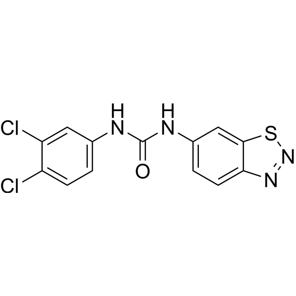
BTdCPUHRI Activator -
HY-17026
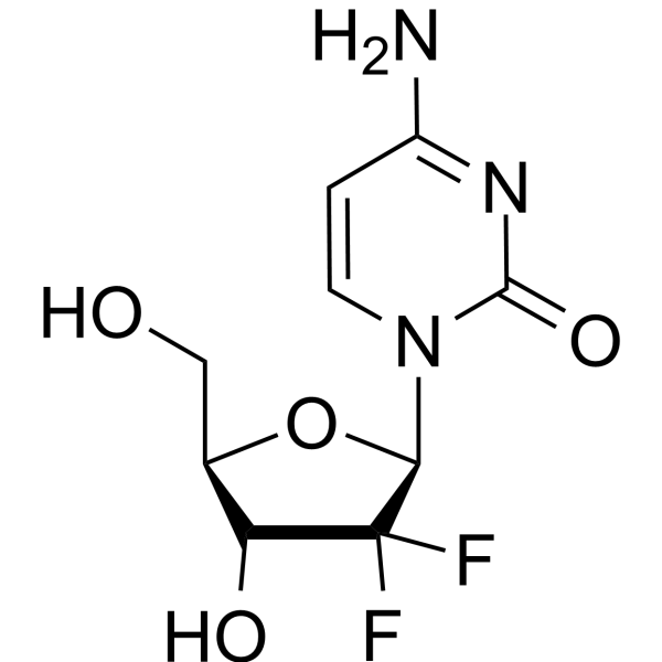
GemcitabineDNA Synthesis Inhibitor -
HY-18982
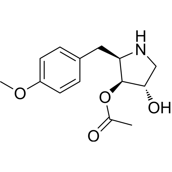
AnisomycinProtein Synthesis Inhibitor -
HY-90006
5-FluorouracilThymidylate Synthase Inhibitor -

HY-N6782
OligomycinH+-ATP-Synthase Inhibitor -
HY-A0004
DecitabineDNMT Inhibitor -

HY-100558
Bafilomycin A1V-ATPase Inhibitor
Publications Citing Use of MedChemExpress (MCE) Halofuginone
More- Cell Metab. 2023 Dec 5;35(12):2216-2230.e8. [Abstract]
- Phytomedicine. 2025 Jul 25:143:156788. [Abstract]
- Proc Natl Acad Sci U S A. 2025 Aug 19;122(33):e2514837122. [Abstract]
- Br J Pharmacol. 2025 Aug 13. [Abstract]
- Br J Pharmacol. 2021 Sep;178(17):3373-3394. [Abstract]
- Anal Chem. 2025 Jun 3;97(21):11099-11109. [Abstract]
- Tissue Eng Regen Med. 2021 Dec;18(6):963-973. [Abstract]
- Hum Gene Ther. 2022 Mar;33(5-6):237-249. [Abstract]
- ACS Infect Dis. 2023 Apr 14;9(4):1004-1021. [Abstract]
- iScience. 2023 Mar 4;26(4):106334. [Abstract]
- Antiviral Res. 2026 Jun:250:106417. [Abstract]
- RNA. 2026 Feb 8:rna.080883.125. [Abstract]
- Fish Shellfish Immunol. 2024 Aug:151:109727. [Abstract]
- J Virol. 2026 May 19;100(5):e0011726. [Abstract]
- J Funct Foods. 2024 Jul.
- Photodiagnosis Photodyn Ther. 2022 Mar:37:102572. [Abstract]
- Indian J Hematol Blood Transfus. 2024 Jul;40(3):407-414. [Abstract]
- bioRxiv. 2026 Sep 13.
- bioRxiv. 2026 Jan 15.
- bioRxiv. 2025 July 11.
- Rutgers University. 2025.
- bioRxiv. 2025 April 12.
- Friedrich-Alexander University Erlangen-Nuremberg. 2023 May 2.
 Customer Validation & Images

-
Cell Imaging/Staining
-
Bio/Physico-chemical Assay
-
Cell Proliferation/Viability Assay
-
Cell Migration/Invasion Assay
-
WB
Voir tous les produits spécifiques à Isoform DNA/RNA Synthesis
MoreVoir tous les produits spécifiques à Isoform Parasite
MoreVoir tous les produits spécifiques à Isoform Calcium Channel
More

Activité biologique
Description
IC50 & Target
|
Plasmodium |
Cellular Effect
|
Cell Line

|
Type | Value | Description | References |
|---|---|---|---|---|
| Huh-7 | IC50 |
0.017 μM
Compound: 2, HF
|
Antiplasmodial activity against liver stage Plasmodium berghei infected in human HuH7 cells co-expressing GFP-Luccon treated for 1 hr prior to infection followed by 24 hrs after compound washout measured after 48 hrs post-infection by Alamar Blue assay
Antiplasmodial activity against liver stage Plasmodium berghei infected in human HuH7 cells co-expressing GFP-Luccon treated for 1 hr prior to infection followed by 24 hrs after compound washout measured after 48 hrs post-infection by Alamar Blue assay
|
[PMID: 23701465] |
In Vitro
Halofuginone competitively inhibits prolyl-tRNA synthetase by occupying both the prolineand tRNA-binding pockets of prolyl-tRNA synthetase[1].
The IC50s of Halofuginone (1-10000 nM; 48 hours) are 114.6 and 58.9 nM in KYSE70 and A549 cells, respectively.
The IC50s of Halofuginone (1-1000 nM; 24 hours) for NRF2 protein are 22.3 and 37.2 nM in KYSE70 and A549 cells, respectively. The IC50 of Halofuginone for global protein synthesis is 22.6 and 45.7 nM in KYSE70 and A549 cells, respectively[1].
Halofuginone increases voltage-gated K+ (Kv) currents in pulmonary artery smooth muscle cells (PASMC) and K+ currents through KCNA5 channels in HEK cells transfected with KCNA5 gene. Halofuginone inhibits receptor-operated Ca2+ entry (ROCE) in HEK cells transfected with calcium-sensing receptor gene and attenuates store-operated Ca2+ entry (SOCE) in PASMC[5].
MedChemExpress (MCE) has not independently confirmed the accuracy of these methods. They are for reference only.
-
Cell Line:KYSE70 cells from human oesophageal cancer harbouring a mutation in the NRF2 gene and A549 cells harbouring theKEAP1 gene mutation
-
Concentration:1, 10, 100, 1000, 10000 nM
-
Incubation Time:48 hours
-
Result:The IC50s were 114.6 and 58.9 nM in KYSE70 and A549 cells, respectively.
-
Cell Line:KYSE70 cells from human oesophageal cancer harbouring a mutation in the NRF2 gene and A549 cells harbouring theKEAP1 gene mutation.
-
Concentration:1, 10, 100, 1000 nM
-
Incubation Time:24 hours
-
Result:The IC50s for NRF2 protein were 22.3 and 37.2 nM in KYSE70 and A549 cells, respectively.
In Vivo
MedChemExpress (MCE) has not independently confirmed the accuracy of these methods. They are for reference only.
-
Animal Model:3-month-old male C57BL/6J (WT) mice[3]
-
Dosage:0.2, 0.5, 1 or 2.5 mg/kg
-
Administration:Injected intraperitoneally every other day for 1 month
-
Result:Attenuated progression of OA in ACLT mice.
-
Animal Model:Male nude mice (BALB/C nu/nu mice) (6-8-week)[1]
-
Dosage:0.25 mg/kg
-
Administration:Intraperitoneally injected; every day; 16 days
-
Result:The combined treatment with Cisplatin significantly suppressed the tumor volume. NRF2 protein levels in tumors were indeed decreased.
Essai clinique
| NCT Number | Sponsor | Condition | Start Date |
Phase
|
|---|---|---|---|---|
| NCT01329991 | Plexxikon| | 2011-05 | PHASE1 |


Chemical Information
-
CAS No. 55837-20-2
-
Appearance Solid
-
Masse moléculaire 414.68
-
Formule C16H17BrClN3O3
-
Color White to off-white
-
SMILES
O=C1N(CC(C[C@@H]2NCCC[C@H]2O)=O)C=NC3=C1C=C(Cl)C(Br)=C3
-
Synonyms
RU-19110
-
Structure Classification
-
Initial Source
-
Livraison
Room temperature in continental US; may vary elsewhere.
-
Stockage
Powder -20°C 3 years 4°C 2 years In solvent -80°C 6 months -20°C 1 month


Publications (23)
-
Journal Impact Factor
-
Most Recent
-
Cell Metab
2023 Dec 5;35(12):2216-2230.e8. PMID: 37979583 -
Phytomedicine
Halofuginone targets Serine/Glycine synthesis to reverse epidermal growth factor receptor Tyrosine Kinase inhibitor resistance in lung adenocarcinoma. [Abstract]2025 Jul 25:143:156788. PMID: 40354707
Halofuginone purchased from MedChemExpress. Usage Cited in: Phytomedicine. 2025 Jul 25:143:156788. [Abstract]
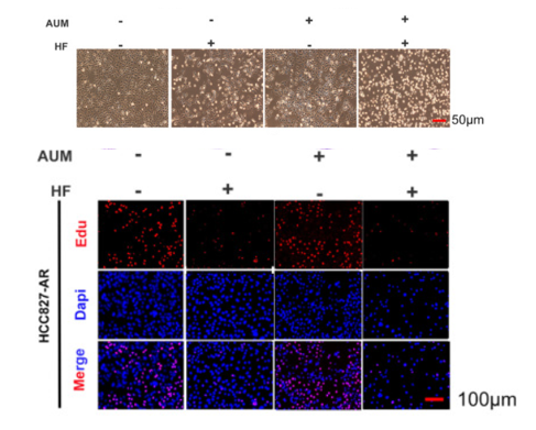
Cell morphology under the microscope after 24 h of 4 μM AUM in combination with 50 nM Halofuginone (HF) in the resistant cell lines and inhibition of colony formation.
-
Proc Natl Acad Sci U S A
Stress granule-mediated ZBP1 activation drives necroptotic cell death in non-obstructive azoospermia and testicular aging. [Abstract]2025 Aug 19;122(33):e2514837122. PMID: 40811463
Halofuginone purchased from MedChemExpress. Usage Cited in: Proc Natl Acad Sci U S A. 2025 Aug 19;122(33):e2514837122. [Abstract]
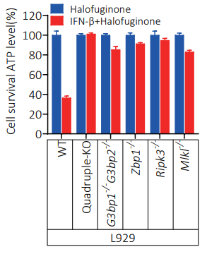
Cultured L929, L929(Pkr-/-Hri-/-Perk-/-Gcn2-/-), L929(G3bp1-/-G3bp2-/-), L929(Zbp1-/-), L929(Ripk3-/-) and L929(Mlkl-/-) cells were treated Halofuginone (500 nM) and Halofuginone (500 nM)+IFN-β(10 ng/ml) for 42 hours; The intracellular ATP levels were measured by Cell Titer-Glo.
-
Br J Pharmacol
2025 Aug 13. PMID: 40803344 -
Br J Pharmacol
2021 Sep;178(17):3373-3394. PMID: 33694155
Halofuginone purchased from MedChemExpress. Usage Cited in: Br J Pharmacol. 2021 Sep;178(17):3373-3394. [Abstract]
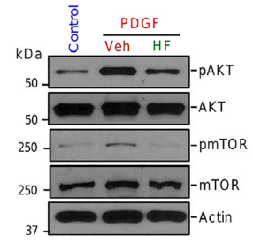
Western blot analysis on p-Akt, Akt, p-mTOR and mTOR in control PASMCs and PASMCs treated with PDGF in the absence (vehicle control, Veh) and presence (Halofuginone; HF) of 1 μM halofuginone (for 24 h).
-
Anal Chem
Exposome-Scale Investigation of Cl-/Br-Containing Chemicals Using High-Resolution Mass Spectrometry, Multistage Machine Learning, and Cloud Computing. [Abstract]2025 Jun 3;97(21):11099-11109. PMID: 40401576 -
Tissue Eng Regen Med
Three-Dimensional Modeling of the Structural Microenvironment in Post-Traumatic War Wounds. [Abstract]2021 Dec;18(6):963-973. PMID: 34363599 -
Hum Gene Ther
Antifibrotic Therapy Augments the Antitumor Effects of Vesicular Stomatitis Virus Via Reprogramming Tumor Microenvironment. [Abstract]2022 Mar;33(5-6):237-249. PMID: 34405694 -
ACS Infect Dis
2023 Apr 14;9(4):1004-1021. PMID: 36919909 -
iScience
EtcPRS Mut as a molecular marker of halofuginone resistance in Eimeria tenella and Toxoplasma gondii. [Abstract]2023 Mar 4;26(4):106334. PMID: 36968087 -
Antiviral Res
Repurposing screen using a robust human rhinovirus infectious clone identifies pyrvinium pamoate with antiviral activity. [Abstract]2026 Jun:250:106417. PMID: 42025967 -
RNA
Tuning tRNA synthetase inhibition reveals parabolic induction of stress granules limited in size and RNA content. [Abstract]2026 Feb 8:rna.080883.125. PMID: 41656003 -
Fish Shellfish Immunol
Loss of Gcn2 exacerbates gossypol induced oxidative stress, apoptosis and inflammation in zebrafish. [Abstract]2024 Aug:151:109727. PMID: 38936520 -
J Virol
Febrifugine dihydrochloride restricts porcine epidemic diarrhea virus replication by modulating the IGF1R-driven PI3K/AKT-apoptosis axis. [Abstract]2026 May 19;100(5):e0011726. PMID: 41983768 -
Halofuginone purchased from MedChemExpress. Usage Cited in: J Funct Foods. 2024 Jul.
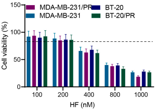
Cell viability measured by MTT assay after treatment with 100, 200, 400, 800, and 1000 nM concentrations of Halofuginone (HF) in parental cells MDA-MB-231 and BT-20 for 24 h, as well as resistant cells MDA-MB-231/PR and BT-20/PR.
Halofuginone purchased from MedChemExpress. Usage Cited in: J Funct Foods. 2024 Jul.
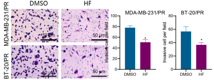
Halofuginone (HF; 200 nM; 24 h) treated MDA-MB-231/PR and BT20/PR cells reduced cell invasion capabilities.
-
Photodiagnosis Photodyn Ther
Halofuginone enhances the anti-tumor effect of ALA-PDT by suppressing NRF2 signaling in cSCC. [Abstract]2022 Mar:37:102572. PMID: 34628069 -
Indian J Hematol Blood Transfus
Halofuginone Inhibits Osteoclastogenesis and Enhances Osteoblastogenesis by Regulating Th17/Treg Cell Balance in Multiple Myeloma Mice with Bone Lesions. [Abstract]2024 Jul;40(3):407-414. PMID: 39011260 -
-
-
-
-
-


Solvant et solubilité
In Vitro:
DMSO : 25 mg/mL (60.29 mM; ultrasonic and warming and heat to 60°C; Hygroscopic DMSO has a significant impact on the solubility of product, please use newly opened DMSO)
Please refer to the solubility information to select the appropriate solvent. Once prepared, please aliquot and store the solution to prevent product inactivation from repeated freeze-thaw cycles.
Storage method and period of stock solution: -80°C, 6 months; -20°C, 1 month. When stored at -80°C, please use it within 6 months. When stored at -20°C, please use it within 1 month.
Please refer to the solubility information to select the appropriate solvent. Once prepared, please aliquot and store the solution to prevent product inactivation from repeated freeze-thaw cycles.
Storage method and period of stock solution: -80°C, 6 months; -20°C, 1 month. When stored at -80°C, please use it within 6 months. When stored at -20°C, please use it within 1 month.
Concentration (start) × Volume (start) = Concentration (final) × Volume (final)
In Vivo:
Select the appropriate dissolution method based on your experimental animal and administration route.
- For the following dissolution methods, please ensure to first prepare a clear stock solution using an In Vitro approach and then sequentially add co-solvents:
- To ensure reliable experimental results, the clarified stock solution can be appropriately stored based on storage conditions. As for the working solution for In Vivo experiments, it is recommended to prepare freshly and use it on the same day.
- The percentages shown for the solvents indicate their volumetric ratio in the final prepared solution. If precipitation or phase separation occurs during preparation, heat and/or sonication can be used to aid dissolution.
Add each solvent one by one: 10% DMSO 40% PEG300 5% Tween-80 45% Saline
Solubility: ≥ 2.5 mg/mL (6.03 mM); Clear solution
This protocol yields a clear solution of ≥ 2.5 mg/mL (saturation unknown).
Taking 1 mL working solution as an example, add 100 μL DMSO stock solution (25.0 mg/mL) to 400 μL PEG300, and mix evenly; then add 50 μL Tween-80 and mix evenly; then add 450 μL Saline to adjust the volume to 1 mL.
Preparation of Saline: Dissolve 0.9 g sodium chloride in ddH₂O and dilute to 100 mL to obtain a clear Saline solution.
Add each solvent one by one: 10% DMSO 90% (20% SBE-β-CD in Saline)
Solubility: ≥ 2.5 mg/mL (6.03 mM); Clear solution
This protocol yields a clear solution of ≥ 2.5 mg/mL (saturation unknown).
Taking 1 mL working solution as an example, add 100 μL DMSO stock solution (25.0 mg/mL) to 900 μL 20% SBE-β-CD in Saline, and mix evenly.
Preparation of 20% SBE-β-CD in Saline (4°C, storage for one week): 2 g SBE-β-CD powder is dissolved in 10 mL Saline, completely dissolve until clear.
In Vivo Dissolution Calculator
Please enter the basic information of animal experiments:
-
-
-
-
Recommended: Prepare an additional quantity of animals to account for potential losses during experiments.
Please enter your animal formula composition:
-
%DMSO +
Recommended: Keep the proportion of DMSO in working solution below 2% if your animal is weak.
-
%+
-
+%Tween-80 + +
-
%Saline +
The co-solvents required include: DMSO, . All of co-solvents are available by MedChemExpress (MCE). , Tween 80. All of co-solvents are available by MedChemExpress (MCE).
Working solution concentration: 0.22 mg/mL
Method for preparing stock solution: mg drug dissolved in μL DMSO. Stock solution concentration: mg/mL.
1. Take μL DMSO stock solution;
2. Add μL .
μL , mix evenly;
3. Then add μL Tween 80, mix evenly;
4. Then add μL
Please ensure that the stock solution in the first step is dissolved to a clear state, and add co-solvents in sequence. You can use ultrasonic heating (ultrasonic cleaner, recommended frequency 20-40 kHz), vortexing, etc. to assist dissolution.


Pureté et documentation
-
Fiche technique (289 KB)
-
SDS (393 KB)
- English - EN (393 KB)
- Français - FR (393 KB)
- Deutsch - DE (393 KB)
- Norwegian - NO (393 KB)
- Español - ES (393 KB)
- Swedish - SV (393 KB)
- Italian - IT (393 KB)
- Korean - KR (393 KB)
- Portuguese - PT (393 KB)
-
Instruction de manipulation (2659 KB)
Références
[1]. Tsuchida K, et al. Halofuginone enhances the chemo-sensitivity of cancer cells by suppressing NRF2 accumulation. Free Radic Biol Med. 2017 Feb;103:236-247. [Content Brief]
[2]. Keller TL, et al. Halofuginone and other Febrifugine derivatives inhibit prolyl-tRNA synthetase. Nat Chem Biol. 2012 Feb 12;8(3):311-7. [Content Brief]
[3]. Cui Z, et al. Halofuginone attenuates osteoarthritis by inhibition of TGF-β activity and H-type vessel formation in subchondral bone. Ann Rheum Dis. 2016 Sep;75(9):1714-21. [Content Brief]
[4]. Tracy L McGaha, et al. Halofuginone, an inhibitor of type-I collagen synthesis and skin sclerosis, blocks transforming-growth-factor-beta-mediated Smad3 activation in fibroblasts. J Invest Dermatol. 2002 Mar;118(3):461-70. [Content Brief]
[5]. Pritesh P Jain, et al. Halofuginone, a Promising Drug for Treatment of Pulmonary Hypertension. Br J Pharmacol. 2021 Mar 10. [Content Brief]


Complete Stock Solution Preparation Table
Please refer to the solubility information to select the appropriate solvent. Once prepared, please aliquot and store the solution to prevent product inactivation from repeated freeze-thaw cycles.
Storage method and period of stock solution: -80°C, 6 months; -20°C, 1 month. When stored at -80°C, please use it within 6 months. When stored at -20°C, please use it within 1 month.
| Optional Solvent | Concentration Solvent Mass | 1 mg | 5 mg | 10 mg | 25 mg |
|---|---|---|---|---|---|
| DMSO | 1 mM | 2.4115 mL | 12.0575 mL | 24.1150 mL | 60.2875 mL |
| 5 mM | 0.4823 mL | 2.4115 mL | 4.8230 mL | 12.0575 mL | |
| 10 mM | 0.2411 mL | 1.2057 mL | 2.4115 mL | 6.0287 mL | |
| 15 mM | 0.1608 mL | 0.8038 mL | 1.6077 mL | 4.0192 mL | |
| 20 mM | 0.1206 mL | 0.6029 mL | 1.2057 mL | 3.0144 mL | |
| 25 mM | 0.0965 mL | 0.4823 mL | 0.9646 mL | 2.4115 mL | |
| 30 mM | 0.0804 mL | 0.4019 mL | 0.8038 mL | 2.0096 mL | |
| 40 mM | 0.0603 mL | 0.3014 mL | 0.6029 mL | 1.5072 mL | |
| 50 mM | 0.0482 mL | 0.2411 mL | 0.4823 mL | 1.2057 mL | |
| 60 mM | 0.0402 mL | 0.2010 mL | 0.4019 mL | 1.0048 mL |


Halofuginone Related Classifications
HY-N1584 Related Classifications
 Powered by Bioz