


Andrographolide
Based on 34 publication(s) in Google Scholar
Andrographolide is a NF-κB inhibitor, which inhibits NF-κB activation through covalent modification of a cysteine residue on p50 in endothelial cells without affecting IκBα degradation or p50/p65 nuclear translocation. Andrographolide has antiviral effects.
For research use only. We do not sell to patients.
- Purity: 99.74%
- CAS No.: 5508-58-7
- Formula: C20H30O5
- Molecular Weight:350.45
-
Storage:Powder -20°C, 3 years , 4°C, 2 years ; In solvent -80°C, 1 year , -20°C, 6 months


-
 Biological Activity
-
Chemical Information
-
Solvent & Solubility
- Protocol
- Purity & Documentation
- References
-
 Help & FAQs
-


Anti-Infection Compound Library
HY-L002
-
Immunology/Inflammation Compound Library
HY-L007
-
NF-κB Signaling Compound Library
HY-L014
-
Stem Cell Signaling Compound Library
HY-L017
-
Natural Product Library
HY-L021
-
FDA-Approved Drug Library
HY-L022
-
Anti-Cancer Compound Library
HY-L025
-
Antiviral Compound Library
HY-L027
-
Autophagy Compound Library
HY-L029
-
Anti-Aging Compound Library
HY-L034
-
Drug Repurposing Compound Library
HY-L035
-
Antioxidant Compound Library
HY-L037
-
Differentiation Inducing Compound Library
HY-L038
-
Oxygen Sensing Compound Library
HY-L045
-
Anti-COVID-19 Compound Library
HY-L052
-
NMPA-Approved Drug Library
HY-L053
-
Medicine Food Homology Compound Library
HY-L055
-
Terpenoids Library
HY-L056
-
Pyroptosis Compound Library
HY-L059
-
Traditional Chinese Medicine Active Compound Library
HY-L065
-
FDA Approved & Pharmacopeial Drug Library
HY-L066
-
Anti-Breast Cancer Compound Library
HY-L074
-
Anti-Pancreatic Cancer Compound Library
HY-L077
-
Anti-Blood Cancer Compound Library
HY-L079
-
Antiparasitic Compound library
HY-L082
-
Anti-Cancer Metabolism Compound Library
HY-L083
-
Anti-Obesity Compound Library
HY-L087
-
Transcription Factor-Targeted Library
HY-L090
-
Targeted Diversity Library
HY-L099
-
Anti-Liver Cancer Compound Library
HY-L101
-
Rare Diseases Drug Library
HY-L102
-
Anti-Cancer Natural Product Library
HY-L107
-
Antidepressant Compound Library
HY-L108
-
Antiviral Traditional Chinese Medicine Active Compound Library
HY-L113
-
Anti-inflammatory Traditional Chinese Medicine Active Compound Library
HY-L114
-
Plant-Sourced Natural Product Library
HY-L115
-
FDA-Approved Anticancer Drug Library
HY-L122
-
Anti-Prostate Cancer Compound Library
HY-L124
-
Non-steroidal Anti-Inflammatory Compound Library
HY-L130
-
Cancer Stem Cells Compound Library
HY-L135
-
Pain-Related Compound Library
HY-L139
-
Off-patent Drug Library
HY-L141
-
Cysteine Targeted Covalent Library
HY-L153
-
Highly Selective Inhibitors Library
HY-L158
-
Highly Selective Activators Library
HY-L159
-
Cell Death Library
HY-L162
-
FDA-Approved Traditional Chinese Medicine Active Compound Library
HY-L163
-
Anti-Hematopathy Compound Library
HY-L171
-
Anti-Ovarian Cancer Compound Library
HY-L173
-
Multi-Target Compound Library
HY-L176
-
Radioprotector Library
HY-L178
-
Bioactive Compound Library Max
HY-L181
-
MCE Bioactive Compound Library
HY-L001V
-
Natural Product Library Plus
HY-L021P
-
Drug Repurposing Compound Library Plus
HY-L035P
-
FDA-Approved Drug Library Plus
HY-L022P
-
FDA-Approved Drug Library Mini
HY-L022M
-
Natural Product and Natural Product-Like Compound Library
HY-L021M
-
Bioactive Compound Library
HY-L001
-
Anti-Fibrosis Compound Library
HY-L185
-
Miao Ethnicity Medicine Compound Library
HY-L190
-
Dietary Supplement Library
HY-L192
-
Heat-Clearing and Detoxification Traditional Chinese Medicine Compound Library
HY-L194
-
Non-Alcoholic Fatty Liver Disease (NAFLD) Compound Library
HY-L199
-
RO5 Drug-like Natural Product Library
HY-L200
-
Cell Proliferation Compound Library
HY-L201
-
Flavor Compound Library
HY-L202
-
High-Throughput Bioactive Compound Library
HY-L205
-
High-Throughput Natural Product Library
HY-L206
-
Ancient Chinese Classical Formulas Traditional Chinese Medicine Active Compound Library
HY-L209
-
Cosmetic Ingredient Compound Library
HY-L221
-
Diarrhea-Related Traditional Chinese Medicine Active Compound Library
HY-L224
-
Mongolian Medicine Compound Library
HY-L238
-
Chinese Baijiu Compound Library
HY-L241
-
Natural Product Diversity Scaffold Library
HY-L245
-
RNA Binding Bioactive Compound Library
HY-L248
-
Lactylation Compound Library
HY-L249
-
Mass Spectrometry Natural Product Library
HY-L262


Publications Citing Use of MedChemExpress (MCE) Andrographolide
More- Cell Res. 2025 Jan;35(1):23-44. [Abstract]
- Adv Sci (Weinh). 2024 Dec 5:e2410360. [Abstract]
- Nucleic Acids Res. 2021 Jan 8;49(D1):D1113-D1121. [Abstract]
- Phytomedicine. 2025 May:140:156484. [Abstract]
- Phytomedicine. 2023 Jan:109:154601. [Abstract]
- Int J Pharm. 2022 Nov 25:628:122361. [Abstract]
- Drug Des Devel Ther. 2023 Feb 20:17:535-550. [Abstract]
- Biofactors. 2025 Nov-Dec;51(6):e70063. [Abstract]
- Front Pharmacol. 2021 Mar 16;12:653035. [Abstract]
- Int Immunopharmacol. 2025 May 7:157:114764. [Abstract]
- Front Microbiol. 2018 Oct 8:9:2407. [Abstract]
- J Cell Mol Med. 2019 Aug;23(8):5518-5531. [Abstract]
- iScience. 2025 Dec 8.
- J Inflamm Res. 2023 Oct 6:16:4413-4423. [Abstract]
- J Biol Chem. 2026 Jan 20:111182. [Abstract]
- Oncol Rep. 2020 Nov;44(5):1939-1948. [Abstract]
- Cell Signal. 2025 Nov:135:112014. [Abstract]
- Arch Pharm (Weinheim). 2022 May;355(5):e2100467. [Abstract]
- Med Oncol. 2022 Nov 9;40(1):11. [Abstract]
- ChemMedChem. 2022 Mar 4;17(5):e202100732. [Abstract]
- Pathogens. 2026 Feb;15(2):169.
- J Pharm Pharmacol. 2025 Aug 4:rgaf052. [Abstract]
- Naunyn Schmiedebergs Arch Pharmacol. 2025 Dec 18. [Abstract]
- Genes (Basel). 2025 Dec 17;16(12):1514. [Abstract]
- Vet Microbiol. 2026 May:316:110992. [Abstract]
- Fitoterapia. 2026 Apr:190:107179. [Abstract]
- Ultrasound Med Biol. 2020 Sep;46(9):2464-2471. [Abstract]
- Gene. 2024 Jun 15:911:148351. [Abstract]
- Biochem Biophys Res Commun. 2024 May 25:723:150179. [Abstract]
- Vet Parasitol. 2022 May;305:109710. [Abstract]
- J Gastrointest Oncol. 2025 Aug 30;16(4):1550-1561. [Abstract]
- J Orthop. 2024 Nov 26:64:108-116. [Abstract]
- Oxid Med Cell Longev. 2022 May 12;2022:4139330. [Abstract]
- Research Square Preprint. 2021 Aug.
 Customer Validation & Images

-
Cell Proliferation/Viability Assay
-
ELISA
-
ELISA
-
WB
-
WB
All Parasite Isoforms
More

Biological Activity
|
p50 |
|
Cell Line
|
Type | Value | Description | References |
|---|---|---|---|---|
| A2058 | IC50 |
26 μM
Compound: 4; AGL
|
Cytotoxicity against human A2058 cells incubated for 48 hrs by MTT assay
Cytotoxicity against human A2058 cells incubated for 48 hrs by MTT assay
|
[PMID: 33289552] |
| A498 | GI50 |
3 μM
Compound: 1
|
Growth inhibition of human A498 cells
Growth inhibition of human A498 cells
|
[PMID: 31151068] |
| A549 | GI50 |
13.37 μM
Compound: 1
|
Growth inhibition of human A549 cells after 72 hrs by SRB assay
Growth inhibition of human A549 cells after 72 hrs by SRB assay
|
[PMID: 23768904] |
| A549 | GI50 |
13.37 μM/L
Compound: 1
|
Growth inhibition of human A549 cells after 72 hrs by SRB assay
Growth inhibition of human A549 cells after 72 hrs by SRB assay
|
[PMID: 23768904] |
| A549 | IC50 |
27.9 μM
Compound: 1
|
Cytotoxicity against human A549 cells assessed as reduction in cell viability by SRB assay
Cytotoxicity against human A549 cells assessed as reduction in cell viability by SRB assay
|
[PMID: 28755636] |
| A549 | IC50 |
32.02 μM
Compound: 1
|
Anticancer activity against human A549 cells assessed as reduction in cell viability measured after 72 hrs by SRB assay
Anticancer activity against human A549 cells assessed as reduction in cell viability measured after 72 hrs by SRB assay
|
[PMID: 32527561] |
| A549 | IC50 |
12.15 μM
Compound: 1
|
Cytotoxicity against human A549 cells assessed as reduction in cell viability by SRB assay
Cytotoxicity against human A549 cells assessed as reduction in cell viability by SRB assay
|
[PMID: 33316411] |
| A549 | IC50 |
9.71 μg/mL
Compound: 1
|
Cytotoxicity against human A549 cells by MTT assay
Cytotoxicity against human A549 cells by MTT assay
|
10.1039/C4MD00566J |
| ACHN | IC50 |
3.03 μg/mL
Compound: 1
|
Cytotoxicity against human ACHN cells by MTT assay
Cytotoxicity against human ACHN cells by MTT assay
|
10.1039/C4MD00566J |
| B16 | IC50 |
7.54 μg/mL
Compound: 1
|
Cytotoxicity against mouse B16 cells by MTT assay
Cytotoxicity against mouse B16 cells by MTT assay
|
10.1039/C4MD00566J |
| BMDM | IC50 |
0.55 μM
Compound: AM-721/20681006
|
Inhibition of GST-tagged recombinant RANKL/M-CSF-induced osteoclastogenesis in C57BL/6 mouse bone marrow derived macrophages assessed as TRAcP enzymatic activity incubated for 5 days with subsequent replenishment of media for 2 days and measured by leucoc
Inhibition of GST-tagged recombinant RANKL/M-CSF-induced osteoclastogenesis in C57BL/6 mouse bone marrow derived macrophages assessed as TRAcP enzymatic activity incubated for 5 days with subsequent replenishment of media for 2 days and measured by leucoc
|
[PMID: 33158578] |
| BMMC | CC50 |
10.31 μM
Compound: 1; AP
|
Cytotoxicity against C57BL/6 mouse BMM cells assessed as reduction in cell viability measured after 48 hrs in presence of M-CSF by CCK8 assay
Cytotoxicity against C57BL/6 mouse BMM cells assessed as reduction in cell viability measured after 48 hrs in presence of M-CSF by CCK8 assay
|
[PMID: 33485256] |
| BMMC | IC50 |
1.01 μM
Compound: 1; AP
|
Anti-osteoporosis activity in C57BL/6 mouse BMM cells assessed as inhibition of M-CSF/RANKL-induced osteoclastogenesis by measuring reduction in multinucleated TRAP+ cells incubated for 5 days with media replenishment for every 48 hrs measured by TRAP-sta
Anti-osteoporosis activity in C57BL/6 mouse BMM cells assessed as inhibition of M-CSF/RANKL-induced osteoclastogenesis by measuring reduction in multinucleated TRAP+ cells incubated for 5 days with media replenishment for every 48 hrs measured by TRAP-sta
|
[PMID: 33485256] |
| BT-549 | IC50 |
23.2 μM
Compound: Andrographolide
|
Antiproliferative activity against human BT-549 cells
Antiproliferative activity against human BT-549 cells
|
[PMID: 34315039] |
| Col2 | ED50 |
13.6 μM
Compound: 1
|
Cytotoxicity against human Col2 cells by SRB assay
Cytotoxicity against human Col2 cells by SRB assay
|
[PMID: 22154665] |
| DU-145 | GI50 |
15.99 μM
Compound: 1
|
Growth inhibition of human DU145 cells after 72 hrs by SRB assay
Growth inhibition of human DU145 cells after 72 hrs by SRB assay
|
[PMID: 23768904] |
| DU-145 | GI50 |
15.99 μM/L
Compound: 1
|
Growth inhibition of human DU145 cells after 72 hrs by SRB assay
Growth inhibition of human DU145 cells after 72 hrs by SRB assay
|
[PMID: 23768904] |
| DU-145 | GI50 |
3 μM
Compound: 1
|
Growth inhibition of human DU145 cells
Growth inhibition of human DU145 cells
|
[PMID: 31151068] |
| HCT-116 | IC50 |
23.93 μM
Compound: 1
|
Cytotoxicity against human HCT116 cells assessed as growth inhibition after 72 hrs by MTS assay
Cytotoxicity against human HCT116 cells assessed as growth inhibition after 72 hrs by MTS assay
|
[PMID: 23628335] |
| HCT-116 | GI50 |
4.5 μM
Compound: 21; AGP
|
Antiproliferative activity against human HCT-116 cells harboring KRAS G13D mutant assessed as cell growth inhibition measured after 96 hrs by MTT assay
Antiproliferative activity against human HCT-116 cells harboring KRAS G13D mutant assessed as cell growth inhibition measured after 96 hrs by MTT assay
|
[PMID: 33228976] |
| HCT-116 | IC50 |
>40 μM
Compound: 4; AGL
|
Cytotoxicity against human HCT-116 cells
Cytotoxicity against human HCT-116 cells
|
[PMID: 33289552] |
| HCT-116 | IC50 |
31.8 μM
Compound: Andrographolide
|
Antiproliferative activity against human HCT-116 cells assessed as cell growth inhibition measured after 48 hrs by MTT assay
Antiproliferative activity against human HCT-116 cells assessed as cell growth inhibition measured after 48 hrs by MTT assay
|
[PMID: 34315039] |
| HCT-116 | IC50 |
19 μg/mL
Compound: Andro
|
Antiproliferative activity against human HCT-116 cells assessed as reduction in cell viability incubated for 48 hrs by WST-1 assay
Antiproliferative activity against human HCT-116 cells assessed as reduction in cell viability incubated for 48 hrs by WST-1 assay
|
[PMID: 38306267] |
| HEK293 | IC50 |
49.6 μM
Compound: 1
|
Inhibition of TNF-alpha-stimulated NF-kappaB p50 (unknown origin) expressed in HEK293 cells preincubated for 4 hrs followed by TNF-alpha challenge measured after 24 hrs by secreted alkaline phosphatase reporter assay
Inhibition of TNF-alpha-stimulated NF-kappaB p50 (unknown origin) expressed in HEK293 cells preincubated for 4 hrs followed by TNF-alpha challenge measured after 24 hrs by secreted alkaline phosphatase reporter assay
|
[PMID: 25615020] |
| HEK293 | CC50 |
26.25 μM
Compound: 1
|
Cytotoxicity against HEK293 cells assessed as reduction in cell viability measured after 24 hrs by Celltiter-glo assay
Cytotoxicity against HEK293 cells assessed as reduction in cell viability measured after 24 hrs by Celltiter-glo assay
|
[PMID: 32712435] |
| HeLa | IC50 |
23 μM
Compound: Andrographolide
|
Antiviral activity against DENV2 infected in human HeLa cells incubated for 1 hrs by plaque assay
Antiviral activity against DENV2 infected in human HeLa cells incubated for 1 hrs by plaque assay
|
[PMID: 34315039] |
| HeLa | IC50 |
10.42 μg/mL
Compound: 1
|
Cytotoxicity against human HeLa cells by MTT assay
Cytotoxicity against human HeLa cells by MTT assay
|
10.1039/C4MD00566J |
| HepG2 | IC50 |
9.7 μM
Compound: 1
|
Antagonist activity at Gal4 DBD-fused human FXR LBD expressed in human HepG2 cells after 8 hrs by luciferase reporter gene assay
Antagonist activity at Gal4 DBD-fused human FXR LBD expressed in human HepG2 cells after 8 hrs by luciferase reporter gene assay
|
[PMID: 31151068] |
| HepG2 | IC50 |
21 μM
Compound: Andrographolide
|
Antiviral activity against DENV2 infected in human HepG2 cells incubated for 1 hrs by plaque assay
Antiviral activity against DENV2 infected in human HepG2 cells incubated for 1 hrs by plaque assay
|
[PMID: 34315039] |
| HepG2 2.2.15 | CC50 |
198 μM
Compound: 1b
|
Cytotoxicity against human HepG2.2.15 cells by MTT assay
Cytotoxicity against human HepG2.2.15 cells by MTT assay
|
[PMID: 24731274] |
| HepG2 2.2.15 | IC50 |
54.1 μM
Compound: 1b
|
Antiviral activity against HBV infected in human HepG2.2.15 cells assessed as inhibition of DNA replication
Antiviral activity against HBV infected in human HepG2.2.15 cells assessed as inhibition of DNA replication
|
[PMID: 24731274] |
| HK-2 | IC50 |
30.6 μM
Compound: Andrographolide
|
Antiproliferative activity against human HK-2 cells
Antiproliferative activity against human HK-2 cells
|
[PMID: 34315039] |
| HL-60 | GI50 |
9.33 μM
Compound: 7
|
Antiproliferative activity against human HL60 cells by trypan blue assay
Antiproliferative activity against human HL60 cells by trypan blue assay
|
[PMID: 18357994] |
| HL-60 | IC50 |
27 μM
Compound: 6
|
Cytotoxicity against human HL60 cells after 18 hrs by annexin-V labeling-based flow cytometry
Cytotoxicity against human HL60 cells after 18 hrs by annexin-V labeling-based flow cytometry
|
[PMID: 22985027] |
| HL-60 | IC50 |
2.4 μM
Compound: 4; AGL
|
Antiproliferative activity against human HL-60 cells incubated for 24 hrs by MTT assay
Antiproliferative activity against human HL-60 cells incubated for 24 hrs by MTT assay
|
[PMID: 33289552] |
| HL-60 | IC50 |
7 μM
Compound: 4; AGL
|
Antiproliferative activity against human HL-60 cells
Antiproliferative activity against human HL-60 cells
|
[PMID: 33289552] |
| HT-29 | IC50 |
9.34 μM
Compound: 1
|
Cytotoxicity against human HT-29 cells assessed as reduction in cell viability by SRB assay
Cytotoxicity against human HT-29 cells assessed as reduction in cell viability by SRB assay
|
[PMID: 28755636] |
| HT-29 | IC50 |
8.91 μM
Compound: 1
|
Anticancer activity against human HT-29 cells assessed as reduction in cell viability measured after 72 hrs by SRB assay
Anticancer activity against human HT-29 cells assessed as reduction in cell viability measured after 72 hrs by SRB assay
|
[PMID: 32527561] |
| HT-29 | IC50 |
>30 μM
Compound: 4; AGL
|
Cytotoxicity against human HT-29 cells incubated for 48 hrs by MTT assay
Cytotoxicity against human HT-29 cells incubated for 48 hrs by MTT assay
|
[PMID: 33289552] |
| HT-29 | IC50 |
10.8 μM
Compound: 1
|
Cytotoxicity against human HT-29 cells assessed as reduction in cell viability by SRB assay
Cytotoxicity against human HT-29 cells assessed as reduction in cell viability by SRB assay
|
[PMID: 33316411] |
| HT-29 | IC50 |
10 μg/mL
Compound: Andro
|
Antiproliferative activity against human HT-29 cells assessed as reduction in cell viability measured after 48 hrs by WST-1 assay
Antiproliferative activity against human HT-29 cells assessed as reduction in cell viability measured after 48 hrs by WST-1 assay
|
[PMID: 38306267] |
| HuCC-A1 | IC50 |
23.61 μM
Compound: 1
|
Anticancer activity against human HuCCa1 cells assessed as reduction in cell viability measured after 72 hrs by SRB assay
Anticancer activity against human HuCCa1 cells assessed as reduction in cell viability measured after 72 hrs by SRB assay
|
[PMID: 32527561] |
| IEC-6 | IC50 |
8.26 μg/mL
Compound: 1
|
Cytotoxicity against rat IEC-6 cells by MTT assay
Cytotoxicity against rat IEC-6 cells by MTT assay
|
10.1039/C4MD00566J |
| K562 | IC50 |
41.85 μM
Compound: Andrographolide
|
Antiproliferative activity against human K562 cells assessed as growth inhibition after 48 hrs by MTS-PMS assay
Antiproliferative activity against human K562 cells assessed as growth inhibition after 48 hrs by MTS-PMS assay
|
[PMID: 20974534] |
| K562 | IC50 |
23.66 μM
Compound: Andrographolide
|
Antiproliferative activity against human K562 cells assessed as reduction in cell viability by MTT assay
Antiproliferative activity against human K562 cells assessed as reduction in cell viability by MTT assay
|
[PMID: 34315039] |
| KB | ED50 |
27.37 μM
Compound: 1
|
Cytotoxicity against human KB cells by SRB assay
Cytotoxicity against human KB cells by SRB assay
|
[PMID: 22154665] |
| KB | GI50 |
13.18 μM
Compound: 1
|
Growth inhibition of human KB cells after 72 hrs by SRB assay
Growth inhibition of human KB cells after 72 hrs by SRB assay
|
[PMID: 23768904] |
| KB | GI50 |
13.18 μM/L
Compound: 1
|
Growth inhibition of human KB cells after 72 hrs by SRB assay
Growth inhibition of human KB cells after 72 hrs by SRB assay
|
[PMID: 23768904] |
| KB | IC50 |
27.37 μM
Compound: 1
|
Cytotoxicity against human KB cells assessed as reduction in cell viability by SRB assay
Cytotoxicity against human KB cells assessed as reduction in cell viability by SRB assay
|
[PMID: 28755636] |
| KB | IC50 |
31.73 μM
Compound: 1
|
Anticancer activity against human KB cells assessed as reduction in cell viability measured after 72 hrs by SRB assay
Anticancer activity against human KB cells assessed as reduction in cell viability measured after 72 hrs by SRB assay
|
[PMID: 32527561] |
| KB | IC50 |
41.53 μM
Compound: 1
|
Cytotoxicity against human KB cells assessed as reduction in cell viability by SRB assay
Cytotoxicity against human KB cells assessed as reduction in cell viability by SRB assay
|
[PMID: 33316411] |
| KBM5 | IC50 |
8.5 μM
Compound: Andrographolide
|
Antiproliferative activity against human KBM5 cells assessed as reduction in cell viability by MTT assay
Antiproliferative activity against human KBM5 cells assessed as reduction in cell viability by MTT assay
|
[PMID: 34315039] |
| L02 | CC50 |
14.68 μM
Compound: 1
|
Cytotoxicity against human L02 cells assessed as reduction in cell viability measured after 24 hrs by Celltiter-glo assay
Cytotoxicity against human L02 cells assessed as reduction in cell viability measured after 24 hrs by Celltiter-glo assay
|
[PMID: 32712435] |
| L132 | IC50 |
53.8 μM
Compound: Andrographolide
|
Antiproliferative activity against human L-132 cells assessed as growth inhibition after 48 hrs by MTS-PMS assay
Antiproliferative activity against human L-132 cells assessed as growth inhibition after 48 hrs by MTS-PMS assay
|
[PMID: 20974534] |
| L5178Y | IC50 |
14.52 μM
Compound: 1
|
Antiproliferative activity against mouse L5178Y cells assessed as reduction in cell viability incubated for 72 hrs by MTT assay
Antiproliferative activity against mouse L5178Y cells assessed as reduction in cell viability incubated for 72 hrs by MTT assay
|
[PMID: 38665830] |
| L5178Y | IC50 |
14.95 μM
Compound: 1
|
Reversal of multidrug resistance in multidrug resistant mouse L5178Y cells transfected with human ABCB1 assessed as reduction in cell viability incubated for 72 hrs by MTT assay
Reversal of multidrug resistance in multidrug resistant mouse L5178Y cells transfected with human ABCB1 assessed as reduction in cell viability incubated for 72 hrs by MTT assay
|
[PMID: 38665830] |
| Lu1 | ED50 |
12.98 μM
Compound: 1
|
Cytotoxicity against human Lu1 cells by SRB assay
Cytotoxicity against human Lu1 cells by SRB assay
|
[PMID: 22154665] |
| MCF7 | ED50 |
15.4 μM
Compound: 1
|
Cytotoxicity against human MCF7 cells by SRB assay
Cytotoxicity against human MCF7 cells by SRB assay
|
[PMID: 22154665] |
| MCF7 | IC50 |
>66 μM
Compound: 1
|
Cytotoxicity against human MCF7 cells assessed as growth inhibition after 72 hrs by MTS assay
Cytotoxicity against human MCF7 cells assessed as growth inhibition after 72 hrs by MTS assay
|
[PMID: 23628335] |
| MCF7 | IC50 |
30.82 μM
Compound: 1
|
Cytotoxicity against human MCF7 cells after 48 hrs by MTT assay
Cytotoxicity against human MCF7 cells after 48 hrs by MTT assay
|
[PMID: 25913115] |
| MCF7 | IC50 |
15.4 μM
Compound: 1
|
Cytotoxicity against human MCF7 cells assessed as reduction in cell viability by SRB assay
Cytotoxicity against human MCF7 cells assessed as reduction in cell viability by SRB assay
|
[PMID: 28755636] |
| MCF7 | GI50 |
3 μM
Compound: 1
|
Growth inhibition of human MCF7 cells
Growth inhibition of human MCF7 cells
|
[PMID: 31151068] |
| MCF7 | IC50 |
8.48 μM
Compound: 1
|
Anticancer activity against human MCF7 cells assessed as reduction in cell viability measured after 72 hrs by SRB assay
Anticancer activity against human MCF7 cells assessed as reduction in cell viability measured after 72 hrs by SRB assay
|
[PMID: 32527561] |
| MCF7 | IC50 |
15 μM
Compound: 4; AGL
|
Antiproliferative activity against human MCF7 cells incubated for 48 hrs by SRB assay
Antiproliferative activity against human MCF7 cells incubated for 48 hrs by SRB assay
|
[PMID: 33289552] |
| MCF7 | IC50 |
29.06 μM
Compound: 1
|
Cytotoxicity against human MCF7 cells assessed as reduction in cell viability by SRB assay
Cytotoxicity against human MCF7 cells assessed as reduction in cell viability by SRB assay
|
[PMID: 33316411] |
| MCF7 | IC50 |
35.1 μM
Compound: Andrographolide
|
Cytotoxicity against human MCF7 cells
Cytotoxicity against human MCF7 cells
|
[PMID: 34315039] |
| MDA-MB-231 | IC50 |
16.55 μM
Compound: 1
|
Cytotoxicity against human MDA-MB-231 cells after 48 hrs by MTT assay
Cytotoxicity against human MDA-MB-231 cells after 48 hrs by MTT assay
|
[PMID: 25913115] |
| MDA-MB-231 | IC50 |
>30 μM
Compound: 4; AGL
|
Cytotoxicity against human MDA-MB-231 cells incubated for 48 hrs by MTT assay
Cytotoxicity against human MDA-MB-231 cells incubated for 48 hrs by MTT assay
|
[PMID: 33289552] |
| MDA-MB-231 | IC50 |
20.3 μM
Compound: Andrographolide
|
Antitumor activity against human MDA-MB-231 cells assessed as tumor growth inhibition
Antitumor activity against human MDA-MB-231 cells assessed as tumor growth inhibition
|
[PMID: 34315039] |
| MDA-MB-361 | IC50 |
31.5 μM
Compound: Andrographolide
|
Antitumor activity against human MDA-MB-361 cells assessed as tumor growth inhibition
Antitumor activity against human MDA-MB-361 cells assessed as tumor growth inhibition
|
[PMID: 34315039] |
| MIA PaCa-2 | IC50 |
21.07 μM
Compound: 1; Andro
|
Antiproliferative activity against human MIA PaCa-2 cells assessed as inhibition of cell growth measured after 48 hrs by MTT assay
Antiproliferative activity against human MIA PaCa-2 cells assessed as inhibition of cell growth measured after 48 hrs by MTT assay
|
[PMID: 38171253] |
| MT2 | IC50 |
140 μM
Compound: Andrographolide
|
Antiviral activity against HIV infected in human MT2 cells
Antiviral activity against HIV infected in human MT2 cells
|
[PMID: 34315039] |
| NCI/ADR-RES | GI50 |
3 μM
Compound: 1
|
Growth inhibition of human MCF7/ADR cells
Growth inhibition of human MCF7/ADR cells
|
[PMID: 31151068] |
| NIH3T3 | IC50 |
44.5 μM
Compound: Andrographolide
|
Antiproliferative activity against mouse NIH/3T3 cells assessed as growth inhibition after 48 hrs by MTS-PMS assay
Antiproliferative activity against mouse NIH/3T3 cells assessed as growth inhibition after 48 hrs by MTS-PMS assay
|
[PMID: 20974534] |
| NIH3T3 | IC50 |
95.28 μM
Compound: 1
|
Antifibrotic activity against mouse NIH/3T3 cells assessed as inhibition of cell proliferation after 72 hrs by MTT assay
Antifibrotic activity against mouse NIH/3T3 cells assessed as inhibition of cell proliferation after 72 hrs by MTT assay
|
[PMID: 30144698] |
| NTUB1 | IC50 |
32.52 μM
Compound: 1
|
Cytotoxicity against human NTUB1 cells after 48 hrs by MTT assay
Cytotoxicity against human NTUB1 cells after 48 hrs by MTT assay
|
[PMID: 25913115] |
| P388 | ED50 |
2.25 μM
Compound: 1
|
Cytotoxicity against mouse P388 cells by SRB assay
Cytotoxicity against mouse P388 cells by SRB assay
|
[PMID: 22154665] |
| P388 | IC50 |
2.25 μM
Compound: 1
|
Cytotoxicity against mouse P388 cells assessed as reduction in cell viability by SRB assay
Cytotoxicity against mouse P388 cells assessed as reduction in cell viability by SRB assay
|
[PMID: 28755636] |
| P388 | IC50 |
6.52 μM
Compound: 1
|
Anticancer activity against mouse P388 cells assessed as reduction in cell viability measured after 48 hrs by SRB assay
Anticancer activity against mouse P388 cells assessed as reduction in cell viability measured after 48 hrs by SRB assay
|
[PMID: 32527561] |
| P388 | IC50 |
2.9 μM
Compound: 4; AGL
|
Antiproliferative activity against mouse P388 cells
Antiproliferative activity against mouse P388 cells
|
[PMID: 33289552] |
| PANC-1 | IC50 |
20.41 μM
Compound: 1; Andro
|
Antiproliferative activity against human PANC-1 cells assessed as inhibition of cell growth measured after 48 hrs by MTT assay
Antiproliferative activity against human PANC-1 cells assessed as inhibition of cell growth measured after 48 hrs by MTT assay
|
[PMID: 38171253] |
| PC-3 | IC50 |
30 μM
Compound: Andrographolide
|
Growth inhibition of human PC3 cells after 48 hrs by MTT assay
Growth inhibition of human PC3 cells after 48 hrs by MTT assay
|
[PMID: 30108837] |
| RAW264.7 | IC50 |
6.96 μM
Compound: 21
|
Antiinflammatory activity in mouse RAW264.7 cells assessed as inhibition of LPS-induced NO production after 24 hrs by Griess method
Antiinflammatory activity in mouse RAW264.7 cells assessed as inhibition of LPS-induced NO production after 24 hrs by Griess method
|
[PMID: 22264489] |
| RAW264.7 | IC50 |
8.81 μM
Compound: 1
|
Antiinflammatory activity in mouse RAW264.7 cells assessed as inhibition of LPS-induced nitric oxide production preincubated for 3 hrs followed by LPS addition and measured after 24 hrs by griess assay
Antiinflammatory activity in mouse RAW264.7 cells assessed as inhibition of LPS-induced nitric oxide production preincubated for 3 hrs followed by LPS addition and measured after 24 hrs by griess assay
|
[PMID: 30419492] |
| RAW264.7 | IC50 |
14.42 μM
Compound: Andrographolide
|
Antiinflammatory activity in mouse RAW264.7 cells assessed as inhibition of LPS-induced NO production preincubated for 3 hrs followed by LPS addition measured after 24 hrs by Griess assay
Antiinflammatory activity in mouse RAW264.7 cells assessed as inhibition of LPS-induced NO production preincubated for 3 hrs followed by LPS addition measured after 24 hrs by Griess assay
|
[PMID: 31009914] |
| RAW264.7 | CC50 |
10 μM
Compound: 1
|
Cytotoxicity against BSO-treated mouse RAW264.7 cells assessed as reduction cell viability measured after 24 hrs by Celltiter-glo assay
Cytotoxicity against BSO-treated mouse RAW264.7 cells assessed as reduction cell viability measured after 24 hrs by Celltiter-glo assay
|
[PMID: 32712435] |
| SK-OV-3 | GI50 |
3 μM
Compound: 1
|
Growth inhibition of human SKOV3 cells
Growth inhibition of human SKOV3 cells
|
[PMID: 31151068] |
| SW-620 | GI50 |
3 μM
Compound: 1
|
Growth inhibition of human SW620 cells
Growth inhibition of human SW620 cells
|
[PMID: 31151068] |
| T47D | IC50 |
32.4 μM
Compound: Andrographolide
|
Antiproliferative activity against human T47D cells
Antiproliferative activity against human T47D cells
|
[PMID: 34315039] |
| THP-1 | IC50 |
6.69 μM
Compound: Andrographolide
|
Antiproliferative activity against human THP1 cells assessed as growth inhibition after 48 hrs by MTS-PMS assay
Antiproliferative activity against human THP1 cells assessed as growth inhibition after 48 hrs by MTS-PMS assay
|
[PMID: 20974534] |
| TZM | CC50 |
1696 μM
Compound: 1, andro
|
Cytotoxicity against human TZM-bl cells after 48 hrs by MTT assay
Cytotoxicity against human TZM-bl cells after 48 hrs by MTT assay
|
[PMID: 22858223] |
| U-251 | GI50 |
3 μM
Compound: 1
|
Growth inhibition of human U251 cells
Growth inhibition of human U251 cells
|
[PMID: 31151068] |
| U-937 | IC50 |
12.87 μM
Compound: Andrographolide
|
Antiproliferative activity against human U937 cells assessed as growth inhibition after 48 hrs by MTS-PMS assay
Antiproliferative activity against human U937 cells assessed as growth inhibition after 48 hrs by MTS-PMS assay
|
[PMID: 20974534] |
| Vero | IC50 |
100 μM
Compound: 2
|
Antiviral activity against West Nile virus Kunjin infected in African green monkey Vero cells assessed as reduction in plague formation incubated for 72 hrs by immunofluorescence assay
Antiviral activity against West Nile virus Kunjin infected in African green monkey Vero cells assessed as reduction in plague formation incubated for 72 hrs by immunofluorescence assay
|
[PMID: 31553187] |
| Vero | CC50 |
>200 μM
Compound: 1
|
Cytotoxicity against african green monkey Vero cells incubated for 96 hrs
Cytotoxicity against african green monkey Vero cells incubated for 96 hrs
|
[PMID: 36055002] |
Andrographolide (AP) concentration-dependently suppresses receptor activator of nuclear factor kappa B ligand (RANKL)-mediated osteoclast differentiation and bone resorption in vitro and reduces the expression of osteoclast-specific markers. Andrographolide attenuates inflammation by inhibition of TNFα-induced NF-κB activation through covalent modification of reduced Cys62 of p50, without affecting IκBα degradation or p50/p65 nuclear translocation. Andrographolide also inhibits the ERK/MAPK signalling pathway without affecting p38 or JNK signalling. Andrographolide inhibits osteoclast differentiation of RAW 264.7 cells in a concentration-dependent manner. Andrographolide suppresses osteoclast formation in a concentration-dependent manner without any obvious cytotoxic effects, in both BMMs and RAW 264.7 cells. Andrographolide treatment substantially reduces the area of bone resorption. Only approximately 30% of the bone resorption observed in the control group is achieved after treatment with 2.5?μM Andrographolide. Osteoclastic bone resorption is almost completely inhibited after treatment with 10?μM Andrographolide[1].
MedChemExpress (MCE) has not independently confirmed the accuracy of these methods. They are for reference only.
MedChemExpress (MCE) has not independently confirmed the accuracy of these methods. They are for reference only.
| NCT Number | Sponsor | Condition | Start Date |
Phase
|
|---|---|---|---|---|
| NCT01329991 | Plexxikon| | 2011-05 | PHASE1 |


Chemical Information
-
CAS No. 5508-58-7
-
Appearance Solid
-
Molecular Weight 350.45
-
Formula C20H30O5
-
Color White to off-white
-
SMILES
O=C1OC[C@@H](O)/C1=C\C[C@@H]2C(CC[C@]3([H])[C@](C)(CO)[C@H](O)CC[C@@]23C)=C
-
Synonyms
Andrographis
-
Structure Classification
-
Initial Source
-
Shipping
Room temperature in continental US; may vary elsewhere.
-
Storage
Powder -20°C 3 years 4°C 2 years In solvent -80°C 1 year -20°C 6 months


Publications (34)
-
Journal Impact Factor
-
Most Recent
-
Cell Res
Tau is a receptor with low affinity for glucocorticoids and is required for glucocorticoid-induced bone loss. [Abstract]2025 Jan;35(1):23-44. PMID: 39743632 -
Adv Sci (Weinh)
Targeted SPP1 Inhibition of Tumor-Associated Myeloid Cells Effectively Decreases Tumor Sizes. [Abstract]2024 Dec 5:e2410360. PMID: 39639496 -
Nucleic Acids Res
COVID19 Drug Repository: text-mining the literature in search of putative COVID19 therapeutics. [Abstract]2021 Jan 8;49(D1):D1113-D1121. PMID: 33166390 -
Phytomedicine
Fangchinoline suppresses nasopharyngeal carcinoma progression by inhibiting SQLE to regulate the PI3K/AKT pathway dysregulation. [Abstract]2025 May:140:156484. PMID: 40090046 -
Phytomedicine
Andrographolide promoted ferroptosis to repress the development of non-small cell lung cancer through activation of the mitochondrial dysfunction. [Abstract]2023 Jan:109:154601. PMID: 36610134 -
Int J Pharm
Norcantharidin liposome emulsion hybrid delivery system enhances PD-1/PD-L1 immunotherapy by agonizing the non-canonical NF-κB pathway. [Abstract]2022 Nov 25:628:122361. PMID: 36332828 -
Drug Des Devel Ther
Andrographolide Inhibits Static Mechanical Pressure-Induced Intervertebral Disc Degeneration via the MAPK/Nrf2/HO-1 Pathway. [Abstract]2023 Feb 20:17:535-550. PMID: 36845666 -
Biofactors
Andrographolide Promotes Ferroptosis in Pancreatic Cancer via Targeting and Activating HSP90/GPX4 Ubiquitination. [Abstract]2025 Nov-Dec;51(6):e70063. PMID: 41292183 -
Front Pharmacol
Positive Effect of Andrographolide Induced Autophagy on Random-Pattern Skin Flaps Survival. [Abstract]2021 Mar 16;12:653035. PMID: 33796027 -
Int Immunopharmacol
Andrographolide attenuates PM2.5-induced blood-brain barrier damage via antioxidant and PI3K/AKT/mTOR/NRF2 pathways. [Abstract]2025 May 7:157:114764. PMID: 40339493 -
Front Microbiol
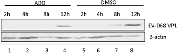
Andrographolide Prevents EV-D68 Replication by Inhibiting the Acidification of Virus-Containing Endocytic Vesicles. [Abstract]2018 Oct 8:9:2407. PMID: 30349523
Andrographolide purchased from MedChemExpress. Usage Cited in: Front Microbiol. 2018 Oct 8:9:2407. [Abstract]
RD cells are pretreated with andrographolide or DMSO vehicle overnight and subsequently infected with EV-D68. Immunoblotting of VP1 expression at 2, 4, 8, 12 h post-infection. There is an observable reduction in EV-D68 VP1 protein expression in ADO-treated cells compared to vehicle-treated cells on immunoblotting.
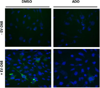
Andrographolide purchased from MedChemExpress. Usage Cited in: Front Microbiol. 2018 Oct 8:9:2407. [Abstract]
Immunofluorescence of VP1 expression at 10 h post-infection. There is an observable reduction in EV-D68 VP1 protein expression in ADO-treated cells compared to vehicle-treated cells on immunofluorescence assays
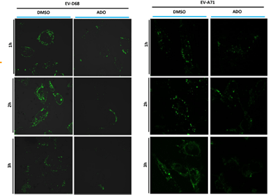
Andrographolide purchased from MedChemExpress. Usage Cited in: Front Microbiol. 2018 Oct 8:9:2407. [Abstract]
Fluorescence intensity peaked at around 2 h post-infection and is quickly quenched by 3 h post-infection in vehicle-treated RD cells. ADO-treated cells exhibit limited fluorescence.
-
J Cell Mol Med
Long non-coding RNA LINC00526 represses glioma progression via forming a double negative feedback loop with AXL. [Abstract]2019 Aug;23(8):5518-5531. PMID: 31240814 -
-
J Inflamm Res
Andrographolide Attenuates Inflammation Due to Intra-Abdominal Sepsis by Enhancing Bacterial Clearance in Mice. [Abstract]2023 Oct 6:16:4413-4423. PMID: 37822531 -
J Biol Chem
Andrographolide targets syndecan4 to impair its interaction with syntenin and inhibits the biogenesis of small extracellular vesicles. [Abstract]2026 Jan 20:111182. PMID: 41570990 -
Oncol Rep
Andrographolide sensitizes human renal carcinoma cells to TRAIL‑induced apoptosis through upregulation of death receptor 4. [Abstract]2020 Nov;44(5):1939-1948. PMID: 33000263 -
Cell Signal
Paederia scandens-derived exosome-like nanoparticles as a delivery system for andrographolide to treat ulcerative colitis. [Abstract]2025 Nov:135:112014. PMID: 40695370 -
Arch Pharm (Weinheim)
Design, synthesis, and characterization of PROTACs targeting the androgen receptor in prostate and lung cancer models. [Abstract]2022 May;355(5):e2100467. PMID: 35128717 -
Med Oncol
Andrographolide elevates tumor necrosis factor-related apoptosis-inducing ligand lethality through reactive oxygen species accumulation and gasdermin E cleavage in breast cancer cells. [Abstract]2022 Nov 9;40(1):11. PMID: 36352155 -
ChemMedChem
Andrographolide Derivatives Target the KEAP1/NRF2 Axis and Possess Potent Anti-SARS-CoV-2 Activity. [Abstract]2022 Mar 4;17(5):e202100732. PMID: 35099120 -
-
J Pharm Pharmacol
Andrographolide attenuates oxidative stress and apoptosis in osteoporosis rats via MEK/ERK and Beclin-1/ATG-5-mediated autophagy pathway. [Abstract]2025 Aug 4:rgaf052. PMID: 40757977 -
Naunyn Schmiedebergs Arch Pharmacol
Andrographolide blocked the progression of endometriosis by promoting ferroptosis via inhibiting anabolism of serine. [Abstract]2025 Dec 18. PMID: 41408483 -
Genes (Basel)
Differential Expression of Key Oncogenic and Tumor Suppressor MicroRNAs Induced by Andrographolide in Androgen-Independent PC3 and Androgen-Dependent LNCaP Prostate Cancer Cells. [Abstract]2025 Dec 17;16(12):1514. PMID: 41465187 -
Vet Microbiol
The Chinese medicine monomer Schisandrin C inhibits PRRSV infection by regulating the OGT-PI3K/AKT/mTOR signaling pathway. [Abstract]2026 May:316:110992. PMID: 41865607 -
Fitoterapia
Characterization of andrographolide-loaded exosome-like nanoparticles from Nauclea officinalis and their modulation of inflammatory and lipid metabolic pathologies in WRL68 cells. [Abstract]2026 Apr:190:107179. PMID: 41850591 -
Ultrasound Med Biol
Can Viscoelasticity Measurements Obtained Through Shear-Wave US Elastography be used to Monitor Hepatic Ischemia-Reperfusion Injury and Treatment Response? An Animal Study. [Abstract]2020 Sep;46(9):2464-2471. PMID: 32553529 -
Gene
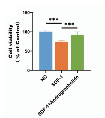
Elucidation of the key therapeutic targets and potential mechanisms of Andrographolide multi-targets against osteoarthritis via network pharmacological analysis and experimental validation. [Abstract]2024 Jun 15:911:148351. PMID: 38462021
Andrographolide purchased from MedChemExpress. Usage Cited in: Gene. 2024 Jun 15:911:148351. [Abstract]
Andrographolide (30 μM, 48 h) reversed SDF-1 (100 ng/mL, 48 h)-induced cell growth inhibition in chondrocytes.
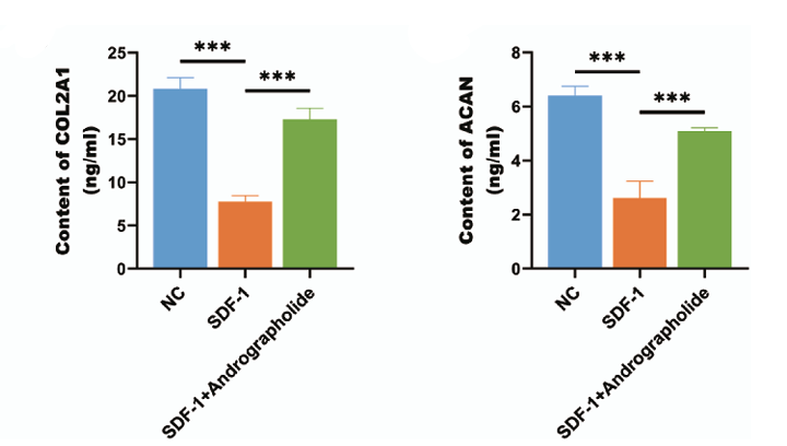
Andrographolide purchased from MedChemExpress. Usage Cited in: Gene. 2024 Jun 15:911:148351. [Abstract]
Andrographolide (30 μM, 48 h) reversed the SDF-1 (100 ng/mL, 48 h)-induced decrease in COL2A1 and ACAN content in chondrocytes.
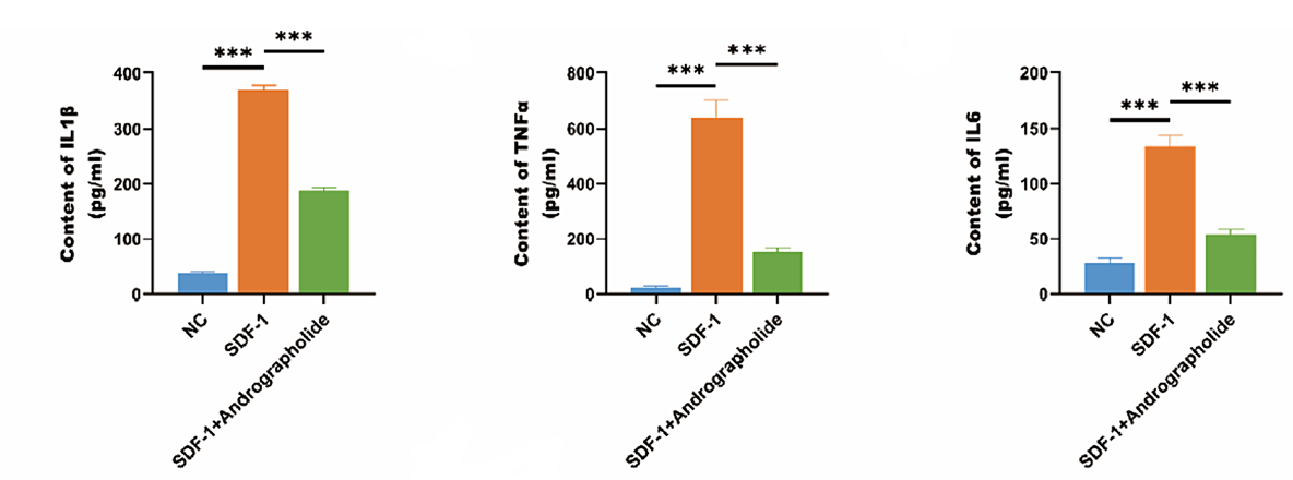
Andrographolide purchased from MedChemExpress. Usage Cited in: Gene. 2024 Jun 15:911:148351. [Abstract]
Andrographolide (30 μM, 48 h) reversed SDF-1 (100 ng/mL, 48 h)-induced expression of TNF-α, IL-6, and IL-1β in chondrocytes.
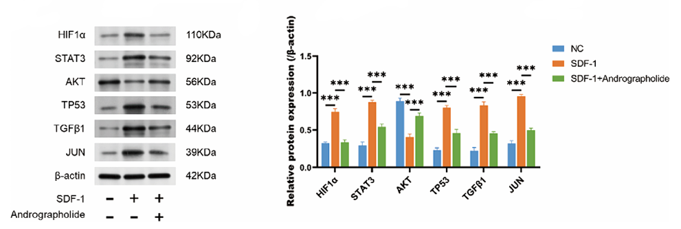
Andrographolide purchased from MedChemExpress. Usage Cited in: Gene. 2024 Jun 15:911:148351. [Abstract]
Andrographolide (30 μM, 48 h) reversed SDF-1 (100 ng/mL, 48 h)-induced expression of STAT3, TP53, JUN, HIF-1α, TGF-β1, and AKT1.
-
Biochem Biophys Res Commun
Andrographolide promotes lymphangiogenesis and lymphatic vessel remodeling to alleviate secondary lymphedema. [Abstract]2024 May 25:723:150179. PMID: 38820623 -
Vet Parasitol
Cryptosporidium parvum downregulates miR-181d in HCT-8 cells via the p50-dependent TLRs/NF-κB pathway. [Abstract]2022 May;305:109710. PMID: 35462275 -
J Gastrointest Oncol
Andrographolide potentiates anti-tumor immunity in colorectal cancer (CRC) by targeting voltage-dependent anion channel (VDAC) and activating the cGAS-STING axis. [Abstract]2025 Aug 30;16(4):1550-1561. PMID: 40950339 -
J Orthop
Andrographolide suppresses fibrogenic phenotype of chondrocytes and ameliorates osteoarthritis by regulating miR-137/BMP7 axis. [Abstract]2024 Nov 26:64:108-116. PMID: 39691644 -
Oxid Med Cell Longev
URB597 and Andrographolide Improve Brain Microvascular Endothelial Cell Permeability and Apoptosis by Reducing Oxidative Stress and Inflammation Associated with Activation of Nrf2 Signaling in Oxygen-Glucose Deprivation. [Abstract]2022 May 12;2022:4139330. PMID: 35602108 -


Solvent & Solubility
DMSO : 100 mg/mL (285.35 mM; Need ultrasonic; Hygroscopic DMSO has a significant impact on the solubility of product, please use newly opened DMSO)
H2O : < 0.1 mg/mL (insoluble)
Please refer to the solubility information to select the appropriate solvent. Once prepared, please aliquot and store the solution to prevent product inactivation from repeated freeze-thaw cycles.
Storage method and period of stock solution: -80°C, 1 year; -20°C, 6 months. When stored at -80°C, please use it within 1 year. When stored at -20°C, please use it within 6 months.
Concentration (start) × Volume (start) = Concentration (final) × Volume (final)
Select the appropriate dissolution method based on your experimental animal and administration route.
- For the following dissolution methods, please ensure to first prepare a clear stock solution using an In Vitro approach and then sequentially add co-solvents:
- To ensure reliable experimental results, the clarified stock solution can be appropriately stored based on storage conditions. As for the working solution for In Vivo experiments, it is recommended to prepare freshly and use it on the same day.
- The percentages shown for the solvents indicate their volumetric ratio in the final prepared solution. If precipitation or phase separation occurs during preparation, heat and/or sonication can be used to aid dissolution.
Add each solvent one by one: 10% DMSO 40% PEG300 5% Tween-80 45% Saline
Solubility: ≥ 2.5 mg/mL (7.13 mM); Clear solution
This protocol yields a clear solution of ≥ 2.5 mg/mL (saturation unknown).
Taking 1 mL working solution as an example, add 100 μL DMSO stock solution (25.0 mg/mL) to 400 μL PEG300, and mix evenly; then add 50 μL Tween-80 and mix evenly; then add 450 μL Saline to adjust the volume to 1 mL.
Preparation of Saline: Dissolve 0.9 g sodium chloride in ddH₂O and dilute to 100 mL to obtain a clear Saline solution.
Add each solvent one by one: 10% DMSO 90% (20% SBE-β-CD in Saline)
Solubility: ≥ 2.5 mg/mL (7.13 mM); Clear solution
This protocol yields a clear solution of ≥ 2.5 mg/mL (saturation unknown).
Taking 1 mL working solution as an example, add 100 μL DMSO stock solution (25.0 mg/mL) to 900 μL 20% SBE-β-CD in Saline, and mix evenly.
Preparation of 20% SBE-β-CD in Saline (4°C, storage for one week): 2 g SBE-β-CD powder is dissolved in 10 mL Saline, completely dissolve until clear.
For the following dissolution methods, please prepare the working solution directly:
It is recommended to prepare fresh solutions and use them promptly within a short period of time.
The percentages shown for the solvents indicate their volumetric ratio in the final prepared solution. If precipitation or phase separation occurs during preparation, heat and/or sonication can be used to aid dissolution.
Add each solvent one by one: 0.5% CMC-Na/saline water
Solubility: 2 mg/mL (5.71 mM); Suspended solution; Need ultrasonic
Please enter the basic information of animal experiments:
-
-
-
-
Recommended: Prepare an additional quantity of animals to account for potential losses during experiments.
Please enter your animal formula composition:
-
%DMSO +
Recommended: Keep the proportion of DMSO in working solution below 2% if your animal is weak.
-
%+
-
+%Tween-80 + +
-
%Saline +
The co-solvents required include: DMSO, . All of co-solvents are available by MedChemExpress (MCE). , Tween 80. All of co-solvents are available by MedChemExpress (MCE).
Working solution concentration: 0.22 mg/mL
Method for preparing stock solution: mg drug dissolved in μL DMSO. Stock solution concentration: mg/mL.
1. Take μL DMSO stock solution;
2. Add μL .
μL , mix evenly;
3. Then add μL Tween 80, mix evenly;
4. Then add μL
Please ensure that the stock solution in the first step is dissolved to a clear state, and add co-solvents in sequence. You can use ultrasonic heating (ultrasonic cleaner, recommended frequency 20-40 kHz), vortexing, etc. to assist dissolution.

Protocol
In vitro osteoclastogenesis assays are preformed to examine the effects of Andrographolide on osteoclast differentiation. Bone marrow macrophages (BMM) cells are prepared. Briefly, cells extracted from the femur and tibiae of a 6-week-old C57/BL6 mouse are incubated in complete cell culture media and 30 ng/mL M-CSF in a T-75 cm2 flask for proliferation. When changing the medium, the cells are washed in order to deplete residual stromal cells. After reaching 90% confluence, cells are washed with PBS three times and trypsinized for 30 min to harvest BMMs. Cells adhering to the bottom of the dish are classified as BMMs; these BMMs are plated in 96-well plates at a density of 8×10³ cells per well in triplicate and incubated in a humidified incubator containing 5% CO2 at 37°C for 24 h. The cells are then treated with various concentrations of Andrographolide (0, 2.5, 5, or 10 μM) plus M-CSF (30 ng/mL) and RANKL (50 ng/mL). After 5 days, cells are fixed and stained for tartrate-resistant acid phosphatase (TRAP) activity. TRAP-positive multinucleated cells with more than five nuclei are counted as osteoclasts[1].
MedChemExpress (MCE) has not independently confirmed the accuracy of these methods. They are for reference only.
Effects of Andrographolide on cell proliferation are determined with a CCK-8. BMMs are plated in 96-well plates at a density of 3×10³ cells per well in triplicate. Twenty-four hours later, the cells are treated with increasing concentrations of Andrographolide (0, 2.5, 5, 10 or 20 μM) for 2 days. Next, 10 μL CCK-8 is added to each well, and the plates are then incubated at 37°C for an additional 2 h. The optical density (OD) is then measured with an ELX800 absorbance microplate reader at a wavelength of 450 nm (650 nm reference). The cell viability is calculated[1].
MedChemExpress (MCE) has not independently confirmed the accuracy of these methods. They are for reference only.
Mice[1]
C57BL/6 mice (8 weeks old) are divided into four groups of seven mice each. Mice are injected i.p. with Andrographolide (5 or 30 mg/kg body weight) or PBS as a control 1 day before injection of LPS (5 μg/g body weight). Andrographolide or PBS is injected intraperitoneally every other day for 8 days. LPS is injected intraperitoneally on days one and four. All mice are killed 8 days after the initial LPS injection, and the left femurs of all animals are scanned with a high-resolution micro-CT at a resolution of 9 μm.
MedChemExpress (MCE) has not independently confirmed the accuracy of these methods. They are for reference only.


Purity & Documentation
-
Data Sheet (284 KB)
-
SDS (393 KB)
- English - EN (393 KB)
- Français - FR (393 KB)
- Deutsch - DE (393 KB)
- Norwegian - NO (393 KB)
- Español - ES (393 KB)
- Swedish - SV (393 KB)
- Italian - IT (393 KB)
- Korean - KR (393 KB)
- Portuguese - PT (393 KB)
-
Handling Instructions (2659 KB)
References
[1]. Zhai ZJ, et al. Andrographolide suppresses RANKL-induced osteoclastogenesis in vitro and prevents inflammatory bone loss in vivo. Br J Pharmacol. 2014 Feb;171(3):663-75. [Content Brief]
[2]. Gupta S, et al. Broad-spectrum antiviral properties of andrographolide. Arch Virol. 2017 Mar;162(3):611-623. [Content Brief]


Complete Stock Solution Preparation Table
Please refer to the solubility information to select the appropriate solvent. Once prepared, please aliquot and store the solution to prevent product inactivation from repeated freeze-thaw cycles.
Storage method and period of stock solution: -80°C, 1 year; -20°C, 6 months. When stored at -80°C, please use it within 1 year. When stored at -20°C, please use it within 6 months.
| Optional Solvent | Concentration Solvent Mass | 1 mg | 5 mg | 10 mg | 25 mg |
|---|---|---|---|---|---|
| DMSO | 1 mM | 2.8535 mL | 14.2674 mL | 28.5347 mL | 71.3369 mL |
| 5 mM | 0.5707 mL | 2.8535 mL | 5.7069 mL | 14.2674 mL | |
| 10 mM | 0.2853 mL | 1.4267 mL | 2.8535 mL | 7.1337 mL | |
| 15 mM | 0.1902 mL | 0.9512 mL | 1.9023 mL | 4.7558 mL | |
| 20 mM | 0.1427 mL | 0.7134 mL | 1.4267 mL | 3.5668 mL | |
| 25 mM | 0.1141 mL | 0.5707 mL | 1.1414 mL | 2.8535 mL | |
| 30 mM | 0.0951 mL | 0.4756 mL | 0.9512 mL | 2.3779 mL | |
| 40 mM | 0.0713 mL | 0.3567 mL | 0.7134 mL | 1.7834 mL | |
| 50 mM | 0.0571 mL | 0.2853 mL | 0.5707 mL | 1.4267 mL | |
| 60 mM | 0.0476 mL | 0.2378 mL | 0.4756 mL | 1.1889 mL | |
| 80 mM | 0.0357 mL | 0.1783 mL | 0.3567 mL | 0.8917 mL | |
| 100 mM | 0.0285 mL | 0.1427 mL | 0.2853 mL | 0.7134 mL |


 Powered by Bioz