



14-3-3 epsilon Antibody (YA2629)
(Synonyms: 14-3-3 epsilon; 14-3-3E; 143E; KCIP-1; MDCR; Protein kinase C inhibitor protein-1; YWHAE)
Based on 1 Customer Validation
14-3-3 epsilon Antibody (YA2629) is a Rabbit-derived and non-conjugated IgG monoclonal antibody, targeting to 14-3-3 epsilon.
-
Host:
Rabbit
-
Isotype:
IgG
-
Application:
WB, IHC-P, ICC/IF, FC
-
Reactivity :
Human, Mouse, Rat
-
Formulation:
Supplied in rabbit IgG in 50 mM Tris-Glycine (pH 7.4), 0.15 M NaCl, 40% Glycerol, 0.01% sodium azide and 0.05% BSA.
-
Conjugation:
Non-conjugated


To place orders, for customer services and technical support, please contact: MedChemExpress USA
Tel: 609-228-6898 E-mail: [email protected] [email protected]





Applications
| Application |
WB

WB: Western Blot
|
IHC-P
IHC-P: Immunohistochemistry-Paraffin
|
ICC/IF
ICC/IF: Immunocytochemistry/
Immunofluorescence |
FC
FC: Flow Cytometry
|
|---|---|---|---|---|
| Dilution Ratio | 1:500-1:1000 | 1:50-1:100 | 1:50-1:200 | 1:50-1:100 |


Product Details
14-3-3 epsilon Antibody (YA2629) is a Rabbit-derived and non-conjugated IgG monoclonal antibody, targeting to 14-3-3 epsilon.
-
Host Rabbit

-
Clonality Recombinant,Monoclonal

-
Species ReactivityHuman, Mouse, Rat
-
Observed Molecular WeightObserved band size: 29 kDa
Note: Due to possible protein modifications or aggregation, the molecular weight should be confirmed by actual measurement, and the predicted value is for reference only.

-
Calculated Molecular Weight Predicted band size: 29 kDa

Entrez Gene: 7531 Human ; 22627 Mouse ; 29753 Rat
SwissProt: P62258 Human ; P62259 Mouse ; P62260 Rat
OMIM: 605066 Human
A synthesized peptide derived from human 14-3-3 epsilon aa55-95.
Endogenous
Affinity Chromatography
Non-conjugated
Unmodified
IgG


Product Properties
-
Appearance
Solution
-
Formulation
Supplied in rabbit IgG in 50 mM Tris-Glycine (pH 7.4), 0.15 M NaCl, 40% Glycerol, 0.01% sodium azide and 0.05% BSA.
-
Concentration
Batch-dependent, Please check the COA for the concentration of each lot. Check Lot Concentration
-
Storage & Stability
Stored at -20°C for 1 year. Avoid repeated freeze / thaw cycles.
-
Shipping
Shipping with blue ice.


Verification Images
-
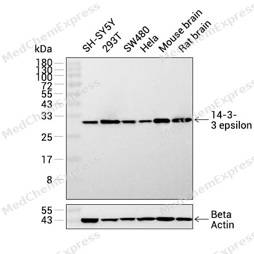 Western blot analysis was performed on extracts from SH-SY5Y (lane 1, 15 μg), 293T (lane 2, 15 μg), SW480 (lane 3, 15 μg), Hela (lane 4, 15 μg), Mouse brain (lane 5, 15 μg), and Rat brain (lane 6, 15 μg) using 14-3-3 epsilon Rabbit mAb.Proteins were transferred to a PVDF membrane and blocked with 5% non-fat milk in TBST at 4°C overnight.The primary antibody (1:1000 dilution) and the loading control antibody (beta-Actin, HY-P83730, 1:20000 dilution) were incubated in 5% non-fat milk in TBST for 1 hour at 37°C.Goat Anti-Rabbit IgG-HRP Secondary Antibody (1:20000 dilution) was then applied for 40 minutes at 37°C.
 -
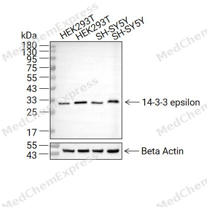 Western blot analysis of extracts from HEK293T (lane 2(20μg) and HEK293T (lane 3(40μg), SH-SY5Y (lane 4(20μg) and SH-SY5Y (lane 5(40μg) using 14-3-3 epsilon (HY-P82884) Rabbit mAb. Proteins were transferred to a PVDF membrane and blocked with 5% non-fat milk in TBST for 2 hour at room temperature. The primary antibody (1/1000) and Loading control antibody (Beta Actin, HY-P80438, 1/10000) was used in 5% non-fat milk in TBST at 4°C overnight. Goat Anti-Rabbit IgG-HRP Secondary Antibody (1/10000) was used for 1 hour at room temperature.
 -
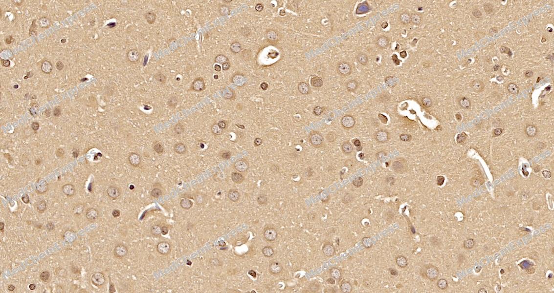 Immunohistochemical analysis of paraffin-embedded rat brain tissue using 14-3-3 epsilon Antibody. The section was pre-treated using heat mediated antigen retrieval with sodium citrate buffer (pH 6.0) for 8 minutes. The tissues were blocked in QuickBlock for 20 minutes at room temperature, washed with ddH2O and PBS, and then probed with the primary antibody at 1/100 dilution in 4℃ overnight. The detection was performed using an HRP conjugated compact polymer system. DAB was used as the chromogen. Tissues were counterstained with hematoxylin and mounted with DPX.
 -
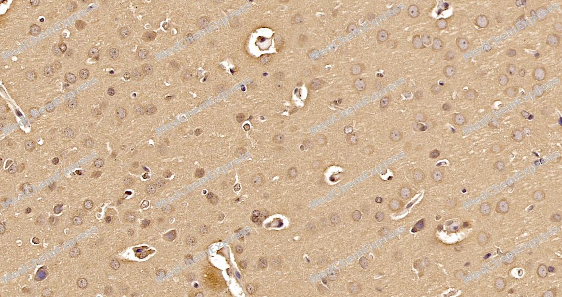 Immunohistochemical analysis of paraffin-embedded rat brain tissue using 14-3-3 epsilon Antibody. The section was pre-treated using heat mediated antigen retrieval with sodium citrate buffer (pH 6.0) for 8 minutes. The tissues were blocked in QuickBlock for 20 minutes at room temperature, washed with ddH2O and PBS, and then probed with the primary antibody at 1/100 dilution in 4℃ overnight. The detection was performed using an HRP conjugated compact polymer system. DAB was used as the chromogen. Tissues were counterstained with hematoxylin and mounted with DPX.
 -
 Flow cytometric analysis of 1X10⁶ HeLa cells labeling 14-3-3 epsilon Antibody (HY-P82884, red). Cells were fixed with 4% paraformaldehyde and permeabilised with 90% methanol. Then stained with the primary antibody at 1/100 dilution for an hour at 4℃. AF488-conjugated Goat Anti-Rabbit IgG H&L (HY-P8002) was used as the secondary antibody at 1/1,000 dilution for 30 minutes at 4℃. Rabbit IgG Isotype Control (HY-P80879, blue) was used as the isotype control, cells without incubation with primary antibody were used as the unlabeled control (black).


Background
-
Function
The 14-3-3 epsilon protein serves as an adapter implicated in the regulation of a diverse array of both general and specialized signaling pathways, binding to numerous partners through the recognition of phosphoserine or phosphothreonine motifs, thereby modulating the activity of the binding partner. Notably, it positively regulates the nuclear export of phosphorylated protein HSF1 to the cytoplasm. Existing as a homodimer, it also forms heterodimers with YWHAZ and interacts with various proteins, including PKA-phosphorylated AANAT, ABL1 in its phosphorylated form, ARHGEF28, BEX3, CDKN1B, the 'Thr-369' phosphorylated form of DAPK2, DENND1A, GAB2, phosphorylated GRB10, KSR1, NDEL1, PI4KB, TBC1D22A, TBC1D22B, the phosphorylated form of SRPK2, TIAM2, the 'Ser-1134' and 'Ser-1161' phosphorylated form of SOS1, ZFP36, SLITRK1, HSF1 in its phosphorylated form, RIPOR2, KLHL22, CRTC1, CRTC2 (probably when phosphorylated at 'Ser-171'), CRTC3 (probably when phosphorylated at 'Ser-162' and/or 'Ser-273'), ATP2B1, ATP2B3, and MEFV. These interactions highlight the multifaceted role of 14-3-3 epsilon in orchestrating various cellular processes and signaling events.
-
Subcellular Localization
Nucleus; Cytoplasm; Melanosome
-
Isoforms & Post-Translational Modification
14-3-3 epsilon has 2 isomers: P62258-1: 29174 Da (predicted); P62258-2: 26504 Da (predicted).
(Microbial infection) Cleaved by poliovirus protease 3C, leading to disruption of the interaction with RIGI -
Subunit
Homodimer. Heterodimerizes with YWHAZ. Interacts with PKA-phosphorylated AANAT.
-
SwissProt ID
-
Synonyms
14-3-3 epsilon; 14-3-3E; 143E; KCIP-1; MDCR; Protein kinase C inhibitor protein-1; YWHAE
-
Research Field
Neuroscience


Documentation
-
Data Sheet (263 KB)
-
SDS (252 KB)
- English - EN (252 KB)
- Français - FR (252 KB)
- Deutsch - DE (252 KB)
- Norwegian - NO (252 KB)
- Español - ES (252 KB)
- Swedish - SV (252 KB)
- Italian - IT (252 KB)
- Korean - KR (252 KB)
- Portuguese - PT (252 KB)
-
User Guide for Antibodies (1077 KB)
References
[1]. Obsilova V, et al. Structural insights into the functional roles of 14-3-3 proteins. Front Mol Biosci. 2022;9:1016071. [Content Brief]
[2]. Muslin AJ, et al. Interaction of 14-3-3 with signaling proteins is mediated by the recognition of phosphoserine. Cell. 1996;84(6):889-897. [Content Brief]
[3]. Yang X, et al. Structural basis for protein-protein interactions in the 14-3-3 protein family. Proc Natl Acad Sci U S A. 2006;103(46):17237-17242. [Content Brief]
[4]. Gardino AK, et al. 14-3-3 proteins as signaling integration points for cell cycle control and apoptosis. Semin Cell Dev Biol. 2011;22(7):688-695. [Content Brief]
[5]. Pennington KL, et al. The dynamic and stress-adaptive signaling hub of 14-3-3: emerging mechanisms of regulation and context-dependent protein-protein interactions. Oncogene. 2018;37(42):5587-5604. [Content Brief]
[6]. Li Z, et al. 14-3-3σ, the double-edged sword of human cancers. Am J Transl Res. 2009;1(4):326-340. [Content Brief]
[7]. Maksymowych WP, et al. Serum 14-3-3η is a novel marker that complements current serological measurements to enhance detection of patients with rheumatoid arthritis. J Rheumatol. 2014;41(11):2104-2113. [Content Brief]
[8]. Aghazadeh Y, et al. The role of the 14-3-3 protein family in health, disease, and drug development. Drug Discov Today. 2016;21(2):278-287. [Content Brief]

 Powered by Bioz
