



AKR1C3 Antibody (YA2338)
(Synonyms: DD3; DDX; HA1753; HAKRB; HAKRe; HSD17B5; KIAA0119; hluPGFS)
Based on 1 Customer Validation
AKR1C3 Antibody (YA2338) is a Rabbit-derived and non-conjugated IgG monoclonal antibody, targeting to AKR1C3.
-
Host:
Rabbit
-
Isotype:
IgG
-
Application:
WB, ICC/IF, IP, FC
-
Reactivity :
Human
-
Formulation:
Supplied in Rabbit IgG in 10mM phosphate buffered saline , pH 7.4, 150mM sodium chloride, 0.05% BSA, 0.02% sodium azide and 50% glycerol.
-
Conjugation:
Non-conjugated


To place orders, for customer services and technical support, please contact: MedChemExpress USA
Tel: 609-228-6898 E-mail: [email protected] [email protected]





Applications
| Application |
WB

WB: Western Blot
|
ICC/IF
ICC/IF: Immunocytochemistry/
Immunofluorescence |
IP
IP: Immunoprecipitation
|
FC
FC: Flow Cytometry
|
|---|---|---|---|---|
| Dilution Ratio | 1:500-1:1000 | 1:50-1:200 | 1:30 | 1:50-1:100 |


Product Details
AKR1C3 Antibody (YA2338) is a Rabbit-derived and non-conjugated IgG monoclonal antibody, targeting to AKR1C3.
-
Host Rabbit

-
Clonality Recombinant, Monoclonal

-
Species ReactivityHuman
-
Observed Molecular WeightObserved band size: 37 kDa
Note: Due to possible protein modifications or aggregation, the molecular weight should be confirmed by actual measurement, and the predicted value is for reference only.

-
Calculated Molecular Weight Predicted band size: 37 kDa

A synthesized peptide derived from human AKR1C3 aa200-250/323.
Endogenous
Affinity Chromatography
Non-conjugated
Unmodified
IgG


Product Properties
-
Appearance
Liquid
-
Formulation
Supplied in Rabbit IgG in 10mM phosphate buffered saline , pH 7.4, 150mM sodium chloride, 0.05% BSA, 0.02% sodium azide and 50% glycerol.
-
Concentration
Batch-dependent, Please check the COA for the concentration of each lot. Check Lot Concentration
-
Storage & Stability
Stored at -20°C for 1 year. Avoid repeated freeze / thaw cycles.
-
Shipping
Shipping with blue ice.


Verification Images
-
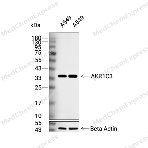 Western blot analysis of extracts from A549 (lane 2(20μg), A549 (lane 3(40μg), using AKR1C3 Antibody. Proteins were transferred to a PVDF membrane and blocked with 5% BSA in TBST for 2 hour at room temperature. The primary antibody and Loading control antibody (Beta Actin, HY-P80438, 1/3000) was used in 5% BSA in TBST at 4°C overnight. Goat Anti-Mouse/Rabbit IgG-HRP Secondary Antibody (HY-P8004/HY-P8001, 1/10,000) was used for 1 hour at room temperature.
 -
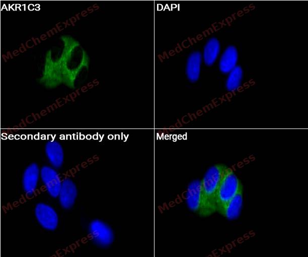 Immunocytochemistry analysis of A549 cells labeling AKR1C3 with AKR1C3 antibody (HY-P82593) at 1/50 dilution. Cells were fixed in 4% paraformaldehyde for 15 minutes at room temperature, permeabilized with 0.1% Triton X-100 for 10 minutes at room temperature, then blocked with QuickBlock™ Blocking Buffer for Immunol Staining for 10 min at room temperature. Cells were then incubated with AKR1C3 antibody (HY-P82593) at 1/50 dilution in QuickBlock™ Blocking Buffer for Immunol Staining at 4 ℃. Alexa Fluor® 488-conjugated AffiniPure Goat Anti-Rabbit IgG H&L(HY-P8002, Green) was used as the secondary antibody at 1/1,000 dilution. PBS instead of the primary antibody was used as the secondary antibody only control. The Nuclear counterstain was DAPI (Blue).
 -
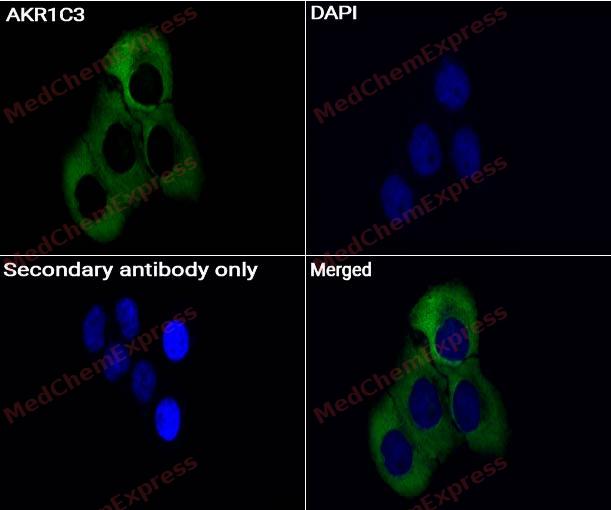 Immunocytochemistry analysis of HeLa cells labeling AKR1C3 with AKR1C3 antibody (HY-P82593) at 1/50 dilution. Cells were fixed in 4% paraformaldehyde for 15 minutes at room temperature, permeabilized with 0.1% Triton X-100 for 10 minutes at room temperature, then blocked with QuickBlock™ Blocking Buffer for Immunol Staining for 10 min at room temperature. Cells were then incubated with AKR1C3 antibody (HY-P82593) at 1/50 dilution in QuickBlock™ Blocking Buffer for Immunol Staining at 4 ℃. Alexa Fluor® 488-conjugated AffiniPure Goat Anti-Rabbit IgG H&L(HY-P8002, Green) was used as the secondary antibody at 1/1,000 dilution. PBS instead of the primary antibody was used as the secondary antibody only control. The Nuclear counterstain was DAPI (Blue).
 -
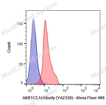 Flow cytometric analysis of 1X10^6 TF-1 cells labeling AKR1C3 Antibody (YA2338) (YA2432) (HY-P82593, red). Cells were fixed with 4% paraformaldehyde. Then stained with the primary antibody at 1/50 dilution for an hour at 4℃. Alexa Fluor® 488-conjugated AffiniPure Goat Anti-Rabbit IgG H&L (HY-P8002) was used as the secondary antibody at 1/1,000 dilution for 30 minutes at 4℃. Rabbit IgG Isotype Control (HY-P80879, blue) was used as the isotype control, cells without incubation with primary antibody were used as the unlabeled control (black).


Background
-
Function
AKR1C3 is a Cytosolic aldo-keto reductase that catalyzes the NADH and NADPH-dependent reduction of ketosteroids to hydroxysteroids. Acts as a NAD(P)(H)-dependent 3-, 17- and 20-ketosteroid reductase on the steroid nucleus and side chain and regulates the metabolism of androgens, estrogens and progesterone. Displays the ability to catalyze both oxidation and reduction in vitro, but most probably acts as a reductase in vivo since the oxidase activity measured in vitro is inhibited by physiological concentration of NADPH. Acts preferentially as a 17-ketosteroid reductase and has the highest catalytic efficiency of the AKR1C enzyme for the reduction of delta4-androstenedione to form testosterone. Reduces prostaglandin (PG) D2 to 11beta-prostaglandin F2, progesterone to 20alpha-hydroxyprogesterone and estrone to 17beta-estradiol. Catalyzes the transformation of the potent androgen dihydrotestosterone (DHT) into the less active form, 5-alpha-androstan-3-alpha,17-beta-diol (3-alpha-diol). Also displays retinaldehyde reductase activity toward 9-cis-retinal[1][2][3][4][5][6][7][8][9][10][11][12].
-
Subcellular Localization
Cytoplasm
-
Expression
Tissue_specificity:Expressed in many tissues including adrenal gland, brain, kidney, liver, lung, mammary gland, placenta, small intestine, colon, spleen, prostate and testis. High expression in prostate and mammary gland. In the prostate, higher levels in epithelial cells than in stromal cells. In the brain, expressed in medulla, spinal cord, frontotemporal lobes, thalamus, subthalamic nuclei and amygdala. Weaker expression in the hippocampus, substantia nigra and caudate -
Isoforms & Post-Translational Modification
P42330 has 2 isomers: P42330-1: 36853 Da (predicted); P42330-2: 23364 Da (predicted).
-
SwissProt ID
-
Synonyms
DD3; DDX; HA1753; HAKRB; HAKRe; HSD17B5; KIAA0119; hluPGFS
-
Research Field
Signal Transduction


Documentation
-
Data Sheet (259 KB)
-
SDS (251 KB)
- English - EN (251 KB)
- Français - FR (251 KB)
- Deutsch - DE (251 KB)
- Norwegian - NO (251 KB)
- Español - ES (251 KB)
- Swedish - SV (251 KB)
- Italian - IT (251 KB)
- Korean - KR (251 KB)
- Portuguese - PT (251 KB)
-
User Guide for Antibodies (1077 KB)

 Powered by Bioz
