



Bim Antibody (YA582)
(Synonyms: BIM, BCL2L11, Bcl-2-like protein 11, Bcl2-L-11, Bcl2-interacting mediator of cell death)
Based on 1 Customer Validation
Bim Antibody (YA582) is a Rabbit-derived and non-conjugated IgG monoclonal antibody, targeting to Bim.
-
Host:
Rabbit
-
Isotype:
IgG
-
Application:
WB, ICC/IF, IHC-P, IP
-
Reactivity :
Human, Mouse
-
Formulation:
Supplied in 1*TBS (pH7.4), 0.05% BSA and 40% Glycerol. Preservative: 0.05% Sodium Azide.
-
Conjugation:
Non-conjugated


To place orders, for customer services and technical support, please contact: MedChemExpress USA
Tel: 609-228-6898 E-mail: [email protected] [email protected]





Applications
| Application |
WB

WB: Western Blot
|
ICC/IF
ICC/IF: Immunocytochemistry/
Immunofluorescence |
IHC-P
IHC-P: Immunohistochemistry-Paraffin
|
IP
IP: Immunoprecipitation
|
|---|---|---|---|---|
| Dilution Ratio | 1:500-1:2000 | 1:50-1:200 | 1:50-1:200 | Use at an assay dependent concentration. |


Product Details
Bim Antibody (YA582) is a Rabbit-derived and non-conjugated IgG monoclonal antibody, targeting to Bim.
-
Host Rabbit

-
Clonality Recombinant,Monoclonal

-
Species ReactivityHuman, Mouse
-
Observed Molecular WeightObserved band size: 25 kDa
Note: Due to possible protein modifications or aggregation, the molecular weight should be confirmed by actual measurement, and the predicted value is for reference only.

-
Calculated Molecular Weight Predicted band size: 22 kDa

Entrez Gene: 10018 Human ; 12125 Mouse ;
SwissProt: O43521 Human ; O54918 Mouse ;
OMIM: 603827 Human
Synthetic peptide corresponding to Human Bim.AA range:1-40.
Endogenous
Protein A affinity purified.
Non-conjugated
Unmodified
IgG


Product Properties
-
Appearance
Solution
-
Formulation
Supplied in 1*TBS (pH7.4), 0.05% BSA and 40% Glycerol. Preservative: 0.05% Sodium Azide.
-
Concentration
Batch-dependent, Please check the COA for the concentration of each lot. Check Lot Concentration
-
Storage & Stability
Stored at -20°C for 1 year. Avoid repeated freeze / thaw cycles.
-
Shipping
Shipping with blue ice.


Verification Images
-
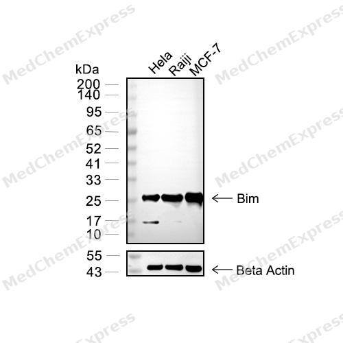 Western blot analysis of extracts from Hela (lane 2(20μg) , Raiji (lane 3(20μg) , MCF-7 (lane 4(20μg),using Bim Antibody (HY-P80032). Proteins were transferred to a PVDF membrane and blocked with 5% BSA in TBST for 2 hour at room temperature. The primary antibody and Loading control antibody (Beta Actin, HY-P80438, 1/3000) was used in 5% BSA in TBST at 4°C overnight. Goat Anti-Mouse/Rabbit IgG-HRP Secondary Antibody (HY-P8004/HY-P8001, 1/10,000) was used for 1 hour at room temperature.
 -
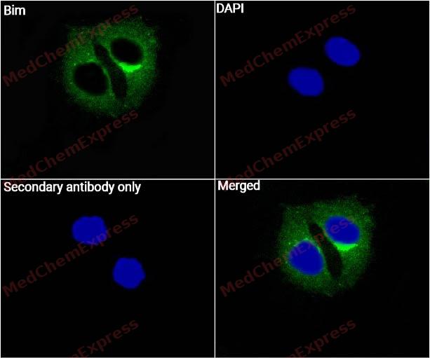 Immunocytochemistry analysis of HepG2 cells labeling Bim with Bim Antibody (HY-P80032) at 1/50 dilution. Cells were fixed in 4% paraformaldehyde for 15 minutes at room temperature, permeabilized with 0.1% Triton X-100 for 10 minutes at room temperature, then blocked with QuickBlock™ Blocking Buffer for Immunol Staining for 10 min at room temperature. Cells were then incubated with Bim Antibody (HY-P0032) at 1/50 dilution in QuickBlock™ Blocking Buffer for Immunol Staining at 4 ℃. Alexa Fluor® 488-conjugated AffiniPure Goat Anti-Rabbit IgG H&L(HY-P8002, Green) was used as the secondary antibody at 1/1,000 dilution. PBS instead of the primary antibody was used as the secondary antibody only control. The Nuclear counterstain was DAPI (Blue).
 -
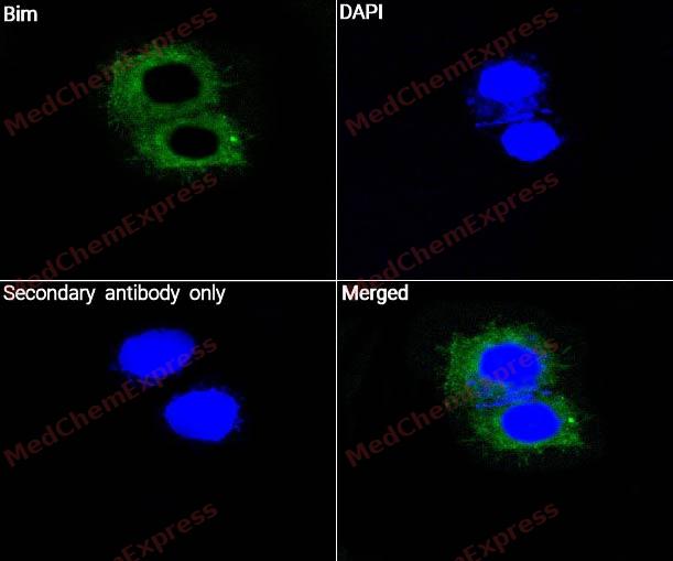 Immunocytochemistry analysis of Hela cells labeling Bim with Bim Antibody (HY-P80032) at 1/50 dilution. Cells were fixed in 4% paraformaldehyde for 15 minutes at room temperature, permeabilized with 0.1% Triton X-100 for 10 minutes at room temperature, then blocked with QuickBlock™ Blocking Buffer for Immunol Staining for 10 min at room temperature. Cells were then incubated with Bim Antibody (HY-P0032) at 1/50 dilution in QuickBlock™ Blocking Buffer for Immunol Staining at 4 ℃. Alexa Fluor® 488-conjugated AffiniPure Goat Anti-Rabbit IgG H&L(HY-P8002, Green) was used as the secondary antibody at 1/1,000 dilution. PBS instead of the primary antibody was used as the secondary antibody only control. The Nuclear counterstain was DAPI (Blue).
 -
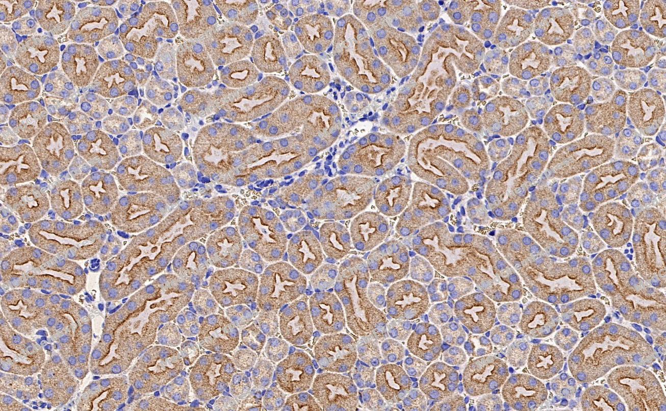 Immunohistochemical analysis of paraffin-embedded Mouse kidney tissue using Bim Antibody. The section was pre-treated using heat mediated antigen retrieval with Tris-EDTA buffer (pH 8.0) for 8 minutes. The tissues were blocked in QuickBlock for 20 minutes at room temperature, washed with ddH2O and PBS, and then probed with the primary antibody (HY-P80032, 1/100) in 4℃ overnight. The detection was performed using an HRP conjugated compact polymer system. DAB was used as the chromogen. Tissues were counterstained with hematoxylin and mounted with DPX.
 -
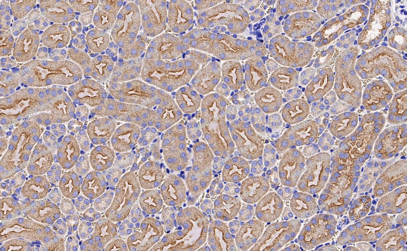 Immunohistochemical analysis of paraffin-embedded Mouse kidney tissue using Bim Antibody. The section was pre-treated using heat mediated antigen retrieval with Tris-EDTA buffer (pH 8.0) for 8 minutes. The tissues were blocked in QuickBlock for 20 minutes at room temperature, washed with ddH2O and PBS, and then probed with the primary antibody (HY-P80032, 1/100) in 4℃ overnight. The detection was performed using an HRP conjugated compact polymer system. DAB was used as the chromogen. Tissues were counterstained with hematoxylin and mounted with DPX.


Background
-
Function
Bim (BCL2L11) is a critical BH3-only protein that functions as a master regulator of intrinsic apoptosis, playing essential roles in immune homeostasis, tumor suppression, and disease pathogenesis. Alternative splicing generates three major isoforms—BimEL, BimL, and BimS—each with distinct biological activities. BimEL is predominantly involved in transcriptional regulation and cellular stress responses, while BimL plays a unique role in lysosomal acidification and autophagy progression by acting as an adaptor for dynein-mediated lysosome positioning; loss of BimL impairs autophagic degradation, leading to p62 accumulation and defective autolysosome formation[1]. In contrast, BimS and BimAD are potent activators of Bax, directly triggering mitochondrial outer membrane permeabilization and caspase activation without requiring anti-apoptotic Bcl-2 family members[6]. Structurally, Bim exists as an intrinsically unstructured protein (IUP), adopting a 'bead on a string' conformation where only the BH3 domain becomes ordered upon binding to pro-survival Bcl-2 proteins, enabling flexible interaction with multiple targets[2]. Notably, Bim exhibits a double-bolt locking mechanism by simultaneously binding both Bcl-XL and Bcl-2 via its canonical BH3 domain and a C-terminal region (residues 181-192), rendering it highly resistant to displacement by current BH3-mimetic drugs, which has significant implications for cancer therapy resistance[5]. In physiological contexts, Bim is indispensable for shaping adaptive immune responses: its absence leads to autoimmunity, while premature induction causes chronic inflammation and tumor progression[3]. Conversely, excessive Bim expression contributes to neurodegenerative disorders such as Alzheimer’s, Parkinson’s, and Huntington’s diseases, and is implicated in type I diabetes through β-cell apoptosis[3]. In cancer, Bim acts as a tumor suppressor; its downregulation or functional inactivation—via mechanisms including deletion polymorphisms in the BIM gene, proteasomal degradation (e.g., via Cul5Wsb2), or viral hijacking (e.g., ASFV CD2v-induced ERK signaling) —promotes survival and therapeutic resistance[4, 7, 8, 10]. Therapeutic strategies targeting Bim include HDAC inhibitors like SB939, which correct aberrant splicing in CML patients with BIM deletion polymorphisms, restoring TKI sensitivity[9], and splice-switching antisense oligonucleotides that rescue pro-apoptotic isoform expression[10]. These findings underscore Bim as a central node in cell death regulation and a promising target for precision medicine in oncology and degenerative diseases.
-
Subcellular Localization
Endomembrane system; Peripheral membrane protein; Mitochondrion; Mitochondrion; Mitochondrion; Mitochondrion
-
Expression
Tissue_specificity:The three main subtypes, BimEL, BimL, and BimS, are widely expressed, but exhibit tissue-specific differences. The Bim-γ subtype is most abundantly expressed in the small intestine and colon, with lower expression levels in the spleen, prostate, testis, heart, liver, and kidney.
Induction:By ER stress in a DDIT3/CHOP-dependent manner -
Subunit
Forms heterodimers with a number of antiapoptotic Bcl-2 proteins, including MCL1, BCL2, BCL2L1 isoform Bcl-X(L), BCL2A1/BFL-1, BHRF1, and BCL2L2/BCLW (PubMed:11997495, PubMed:18812174, PubMed:27013495). Does not heterodimerize with proapoptotic proteins such as BAD, BOK or BAK. Identified in a complex containing BCL2L11, DYNLL1 and BCL2L1 isoform Bcl-X(L); BH3 integrity is required for BCL2L1-binding. Interacts with YWHAZ. When phosphorylated, interacts with TRIM2; this interaction is associated with ubiquitination and degradation (PubMed:21478148). Interacts with MCL1; may sequester BCL2L11 to prevent its pro-apoptotic activity (PubMed:17389404, PubMed:27013495). Interacts with GIMAP5 (PubMed:16509771). Interacts with BCL2L10/BCL-B (PubMed:22498477, PubMed:23235460, PubMed:23563182)
-
SwissProt ID
-
Synonyms
BIM, BCL2L11, Bcl-2-like protein 11, Bcl2-L-11, Bcl2-interacting mediator of cell death
-
Research Field
Cell Biology


Documentation
-
Data Sheet (264 KB)
-
SDS (251 KB)
- English - EN (251 KB)
- Français - FR (251 KB)
- Deutsch - DE (251 KB)
- Norwegian - NO (251 KB)
- Español - ES (251 KB)
- Swedish - SV (251 KB)
- Italian - IT (251 KB)
- Korean - KR (251 KB)
- Portuguese - PT (251 KB)
-
User Guide for Antibodies (1077 KB)
References
[1]. Ruppert SM, et al. The major isoforms of Bim contribute to distinct biological activities that govern the processes of autophagy and apoptosis in interleukin-7 dependent lymphocytes. Biochim Biophys Acta. 2012 Oct;1823(10):1877-93. [Content Brief]
[2]. Hinds MG, et al. Bim, Bad and Bmf: intrinsically unstructured BH3-only proteins that undergo a localized conformational change upon binding to prosurvival Bcl-2 targets. Cell Death Differ. 2007 Jan;14(1):128-36. [Content Brief]
[3]. Sionov RV, et al. Regulation of Bim in Health and Disease. Oncotarget. 2015 Sep 15;6(27):23058-134. [Content Brief]
[4]. Vaysse-Zinkhöfer W, et al. Cul5Wsb2 uses BCL2 proteins as co-receptors to target Bim for degradation. bioRxiv [Preprint]. 2025 Oct 11:2025.08.14.670414. [Content Brief]
[6]. Costa DB, et al. BIM mediates EGFR tyrosine kinase inhibitor-induced apoptosis in lung cancers with oncogenic EGFR mutations. PLoS Med. 2007 Oct;4(10):1669-79; discussion 1680. [Content Brief]
[7]. Marani M, et al. Identification of novel isoforms of the BH3 domain protein Bim which directly activate Bax to trigger apoptosis. Mol Cell Biol. 2002 Jun;22(11):3577-89. [Content Brief]
[8]. Zeng J, et al. The ASFV CD2v protein inhibits apoptosis by inducing proteasomal degradation of BimEL via activation of the TPL2-MEK-ERK signaling pathway. J Virol. 2026 Mar 24;100(3):e0195225. [Content Brief]
[9]. Liu Q, et al. Bim escapes displacement by BH3-mimetic anti-cancer drugs by double-bolt locking both Bcl-XL and Bcl-2. Elife. 2019 Mar 12;8:e37689. [Content Brief]
[10]. Mouhamad S, et al. B cell receptor-mediated apoptosis of human lymphocytes is associated with a new regulatory pathway of Bim isoform expression. J Immunol. 2004 Feb 15;172(4):2084-91. [Content Brief]
[11]. Hermans A, et al. A 3D-Printed and Freely Available Device to Measure the Zebrafish Optokinetic Response Before and After Injury. Zebrafish. 2024 Apr;21(2):144-148. [Content Brief]
[12]. Rauzan M, et al. The HDAC inhibitor SB939 overcomes resistance to BCR-ABL kinase Inhibitors conferred by the BIM deletion polymorphism in chronic myeloid leukemia. PLoS One. 2017 Mar 16;12(3):e0174107. [Content Brief]
[13]. Huseby S, et al. Cyclic AMP induces IPC leukemia cell apoptosis via CRE-and CDK-dependent Bim transcription. Cell Death Dis. 2011 Dec 8;2(12):e237. [Content Brief]
[14]. Abrams MT, et al. Inhibition of glucocorticoid-induced apoptosis by targeting the major splice variants of BIM mRNA with small interfering RNA and short hairpin RNA. J Biol Chem. 2004 Dec 31;279(53):55809-17. [Content Brief]
[15]. Egle A, et al. Bim is a suppressor of Myc-induced mouse B cell leukemia. Proc Natl Acad Sci U S A. 2004 Apr 20;101(16):6164-9. [Content Brief]
[16]. Loo LS. Dynamics of BCL-2 family of proteins in early pancreatic progenitors and β-cells.
[17]. Ahmad JN, et al. cAMP signalling of Bordetella adenylate cyclase toxin through the SHP-1 phosphatase activates the BimEL-Bax pro-apoptotic cascade in phagocytes. Cell Microbiol. 2016 Mar;18(3):384-98. [Content Brief]
[18]. Aguiló N, et al. Bim is a crucial regulator of apoptosis induced by Mycobacterium tuberculosis. Cell Death Dis. 2014 Jul 17;5(7):e1343. [Content Brief]
[19]. Liu J, et al. Overcoming imatinib resistance conferred by the BIM deletion polymorphism in chronic myeloid leukemia with splice-switching antisense oligonucleotides. Oncotarget. 2017 Sep 6;8(44):77567-77585. [Content Brief]
[20]. Butt AJ, et al. A novel plant toxin, persin, with in vivo activity in the mammary gland, induces Bim-dependent apoptosis in human breast cancer cells. Mol Cancer Ther. 2006 Sep;5(9):2300-9. [Content Brief]

 Powered by Bioz
