



Ezrin Antibody (YA3808)
(Synonyms: CVL; CVIL; VIL2; HEL-S-105)Based on 1 Customer Validation
Ezrin Antibody (YA3808) is a Mouse-derived and non-conjugated IgG1 monoclonal antibody, targeting to Ezrin.
-
Host:
Mouse
-
Isotype:
IgG1
-
Application:
WB, ICC/IF, FC, ELISA
-
Reactivity :
Human, Mouse, Monkey
-
Formulation:
Supplied in PBS with 0.05% sodium azide
-
Conjugation:
Non-conjugated


To place orders, for customer services and technical support, please contact: MedChemExpress USA
Tel: 609-228-6898 E-mail: [email protected] [email protected]





Applications
| Application |
WB

WB: Western Blot
|
ICC/IF
ICC/IF: Immunocytochemistry/
Immunofluorescence |
FC
FC: Flow Cytometry
|
ELISA
ELISA: Enzyme Linked Immunosorbent Assay
|
|---|---|---|---|---|
| Dilution Ratio | 1:500-1:2000 | 1:100-1:400 | 1:200-1:400 | 1:10000 |


Product Details
Ezrin Antibody (YA3808) is a Mouse-derived and non-conjugated IgG1 monoclonal antibody, targeting to Ezrin.
-
Host Mouse

-
Clonality Monoclonal

-
Species ReactivityHuman, Mouse, Monkey
-
Observed Molecular WeightObserved band size: 81 kDa
Note: Due to possible protein modifications or aggregation, the molecular weight should be confirmed by actual measurement, and the predicted value is for reference only.

-
Calculated Molecular Weight Predicted band size: 69 kDa

Purified recombinant fragment of human EZR (AA: 292-464) expressed in E. Coli.
affinity purified.
Non-conjugated
Unmodified
IgG1


Product Properties
-
Appearance
Liquid
-
Formulation
Supplied in PBS with 0.05% sodium azide
-
Concentration
Batch-dependent, Please check the COA for the concentration of each lot. Check Lot Concentration
-
Storage & Stability
Stored at -20°C for 1 year. Avoid repeated freeze / thaw cycles.
-
Shipping
Shipping with blue ice.


Verification Images
-
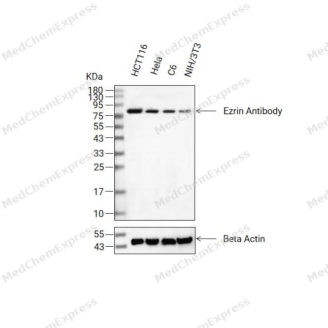 Western blot analysis of extracts from HCT116(lane 2(20ug) , Hela(lane 3(20ug) and C6(lane 4(20ug),NIH/3T3(lane 5(20ug) using Ezrin Antibody (HY-P84111) Mouse mAb. Proteins were transferred to a PVDF membrane and blocked with 5% non-fat milk in TBST for 2 hour at room temperature. The primary antibody (1/1000) and Loading control antibody (Beta Actin, HY-P83730, 1/10000) was used in 5% non-fat milk in TBST at 4°C overnight. Goat Anti-Mouse/Rabbit IgG-HRP Secondary Antibody (1/10000) was used for 1 hour at room temperature.
 -
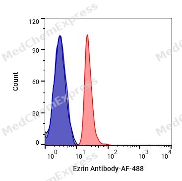 Flow Cytometry analysis of Hela cells labelling Ezrin (red) with Ezrin Antibody (anti-Ezrin) (HY-P84111). Cells were fixed with 4% paraformaldehyde and permeabilised with 90% methanol. Then cells were stained with the primary antibody at 1/200 dilution for an hour at 4℃. Goat Anti-Mouse IgG H&L (AF488) (HY-P8005) was used as the secondary antibody at 1/1,000 dilution for 30 minutes at 4℃. Mouse IgG Isotype Control (HY-P80757, blue) was used as the isotype control, cells without incubation with primary antibody were used as the unlabeled control (black).
 -
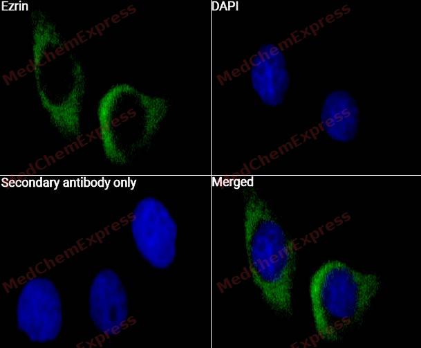 Immunocytochemistry analysis of Hela cells labeling Ezrin with Ezrin Antibody (HY-P84111) at 1/50 dilution. Cells were fixed in 4% paraformaldehyde for 15 minutes at room temperature, permeabilized with 0.1% Triton X-100 for 10 minutes at room temperature, then blocked with QuickBlock™ Blocking Buffer for Immunol Staining for 10 min at room temperature. Cells were then incubated with Ezrin Antibody (HY-P84111) at 1/50 dilution in QuickBlock™ Blocking Buffer for Immunol Staining at 4 ℃. Alexa Fluor® 488-conjugated AffiniPure Goat Anti-Mouse IgG H&L(HY-P8005, Green) was used as the secondary antibody at 1/1,000 dilution. PBS instead of the primary antibody was used as the secondary antibody only control. The Nuclear counterstain was DAPI (Blue).
 -
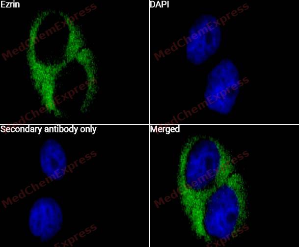 Immunocytochemistry analysis of A549 cells labeling Ezrin with Ezrin Antibody (HY-P84111) at 1/50 dilution. Cells were fixed in 4% paraformaldehyde for 15 minutes at room temperature, permeabilized with 0.1% Triton X-100 for 10 minutes at room temperature, then blocked with QuickBlock™ Blocking Buffer for Immunol Staining for 10 min at room temperature. Cells were then incubated with Ezrin Antibody (HY-P84111) at 1/50 dilution in QuickBlock™ Blocking Buffer for Immunol Staining at 4 ℃. Alexa Fluor® 488-conjugated AffiniPure Goat Anti-Mouse IgG H&L(HY-P8005, Green) was used as the secondary antibody at 1/1,000 dilution. PBS instead of the primary antibody was used as the secondary antibody only control. The Nuclear counterstain was DAPI (Blue).


Background
-
Function
Ezrin is a Probably involved in connections of major cytoskeletal structures to the plasma membrane. In epithelial cells, required for the formation of microvilli and membrane ruffles on the apical pole. Along with PLEKHG6, required for normal macropinocytosis
-
Subcellular Localization
Apical cell membrane; Peripheral membrane protein; Cytoplasmic side; Cell projection; Cell projection, microvillus membrane; Peripheral membrane protein; Cytoplasmic side; Cell projection, ruffle membrane; Peripheral membrane protein; Cytoplasmic side; Cytoplasm, cell cortex; Cytoplasm, cytoskeleton; Cell projection, microvillus
-
Expression
Tissue_specificity:It is expressed in the cerebral cortex, basal ganglia, hippocampus, pituitary gland, and optic nerve. Expression is weaker in the brainstem and diencephalon. Expression is stronger in the frontal gray matter than in white matter (protein level) . It is a component of the microvilli of intestinal epithelial cells. It is preferentially expressed in astrocytes of the hippocampus, frontal cortex, thalamus, parahippocampal cortex, amygdala, insula, and corpus callosum. Expression was not detected in neurons in most of the tissues studied. -
Subunit
Monomer. Homodimer (PubMed:27364155). Interacts with MCC, PLEKHG6, PODXL, SCYL3/PACE1, NHERF1 and TMEM8B (PubMed:12651155, PubMed:15498789, PubMed:17616675, PubMed:17881735, PubMed:19555689, PubMed:9314537).
-
SwissProt ID
-
Synonyms
CVL; CVIL; VIL2; HEL-S-105


Documentation
-
Data Sheet (234 KB)
-
SDS (252 KB)
- English - EN (252 KB)
- Français - FR (252 KB)
- Deutsch - DE (252 KB)
- Norwegian - NO (252 KB)
- Español - ES (252 KB)
- Swedish - SV (252 KB)
- Italian - IT (252 KB)
- Korean - KR (252 KB)
- Portuguese - PT (252 KB)
-
User Guide for Antibodies (1077 KB)

 Powered by Bioz
