



GluA4 Antibody (YA1889)
(Synonyms: AMPA4; GluA4; GluR4; GLUR4C; GLURD; Gria4)
Based on 1 Customer Validation
GluA4 Antibody (YA1889) is a Rabbit-derived and non-conjugated IgG monoclonal antibody, targeting to GluA4.
-
Host:
Rabbit
-
Isotype:
IgG
-
Application:
WB, FC
-
Reactivity :
Human, Mouse, Rat
-
Formulation:
Supplied in rabbit IgG in 50 mM Tris-Glycine, pH 7.4, 0.15 M NaCl, 40% glycerol, 0.01% sodium azide and 0.05% BSA.
-
Conjugation:
Non-conjugated


To place orders, for customer services and technical support, please contact: MedChemExpress USA
Tel: 609-228-6898 E-mail: [email protected] [email protected]





Applications
| Application |
WB

WB: Western Blot
|
FC
FC: Flow Cytometry
|
|---|---|---|
| Dilution Ratio | 1:1000-1:2000 | 1:50-1:100 |


Product Details
GluA4 Antibody (YA1889) is a Rabbit-derived and non-conjugated IgG monoclonal antibody, targeting to GluA4.
-
Host Rabbit

-
Clonality Recombinant,Monoclonal

-
Species ReactivityHuman, Mouse, Rat
-
Observed Molecular WeightObserved band size: 101 kDa
Note: Due to possible protein modifications or aggregation, the molecular weight should be confirmed by actual measurement, and the predicted value is for reference only.

-
Calculated Molecular Weight Predicted band size: 101 kDa

Entrez Gene: 2893 Human ; 14802 Mouse ; 29629 Rat
SwissProt: P48058 Human ; Q9Z2W8 Mouse ; P19493 Rat
OMIM: 617864 Human
A synthesized peptide derived from human GluA4 aa850-902/902.
Endogenous
Affinity Chromatography
Non-conjugated
Unmodified
IgG


Product Properties
-
Appearance
Solution
-
Formulation
Supplied in rabbit IgG in 50 mM Tris-Glycine, pH 7.4, 0.15 M NaCl, 40% glycerol, 0.01% sodium azide and 0.05% BSA.
-
Concentration
Batch-dependent, Please check the COA for the concentration of each lot. Check Lot Concentration
-
Storage & Stability
Stored at -20°C for 1 year. Avoid repeated freeze / thaw cycles.
-
Shipping
Shipping with blue ice.


Verification Images
-
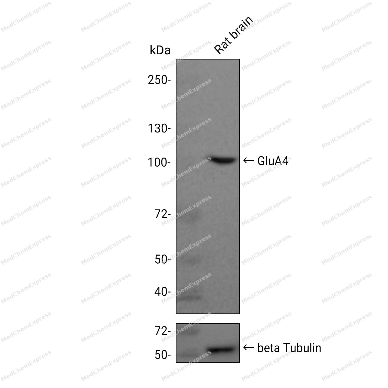 Western blot analysis was performed on extracts from Rat brain (lane 1, 15 μg) using GluA4 Rabbit mAb. Proteins were transferred to a PVDF membrane and blocked with 5% non - fat milk in TBST at 4°C overnight. The primary antibody (1:1000 dilution) and the loading control antibody (beta-Tubulin(HRP), HY-P80955A, 1:5000 dilution) was incubated in 5% non-fat milk in TBST for 1 hour at 37°C. Goat Anti - Rabbit IgG - HRP Secondary Antibody (1:20000 dilution) was then applied for 40 minutes at 37°C.
 -
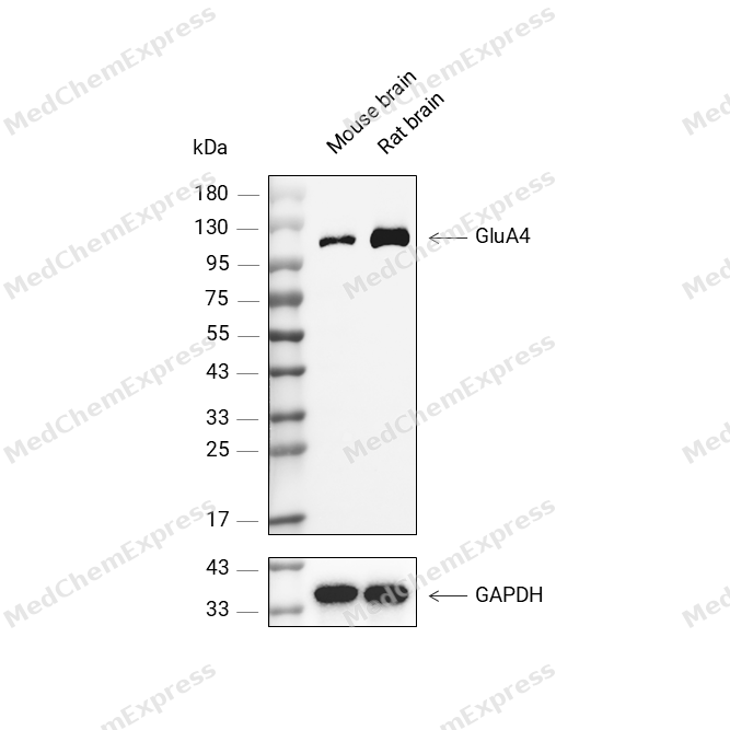 Western blot analysis was performed on protein extracts (25 μg) from Mouse brain (lane 2) and Rat brain (lane 3) using GluA4 antibody. Proteins were transferred onto a 0.45 μm PVDF membrane using the Trans-Blot® Turbo™ system for 13 min. The membrane was then blocked with 5% nonfat milk in TBST (HY-K1025) for 1 h at room temperature. The primary antibody (1:1000) and loading control antibody GAPDH Antibody (HRP) (HY-P80954A) (1:5000) were diluted in 5% nonfat milk in TBST and incubated with the membrane overnight at 4°C. After washing, the membrane of primary antibody was incubated with HRP-conjugated goat anti-rabbit/mouse IgG secondary antibody (HY-P8001/HY-P8004) (1:5000) diluted in 5% nonfat milk in TBST for 1 h at room temperature. Protein bands were visualized using an Ultra High Sensitivity ECL detection kit (HY-K1005).
 -
 Flow cytometric analysis of 1X10⁶ SH-SY5Y cells labeling GluA4 Antibody (HY-P82144, red). Cells were fixed with 4% paraformaldehyde and permeabilised with 90% methanol. Then stained with the primary antibody at 1/100 dilution for an hour at 4℃. AF488-conjugated Goat Anti-Rabbit IgG H&L (HY-P8002) was used as the secondary antibody at 1/1,000 dilution for 30 minutes at 4℃. Rabbit IgG Isotype Control (HY-P80879, blue) was used as the isotype control, cells without incubation with primary antibody were used as the unlabeled control (black).


Background
-
Function
GluA4 is an Ionotropic glutamate receptor that functions as a ligand-gated cation channel, gated by L-glutamate and glutamatergic agonists such as alpha-amino-3-hydroxy-5-methyl-4-isoxazolepropionic acid (AMPA), quisqualic acid, and kainic acid. L-glutamate acts as an excitatory neurotransmitter at many synapses in the central nervous system and plays an important role in fast excitatory synaptic transmission. Binding of the excitatory neurotransmitter L-glutamate induces a conformation change, leading to the opening of the cation channel, and thereby converts the chemical signal to an electrical impulse upon entry of monovalent and divalent cations such as sodium and calcium. The receptor then desensitizes rapidly and enters a transient inactive state, characterized by the presence of bound agonist. In the presence of CACNG8, shows resensitization which is characterized by a delayed accumulation of current flux upon continued application of L-glutamate[1].
-
Subcellular Localization
Cell membrane; Multi-pass membrane protein; Postsynaptic cell membrane; Multi-pass membrane protein; Cell projection, dendrite; Postsynaptic cell membrane; Multi-pass membrane protein
-
Isoforms & Post-Translational Modification
P48058 has 2 isomers: P48058-1: 100871 Da (predicted); P48058-2: 49146 Da (predicted).
Palmitoylated. Depalmitoylated upon L-glutamate stimulation. Cys-611 palmitoylation leads to Golgi retention and decreased cell surface expression. In contrast, Cys-837 palmitoylation does not affect cell surface expression but regulates stimulation-dependent endocytosis;Phosphorylated at Ser-862 by PRKCG; phosphorylation increases plasma membrane-associated GRI4 expression -
Subunit
Homotetramer or heterotetramer of pore-forming glutamate receptor subunits. Tetramers may be formed by the dimerization of dimers. Interacts with EPB41L1 via its C-terminus. Isoform 3 interacts with PICK1. Found in a complex with GRIA1, GRIA2, GRIA3, CNIH2, CNIH3, CACNG2, CACNG3, CACNG4, CACNG5, CACNG7 and CACNG8. Interacts with CACNG5 and PRKCG (By similarity). Found in a complex with GRIA1, GRIA2, GRIA3, DLG4, CACNG8 and CNIH2 (By similarity)
-
SwissProt ID
-
Synonyms
AMPA4; GluA4; GluR4; GLUR4C; GLURD; Gria4
-
Research Field
Neuroscience


Documentation
-
Data Sheet (234 KB)
-
SDS (251 KB)
- English - EN (251 KB)
- Français - FR (251 KB)
- Deutsch - DE (251 KB)
- Norwegian - NO (251 KB)
- Español - ES (251 KB)
- Swedish - SV (251 KB)
- Italian - IT (251 KB)
- Korean - KR (251 KB)
- Portuguese - PT (251 KB)
-
User Guide for Antibodies (1077 KB)

 Powered by Bioz
