



Islet 1 Antibody (YA1244)
(Synonyms: Insulin gene enhancer protein ISL 1 antibody; Insulin gene enhancer protein ISL-1 antibody; Insulin related protein antibody; ISL 1 antibody; ISL LIM homeobox 1 antibody; ISL1 antibody; ISL1 transcription factor LIM homeodomain antibody; ISL1 transcription factor, LIM/homeodomain (islet 1) antibody; ISL1 transcription factor, LIM/homeodomain antibody; ISL1_HUMAN antibody; Islet-1 antibody; Islet1 antibody; )
Based on 1 Customer Validation
Islet 1 Antibody (YA1244) is a Rabbit-derived and non-conjugated IgG monoclonal antibody, targeting to Islet 1.
-
Host:
Rabbit
-
Isotype:
IgG
-
Application:
WB, IHC-P, IHC-F, ICC/IF, IP, FC
-
Reactivity :
Human, Mouse, Rat, Monkey, Pig
-
Formulation:
Supplied in 1*TBS (pH7.4), 0.05% BSA, 40% Glycerol. Preservative: 0.05% Sodium Azide.
-
Conjugation:
Non-conjugated


To place orders, for customer services and technical support, please contact: MedChemExpress USA
Tel: 609-228-6898 E-mail: [email protected] [email protected]





Applications
| Application |
WB

WB: Western Blot
|
ICC/IF
ICC/IF: Immunocytochemistry/
Immunofluorescence |
IHC-P
IHC-P: Immunohistochemistry-Paraffin
|
FC
FC: Flow Cytometry
|
IHC-F
IHC-F: Immunohistochemistry-Frozen
|
IP
IP: Immunoprecipitation
|
|---|---|---|---|---|---|---|
| Dilution Ratio | 1:2000 | 1:100 | 1:1000 | 1:1000 | 1:500 | 1-2μg/sample |


Product Details
Islet 1 Antibody (YA1244) is a Rabbit-derived and non-conjugated IgG monoclonal antibody, targeting to Islet 1.
-
Host Rabbit

-
Clonality Recombinant,Monoclonal

-
Species ReactivityHuman, Mouse, Rat, Monkey, Pig
-
Observed Molecular WeightObserved band size: 45 kDa
Note: Due to possible protein modifications or aggregation, the molecular weight should be confirmed by actual measurement, and the predicted value is for reference only.

-
Calculated Molecular Weight Predicted band size: 39 kDa

Entrez Gene: 3670 Human ; 16392 Mouse ; 64444 Rat
SwissProt: P61371 Human ; P61372 Mouse ; P61374 Rat
OMIM: 600366 Human
Synthetic peptide within Human Islet 1 aa 262-305 / 349.
Endogenous
Affinity Purified
Non-conjugated
Unmodified
IgG


Product Properties
-
Appearance
Solution
-
Formulation
Supplied in 1*TBS (pH7.4), 0.05% BSA, 40% Glycerol. Preservative: 0.05% Sodium Azide.
-
Concentration
Batch-dependent, Please check the COA for the concentration of each lot. Check Lot Concentration
-
Storage & Stability
Stored at -20°C for 1 year. Avoid repeated freeze / thaw cycles.
-
Shipping
Shipping with blue ice.


Verification Images
-
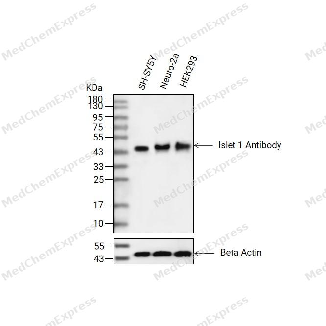 Western blot analysis of extracts from SH-SY5Y(lane 2(20ug) , Neuro-2a(lane 3(20ug) and HEK293(lane 4(20ug) using Islet 1 Antibody (HY-P81499) Rabbit mAb. Proteins were transferred to a PVDF membrane and blocked with 5% non-fat milk in TBST for 2 hour at room temperature. The primary antibody (1/1000) and Loading control antibody (Beta Actin, HY-P83730, 1/10000) was used in 5% non-fat milk in TBST at 4°C overnight. Goat Anti-Mouse/Rabbit IgG-HRP Secondary Antibody (1/10000) was used for 1 hour at room temperature.
 -
 Immunohistochemical analysis of paraffin-embedded human thyroid gland tissue using Islet 1 Antibody (HY-P81499, 1/1000). The section was pretreated using heat mediated antigen retrieval with Tris-EDTA buffer (pH 8.0) for 10 minutes. The tissues were blocked with quick block buffer for 0.5 hours at room temperature, washed with PBS and PBST, and then incubated with the primary antibody overnight at 4℃. The detection was performed using an HRP conjugated compact polymer system. DAB was used as the chromogen. Tissues were counterstained with hematoxylin and mounted with DPX.
 -
 Immunohistochemical analysis of paraffin-embedded human adrenal gland tissue using Islet 1 Antibody (HY-P81499, 1/1000). The section was pretreated using heat mediated antigen retrieval with Tris-EDTA buffer (pH 8.0) for 10 minutes. The tissues were blocked with quick block buffer for 0.5 hours at room temperature, washed with PBS and PBST, and then incubated with the primary antibody overnight at 4℃. The detection was performed using an HRP conjugated compact polymer system. DAB was used as the chromogen. Tissues were counterstained with hematoxylin and mounted with DPX.
 -
 Immunohistochemical analysis of paraffin-embedded human pancreas tissue using Islet 1 Antibody (HY-P81499, 1/1000). The section was pretreated using heat mediated antigen retrieval with Tris-EDTA buffer (pH 8.0) for 10 minutes. The tissues were blocked with quick block buffer for 0.5 hours at room temperature, washed with PBS and PBST, and then incubated with the primary antibody overnight at 4℃. The detection was performed using an HRP conjugated compact polymer system. DAB was used as the chromogen. Tissues were counterstained with hematoxylin and mounted with DPX.
 -
 Immunohistochemical analysis of paraffin-embedded mouse pancreas tissue using Islet 1 Antibody (HY-P81499, 1/1000). The section was pretreated using heat mediated antigen retrieval with Tris-EDTA buffer (pH 8.0) for 10 minutes. The tissues were blocked with quick block buffer for 0.5 hours at room temperature, washed with PBS and PBST, and then incubated with the primary antibody overnight at 4℃. The detection was performed using an HRP conjugated compact polymer system. DAB was used as the chromogen. Tissues were counterstained with hematoxylin and mounted with DPX.
 -
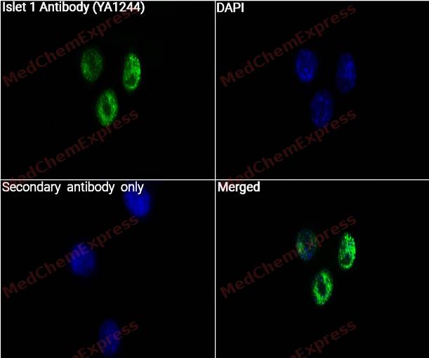 Immunocytochemistry analysis of SH-SY5Y cells labeling Islet 1 with Islet 1 Antibody (YA1244) (HY-P81499) at 1/50 dilution. Cells were fixed in 4% paraformaldehyde for 15 minutes at room temperature, permeabilized with 0.1% Triton X-100 for 10 minutes at room temperature, then blocked with QuickBlock™ Blocking Buffer for Immunol Staining for 10 min at room temperature. Cells were then incubated with Islet 1 Antibody (YA1244) ((HY-P81499) at 1/50 dilution in QuickBlock™ Blocking Buffer for Immunol Staining at 4 ℃. Alexa Fluor® 488-conjugated AffiniPure Goat Anti-Rabbit IgG H&L(HY-P8002, Green) was used as the secondary antibody at 1/1,000 dilution. PBS instead of the primary antibody was used as the secondary antibody only control. The Nuclear counterstain was DAPI (Blue).
 -
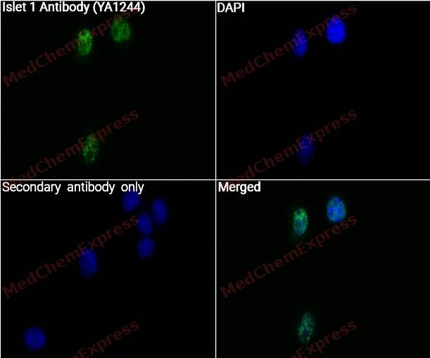 Immunocytochemistry analysis of SH-SY5Y cells labeling Islet 1 with Islet 1 Antibody (YA1244) (HY-P81499) at 1/100 dilution. Cells were fixed in 4% paraformaldehyde for 15 minutes at room temperature, permeabilized with 0.1% Triton X-100 for 10 minutes at room temperature, then blocked with QuickBlock™ Blocking Buffer for Immunol Staining for 10 min at room temperature. Cells were then incubated with Islet 1 Antibody (YA1244) ((HY-P81499) at 1/100 dilution in QuickBlock™ Blocking Buffer for Immunol Staining at 4 ℃. Alexa Fluor® 488-conjugated AffiniPure Goat Anti-Rabbit IgG H&L(HY-P8002, Green) was used as the secondary antibody at 1/1,000 dilution. PBS instead of the primary antibody was used as the secondary antibody only control. The Nuclear counterstain was DAPI (Blue).


Background
-
Function
Islet 1 is a DNA-binding transcriptional activator. Recognizes and binds to the consensus octamer binding site 5'-ATAATTAA-3' in promoter of target genes. Plays a fundamental role in the gene regulatory network essential for retinal ganglion cell (RGC) differentiation. Cooperates with the transcription factor POU4F2 to achieve maximal levels of expression of RGC target genes and RGC fate specification in the developing retina. Involved in the specification of motor neurons in cooperation with LHX3 and LDB1. Binds to insulin gene enhancer sequences. Essential for heart development. Marker of one progenitor cell population that give rise to the outflow tract, right ventricle, a subset of left ventricular cells, and a large number of atrial cells as well, its function is required for these progenitors to contribute to the heart. Controls the expression of FGF and BMP growth factors in this cell population and is required for proliferation and survival of cells within pharyngeal foregut endoderm and adjacent splanchnic mesoderm as well as for migration of cardiac progenitors into the heart
-
Subcellular Localization
Nucleus
-
Expression
Tissue_specificity:This gene is expressed in some neurons of the adrenal medulla and dorsal root ganglion, the nuclear layer and ganglion cell layer of the retina, the pineal gland, and certain areas of the brain.
Positive sample: SH-SY5Y cell lysate, Neuro-2a cell lysate, PC-12 cell lysate, SH-SY5Y, Neuro-2a, human pancreas tissue, mouse embryo tissue, rat pancreas tissue. -
Subunit
At neuronal promoters, displaces LDB1 from LHX3 LIM domain to form a ternary complex in which ISL1 contacts both LHX3 and LDB1; allosteric structural changes in the DNA binding domain of LHX3, induced by the ISL1:LHX3 interaction, may explain differences in sequence specificity of the different complexes. Interacts with LHX3. Interacts (via C-terminus) with POU4F2 (via C-terminus) isoform 1. Interacts with POU3F2. Interacts with POU4F3. Interacts (via N-terminal domain) with MLIP; the interaction represses ISL1 transactivator activity. Interacts with GCN5/KAT2A. Interactions of ISL1 with MLIP1 or KAT2A may be mutually exclusive (By similarity)
-
SwissProt ID
-
Synonyms
Insulin gene enhancer protein ISL 1 antibody; Insulin gene enhancer protein ISL-1 antibody; Insulin related protein antibody; ISL 1 antibody; ISL LIM homeobox 1 antibody; ISL1 antibody; ISL1 transcription factor LIM homeodomain antibody; ISL1 transcription factor, LIM/homeodomain (islet 1) antibody; ISL1 transcription factor, LIM/homeodomain antibody; ISL1_HUMAN antibody; Islet-1 antibody; Islet1 antibody;
-
Research Field
Neuroscience


Documentation
-
Data Sheet (234 KB)
-
SDS (254 KB)
- English - EN (254 KB)
- Français - FR (254 KB)
- Deutsch - DE (254 KB)
- Norwegian - NO (254 KB)
- Español - ES (254 KB)
- Swedish - SV (254 KB)
- Italian - IT (254 KB)
- Korean - KR (254 KB)
- Portuguese - PT (254 KB)
-
User Guide for Antibodies (1077 KB)

 Powered by Bioz
